Shoulder pain is a common symptom for which people seek medical attention. Pain can be associated with problems in the shoulder joint and surrounding tissues, or it can be referred and the source of pain can be localized in other structures, such as the neck, diaphragm or heart. When assessing shoulder pain, it is important to identify any “red flags” that indicate more in-depth evaluation is required. Treatment of pain in the shoulder (shoulder joint) depends on its genesis, and the treatment arsenal includes various methods of influencing the pain syndrome. Blocking pain in the shoulder joint with medication injections can reduce both pain and inflammation.
Risk factors
- Physical factors associated with occupation, including repetitive motion and vibration from machines.
- Work-related psychosocial factors may also be risk factors for shoulder pain, including stress, social support, and job satisfaction.
- Athletes who have overhead arm movements or high-impact contact sports are prone to shoulder pain.
- Occupations in which people are particularly susceptible to shoulder pain include: cashiers, clothing manufacturers, masons/construction workers, air tool operators, welders, meat/food workers, hairdressers, plasterers, painters and decorators, assembly and production line workers and workers who use keyboards for long periods of time - for example, IT. specialists, secretaries.
Cause of pain syndrome
The above diseases are the cause of pain syndrome. However, the nature of pain may have deeper roots. Pain that occurs in the shoulder joint can bring pain and suffering into the daily life of people whose musculoskeletal system has undergone inflammatory processes. During the inflammatory process, layers of connective tissue are formed in the joint capsule, which are capable of sticking together. This is what causes sharp pain, and movements in this place become limited. It becomes problematic to make circular movements, raise the arm and even move it away from the body. Carrying out a blockade procedure for a sore shoulder joint will help restore mobility and get rid of pain.
Causes of shoulder pain
Patients seeking medical attention with shoulder pain often have a combination of various pathological conditions.
Internal shoulder pain:
- Rotator cuff injury
- Rotator cuff tears.
- “Subacromial pain,” which can be caused by impingement if the humeral head does not descend enough to slide under the acromion at arm height. This pain may be due to conditions such as subacromial bursitis, tendinitis or tendinopathy.
- Calcific tendinitis.
- Glenohumeral pathologies: adhesive capsulitis (“frozen shoulder”), arthritis.
- Acromioclavicular disorders.
- Biceps tendinitis.
- Infection (rare).
- Shoulder instability - associated with hypermobility, including subluxation or dislocation.
External shoulder pain:
- Referred pain: neck pain, myocardial ischemia, diaphragmatic pain (eg, gallbladder disease, subphrenic abscess).
- Polymyalgia rheumatica.
- Oncology: apical lung cancer, metastases.
The four most common causes of shoulder pain and disability are rotator cuff injuries, glenohumeral pathologies, acromioclavicular joint disease, and referred neck pain.
How is the procedure performed?
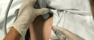
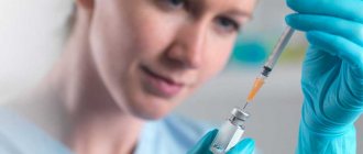
The manipulation is simple and safe. It is carried out in a medical center. Venous blood is taken from the patient, approximately 20–1000 ml. The test tube with the biomaterial is placed in a centrifuge for 20 minutes. As a result of centrifugation, the composition in the test tube is stratified.
The result is an enriched and purified substance. It is drawn into a syringe and injected directly into the inflamed area. All actions are carried out under sterile conditions, which eliminates infection. The injected substance is also sterile.
Stages of preparing PRP substance from the patient’s blood
The patient will be asked to move - bend, straighten the limb. This is done to ensure that the medicine is evenly distributed in the cavity. Then, the patient is sent home. Such introductions will be needed from 3 to 7.
Symptoms/red flags
- History of malignancy or symptoms/signs consistent with neoplasia, such as weight loss, deformity, swelling or swelling, abdominal discomfort/swelling.
- Skin erythema may indicate a tumor or infection.
- Symptoms/signs of systemic disease that may indicate polymyalgia rheumatica/giant cell arteritis.
- Fever may indicate a malignant disease or infection.
- A history of trauma or recent convulsion/electrical injury may indicate unreduced dislocation.
- A change in shoulder contour with loss of range of rotation suggests dislocation.
- The presence of significant sensory or motor deficits suggests neurological involvement.
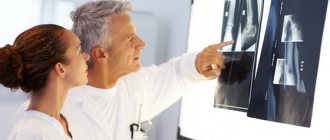
Diagnostics
Laboratory research
- Blood tests are necessary to diagnose systemic and oncological processes
- Ultrasonography is the preferred method for imaging the shoulder.
- X-ray allows you to determine bone damage, shoulder dislocation, and arthrosis of the shoulder joint.
- MRI can determine the presence of damage to soft tissue structures or instability of the shoulder. In addition, if the nature of the pain is referred from the neck, MRI of the cervical spine can help in diagnosing the cause of the pain.
- EMNG and EMG - these neurophysiology methods are necessary in cases where there are signs of damage to nerves or muscle function.
Treatment
Depending on the problem that is causing your shoulder pain, medical intervention may be required, but there are also home remedies and treatments that can be effective.
- Eliminate loads. Sometimes all that is needed to reduce pain is to eliminate stress and allow the body to heal on its own. This is especially true if the pain is caused by repetitive motion or other muscle injury. Unloading can be combined with the use of heat or cold locally.
- Maintain correct posture. Poor posture can lead to serious shoulder pain. In addition, it is advisable to use an orthopedic pillow and an orthopedic mattress.
- Practice scapular retraction exercises. The right exercises will help the patient overcome discomfort and get on the path to healing. Many exercises stretch the area, which promotes flexibility, and most can be done anywhere.
- Massage can provide significant relief from any problems associated with muscle pain, including shoulder pain. Many people report rapid relief after several massage sessions.
- Medicines. For minor pain, over-the-counter medications may be used. For more intense pain, strong medications such as steroids may be used.
- Treatment of major diseases. If shoulder blade pain is caused by arthritis, herpes zoster, or another condition, your doctor may prescribe medications, exercises, etc. If the problem is nerve compression or a fracture, more serious medical intervention may be required.
Types of joint injections
Three types are used:
- paraarticular;
- periarticular;
- intra-articular.
Paraarticular
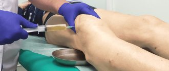
The method involves injecting medication into the tissue of the damaged joint. At the moment, this is one of the most effective ways to help the patient relieve pain and relieve inflammation. In this case, intradermal and subcutaneous administration is performed using thin and short needles. The injections are practically painless, and the procedure itself is completed quickly.
Periarticular
blockades are indispensable for osteoarthritis affecting a large area, arthritis and periarthrosis. The drug is injected into the tendon and muscle tissue.
Intra-articular
The method involves introducing a substance into the joint tissue.
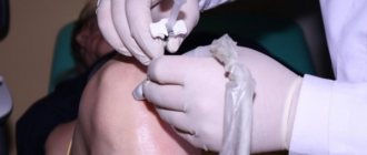
Block pain in the shoulder joint
Along with other conservative treatment methods (physiotherapy, massage, exercise therapy), blockades are widely used to relieve shoulder pain.
As a rule, the effect after the blockade occurs within a few days, if successful, it can last for months, but in some cases the symptoms decrease only within 2-3 weeks. The lack of effect may be due to the fact that the doctor did not reach the necessary area or an incorrect diagnosis.
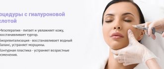
INTRA-ARTICULAR INJECTIONS OF HYALURONIC ACID
The class of chondroreparants includes synthetic joint fluid substitutes based on hyaluronic acid. Injections of chondroreparants into the affected joint restore cartilage tissue and restore mobility and elasticity to the joints. The recovery process starts and the pain subsides. In our medical center, injections of chondrorepairs Hyalrepair, Noltrexin, Ripart long are performed. The required drug is selected by the attending physician in accordance with the clinical picture of the disease. We can also perform an intra-articular injection of the drug brought by the patient; please inquire about this possibility by phone. Treatment with injections of hyaluronic acid into the joint is on average carried out in a course of 2-3 injections with an interval of 7-10 days.
Chondroreparant Hyalrepair® (Hialripayer)
The medical product HYALRIPAYER-10 CHONDROREPARANT® contains a unique form of modified (stabilized) highly purified hyaluronic acid obtained by bacterial fermentation, with the addition of a copolymer of hyaluronic acid with sodium ascorbyl phosphate, L-glutathione, L-cysteine. The structural modification of hyaluronic acid (polymer cross-linking) determines a prolonged reconstructive effect on cartilaginous, ligamentous and connective tissue structures of the musculoskeletal system damaged due to injuries or degenerative processes. The combination of hyaluronic acid salts and an antioxidant complex triggers the processes of blocking free radical forms of oxygen and the formation of its own hyaluronic acid, replenishing the connective tissue matrix.
HYALRIPAYER-10 CHONDROREPARANT® allows you to restore the effective concentration of hyaluronic acid in the matrix, stimulating fibroblasts and chondrocytes. Intermediate products of hyaluronic acid degradation have angiogenic, immunostimulating and anti-inflammatory effects. When administered, the catabolic products of HYALRIPAYER-10 CHONDROREPARANT® and the released amino acids, together with ascorbyl phosphate, create a physiologically favorable environment to protect the cell wall and intercellular structures from peroxide destruction and trigger the synthesis of the main components of the intercellular matrix of cartilage and connective tissue structures, additionally providing an analgesic effect.
Contraindications and precautions when performing injections into the shoulder joint
Contraindications
- Infection: septic arthritis, adjacent osteomyelitis, periarticular cellulitis, severe dermatitis, soft tissue infection, sepsis, bacteremia.
- Increased risk of joint infection - for example, immunosuppression, broken skin over the injection site.
- Trauma: hemarthrosis, fracture.
- Very unstable joint
- Joint surgery scheduled during the day
- Joint prosthesis
- History of allergies to injectable drugs.
- Poorly controlled diabetes mellitus
- Uncontrolled bleeding disorder or coagulopathy
Cautions
- Diabetic patients: Monitor blood glucose levels closely for two weeks after injection.
- Poor clotting
- Immunosuppression.
- Psychogenic pain, severe anxiety.
- Neurogenic disease.
- Active infections (eg, tuberculosis).
- Hypothyroidism.
Advantages of plasma therapy in orthopedics
The new technique has gained popularity due to its special properties:
- no allergic reaction;
- guarantee of sterility of the biomaterial, exclusion of foreign infections;
- the drug acts only at the injection site, without entering the systemic bloodstream;
- recovery occurs 2-3 times faster than with drug treatment;
- minimum complications;
- no side effects.
PRP therapy for joints differs significantly from the effects of non-steroidal, hormonal drugs. Glucocorticoids, which effectively relieve inflammation, have a destructive effect on cartilage and the body as a whole. PRP therapy restores their structure without affecting the internal organs.
Plasma therapy and transplantation
Will the new technique help cancel or postpone the operation? What do doctors say about this?
The operation to implant a prosthesis is far from a harmless procedure. Like any surgical intervention, it requires preparation and long postoperative rehabilitation. The process does not always go without complications and repeated interventions.
In addition, replacing a joint will not always completely solve the problem. Surgeons are usually silent about this
— after replacing one hip joint, due to the difference in load, there is strong pressure on the second joint. As a rule, within a year or two, the patient will need to replace the second joint.
If the disease is not advanced, a course of plasma therapy along with other methods will eliminate the need for surgery. In advanced severe cases, injections will help postpone transplantation for a certain period. Whether a replacement is needed or not in such a situation is decided by the attending physician.
Any joint disease has serious complications, so you should not delay treatment.
Read more about joint treatment in our clinic
For people who have contraindications to surgery, PRP joint therapy provides a chance to reduce symptoms for a long period. The safety of the method has been proven - according to WHO, more than 20 thousand patients are treated every year around the world.
Side effects
This is often the result of poor technique, too much dosage, too little dosage, or improper mixing and dissolution of medications.
Local effects
- Infection (1/10000)
- Post-injection increase in pain (2-5%)
- Skin discoloration that goes away over time
- Atrophy of subcutaneous fat tissue
- Bleeding (rare).
- Soft tissue calcification with repeated injections into one area of the skin
- Joint damage; Cartilage damage and osteoporosis: It is advisable to avoid repeated injections (no more than four injections at each site per year).
- Tendon atrophy and rupture (<1%): Avoid direct tendon injection.
- Pericapsular calcification (>40%).
- Avascular necrosis.
Intra-articular drug Noltrexsin (Noltreksin)
Noltrexsin acts on two levels: physical - it protects articular cartilage from destruction, and biochemical, blocking substances that contribute to the destruction of cartilage.
Noltrexsin restores the viscosity of the synovial fluid in the joint, stabilizes the condition of the articular surfaces by expanding the joint space and protecting the surfaces of hyaline cartilage. Mechanical stabilization entails reducing or stopping the processes of cartilage damage.
The drug actively binds substances in the structure of its own polymer macromolecule that contribute to the destruction of cartilage, and silver ions suppress the development of infection in the joint. Noltrexsin can act as an endoprosthesis for a long time. When the drug interacts with synovial fluid, the aqueous composition of Noltrexsin is completely replaced by tissue fluid. As a result, a new compound is formed in the joint cavity - a three-dimensional biopolymer.
The physiologically active centers of the Noltrexsin molecule adsorb fragments of synovial fluid, restoring the original biochemical environment of the joint and its normal functioning.
Water, which makes up more than 95% of Noltrexsin, plays the role of a natural environment for replacement reactions that constantly occur in the joint, during which the intra-articular fluid necessary for the normal functioning of the joint is adjusted.
Indications for injection
- Injections into the glenohumeral joint are indicated for diseases such as osteoarthritis, adhesive capsulitis, RA, and rotator cuff injuries.
- Acromioclavicular joint blocks are recommended for osteoarthritis (a common cause of shoulder pain in people over 50) and distal clavicular osteolysis.
- Subacromial injections may be prescribed for conditions such as adhesive capsulitis, subacromial bursitis (can occur with gout, reactive arthritis, trauma or RA), impingement syndrome, and rotator cuff tendonitis. Subacromial corticosteroid injections can reduce subacromial pain for up to nine months. They are also likely to be more effective than NSAID medications.
- Bicipital groove: bicipital tendonitis.
The essence of the method
The name PRP is an abbreviation for Platelet rich plasma (blood plasma enriched with platelets). We are talking about a substance with a high platelet content. But where do they get this composition and how do they use it? This will be discussed further below.
Some historical facts
The method is essentially not new. Active searches were conducted back in the 20th century. The first sources on the topic of using platelet-rich plasma to eliminate pathologies of the musculoskeletal system appeared in the 70s of the last century.
More recent articles by American researchers were published in 1997-1998. They talked about the use of the enriched composition in maxillofacial surgery. The new technique quickly moved to Europe. In the 21st century, it is used in almost all areas of medicine, as well as cosmetology.
What is PRP therapy
Treatment of joints with the PRP method is based on the functional properties of human blood platelets. The elements are known as clotting factors. They promote the formation of a blood clot when a blood vessel is damaged. Thanks to them, wounds stop bleeding.
But the main feature is that these blood elements help the regeneration of connective tissue cells. This fact became the reason for their use in the treatment of joint diseases. They stimulate fibroblast cells to actively synthesize collagen and elastogen. These substances are part of the bone and cartilage system, skin, and ligaments.
Enriched blood plasma contains up to 1000–2500×1009/l. platelets. In their usual form they are no more than 180–320×109/l. Venous blood is used for manipulation. Its separation and enrichment is achieved using a centrifuge.
Blood components have different weights and sizes. As a result of centrifugation, the biomaterial in the test tube is stratified. The heavier, red elements settle to the bottom, and the enriched plasma forms the top layer. The result is a concentrated composition.
Blood plasma prepared for the procedure
Growth factors
These blood cells contain bioactive substances - growth factors. They have a protein structure. There are several of them, which explains the multifaceted effect of the medicine.
- insulin-like IGF
– regulates cell growth, migration and development; - platelet PDGF
– ensures cellular regeneration (survival); - transforming TGF
– inhibits tissue necrosis, suppresses inflammation, stimulates collagen production; - endothelial f.
R. blood vessels VEGF – affects vascular permeability, increases blood supply to organs; - epidermal EGF
– triggers self-healing at the cellular level. Stimulates cell division and renewal; - f.r.
fibroblasts FGF – increases the concentration of fibroblasts, accelerates the development of blood vessels.
The destruction of cartilage is difficult to stop. They do not contain blood vessels through which nutrients are delivered. Everything you need comes from the synovial fluid that fills the joint cavity. Therefore, administering the medicine directly to the diseased organ is the most effective method of treatment.