Techniques
- Bone scintigraphy in the “Whole Body” mode allows simultaneous visualization of all parts of the skeleton and is used for early detection of focal changes in the bones.
- 3-phase bone scintigraphy makes it possible to assess blood flow in the affected areas and to most accurately assess the extent of bone damage.
- SPECT, SPECT/CT helps to identify and more accurately localize lesions less than 1.5 cm in size, and to carry out differential diagnosis of benign and malignant lesions of the osteoarticular system.
The use of various methods of radiation diagnostics for injuries of the musculoskeletal system
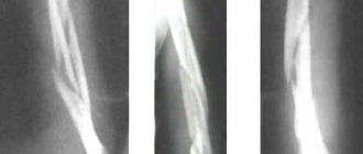
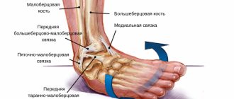
Technological progress in medical imaging has offered traumatologists a wide range of methods, different both in nature and in their information content. Therefore, the task of choosing the optimal examination algorithm fell on the shoulders of the radiologist. The initial evaluation of musculoskeletal lesions should begin with conventional radiography. MSCT allows you to obtain a transverse image of bones and joints, differentiate bone and soft tissue structures, and also identify minor differences in the density of normal and pathologically altered tissues. The possibility of multiplanar and three-dimensional image reconstructions has found wide practical application among traumatologists. Magnetic resonance imaging is perhaps the only method for a comprehensive assessment of the musculoskeletal system. The MRI technique largely depends on the type of tomograph, the strength of its magnetic field, design features, set of coils, etc. Ultrasound diagnosis of damage to the tendon-ligamentous apparatus has been widely developed with the use of high-frequency linear sensors of more than 7.5-17.5 MHz. An important advantage of ultrasound is direct contact with the patient, which allows focusing on the areas of greatest pain. Finally, ultrasound is the method of choice for kinematic studies to identify and evaluate loose intra-articular bodies, muscle, tendon and ligament tears, tendon dislocations, etc. Among radionuclide methods, the most widely used is scintigraphy of skeletal bones using technetium-99m. The method is highly sensitive in the diagnosis of osteoblastic processes. The main principle of complex diagnosis of musculoskeletal system injuries is the syndromic approach. Despite the technical differences in radiological diagnostic methods, radiologists use similar semiotic signs of damage to bones, muscles, their tendons and ligaments. Damage to bone tissue. Damage to skeletal bones includes a complex of various pathomorphological changes associated with both a violation of the integrity of the spongy and cortical bone, and with injury to the bone marrow and periosteum. Studies of patients with acute trauma show that bone injuries are detected in more than 80% of cases. Considering that fractures make up only 10–15% of the structure of skeletal injuries, about 70% of bone pathology remains outside the scope of traditional radiography. The most common radiological finding is bone bruises. They manifest themselves as a hidden minor violation of the integrity of bone trabeculae with hemorrhage, hyperemia and edema of the bone marrow. However, X-ray studies, including CT, cannot detect bone contusion. The diagnosis of a bone bruise is verified by dynamic MRI, the semiotic signs of which disappear 3-4 months after the injury. Only 5–10% of bruises are the only finding of radiological diagnostics. Basically, contusion aggravates the course of other injuries to the bones and tendon-ligamentous apparatus. Another X-ray negative nosology is “subtendinous edema” of the bone marrow, caused by pathology of the adjacent tendon. The MR semiotics of subtendinous edema is similar to the signs of a bone contusion. However, the swelling is localized under the damaged tendon. According to the mechanism of occurrence, it is not caused by ligament rupture or direct bone injury. The timing of detection of bone marrow edema is determined by the duration of the acute phase of tendon damage. X-ray and CT semiotics of bone fractures are identical. MP and 3D reconstructions of computed tomographic images make it possible to study fractures in any projection and volume, which increases the information content of the method. Spiral CT is the most informative in diagnosing fractures. It allows you to diagnose fractures without displacement of fragments, avulsin fractures and additional fracture lines not identified by radiography. The use of computed tomography solves dozens of practical diagnostic issues that determine the patient’s treatment strategy and the prognosis of the disease. They relate to the degree and nature of damage to the articular surfaces, the possibility of prompt access to damaged joints, etc. With an accuracy of 1 mm and 1 degree, the method determines the mutual displacement and rotation of bones and fragments. CT data on the number, shape, size and displacement of fragments are used in choosing osteosynthesis tactics. On MRI, the fracture line is delimited by uneven edges of bone fragments that are hypointense in all VIPs, surrounded by an area of edema and hemorrhage with corresponding semiotic signs. Magnetic resonance imaging additionally makes it possible to diagnose fractures without displacement of the fragment, damage to synchondrosis, and damage to sesamoid bones. One of the types of bone fractures are “hidden” fractures that are not initially detected by x-ray methods. Morphologically, a hidden fracture is characterized by a violation of the integrity of the cancellous bone and minor damage to its cortical layer. The fracture is visualized only with MRI in all VIPs in the form of a linear structure of irregular shape, reduced intensity, surrounded by an area of edema. X-ray and spiral CT of patients with hidden fractures 1.5–2 months after injury can detect minor cloud-like osteosclerotic changes along the fracture line. The main advantage of MRI is the ability to detect violations of the integrity of articular cartilage tissue. The trochlea of the talus and the medial condyle of the femur are the most typical sites of occurrence of osteochondritis dissecans. The classification of osteochondritis dissecans was proposed by AL Berdt and M. Harty (1959), based on the amount of damage to the articular cartilage and the degree of freedom of the osteochondral fragment. Damage to soft tissues. The proportion of damage to soft tissues of the musculoskeletal system is up to 20% for cartilage, up to 25% for tendons, up to 40% for fibrocartilaginous structures, and up to 90% for ligaments. In 60% of cases of tendon injuries, tenosynovitis syndrome is diagnosed. In a quarter of cases, it accompanies tendinosis and ruptures of the own tendons. CT, MRI and ultrasound signs of tenosynovitis include an increase in tendon diameter. Fluid collection on axial scans appears as an eccentric halo, hypodense on CT, anechoic on ultrasound, and hyperintense on MRI on T2-VIP and STIR tomograms. Sagittal and coronal sections demonstrate the extent of fluid along the tendon. Perifocal swelling of the soft tissues leads to heterogeneous compaction of fatty tissue and a decrease in the clarity of the tendon contours. Traumatic tendinosis is diagnosed in approximately 15% of cases. In the absence of tenosynovitis, according to CT data, tendinosis is determined in the form of local thickening of the tendon, with a long-term process with isolated calcifications. On MRI scans, tendinosis is visualized in T2-VIP. There is local heterogeneity of the tendon structure with small foci of increased signal against the background of slight thickening of the tendon. The method of choice for assessing tendons is ultrasound. The echographic semiotics of tendinosis includes a violation of the fibrillar pattern of tendons with hypoechoic foci and isolated calcifications. The share of ruptures in the structure of tendon injuries is about 12%. There are three types of breaks. Type I corresponds to a partial tear. According to MRI data, pronounced heterogeneity of the structure and increased signal are determined in all VIPs. Ultrasound reveals violations of the integrity of the fibrils, an increase in the distance between them, and uneven contours of the tendon. CT scan allowed only to identify tendon thickening. In a type II partial tear, thinning of the tendon occurs. CT, MRI and ultrasound detect local thickening of the tendon proximal and distal to the affected segment, which is reduced in diameter. T2-VIP MR tomograms show a pronounced heterogeneity of the tendon structure with areas of increased signal. Ultrasound additionally makes it possible to detect fibrillary pattern disturbances. Type III lesions are complete tendon ruptures and appear as absent tendons on axial scans. The rupture site is filled with adipose tissue and hematoma. Ultrasound with kinematic tests allows you to accurately localize the ends of the tendon and determine the true size of the gap between them. When studying ligamentous injuries, most radiologists use pathological classification. Stage I is characterized by minor damage with the presence of microcracks. In stage II lesions, partial macrotears occur. Complete ligament rupture occurred in stage III. In the vast majority (up to 85%), ligament ruptures are multiple. The capabilities of radiography in diagnosing damage to the ligamentous apparatus are limited by identifying such indirect signs as a violation of the comparison of articular surfaces and displacement of fragments. The information content of CT is limited by partial visualization of the largest ligaments, detection of avulsive fractures, edema and hemorrhages in the periarticular tissues. MRI allows visualization of all ligaments whose damage is of clinical significance. Direct semiotic signs of ligament ruptures are: complete break of fibers, their wavy appearance, thinning or thickening of the ligament and blurred contours. Indirect signs are considered local swelling of the bone marrow at the attachment points of the ligaments, and tenosynovitis of the adjacent tendon. Ultrasound is inferior to MRI in diagnosing ligament injuries. Normally, only the largest ligaments are visualized. The semiotics of ligament damage included: thickening of the ligament, decreased echogenicity, deformation and break of the structure. In the diagnosis of damage to fibrocartilaginous structures such as the menisci of the knee joints, the cartilaginous lip of the shoulder and hip joints, MRI and ultrasound methods are most widely used. Manifestations of damage to the menisci and labrum on MRI are an increase in the intensity of the MR signal in the structure due to fiber disintegration, impregnation with synovial fluid, possible fragmentation, or complete absence at the anatomical site. In the first degree, there is a marginal defect that does not communicate with the articular surface. The second is characterized by the presence of a linear defect communicating with the articular surface, but without elements of fragmentation. Ultimately, fragmentation and dislocation of parts of the meniscus or lip are visualized. The methods of choice for assessing muscle tissue damage are MRI and ultrasound. Most often, damage to muscle fibers occurs in the anterior and posterior thigh muscle groups, as well as in the muscles of the shoulder girdle. There are 3 degrees of damage. The first degree is characterized by disintegration of myofibrillar structures, their imbibition with blood and intentional fluid, without signs of separation of muscle fibers from the tendon bundle. In the second degree of injury, small intermyofibrillar hematomas are additionally detected, as well as hematomas at the site of separation of part of the fibers from the tendon bundle, without signs of damage to the tendon fibers. The third degree is characterized by the presence of a massive hematoma at the site of separation of muscle fibers from the tendon bundle. Interfascial leaks of liquid contents, disruption of the integrity of muscle fibers at the site of attachment, and their thickening proximal or distal to the rupture site are visualized. Thus, trauma to the musculoskeletal system in most cases requires complex radiation diagnostics. Each of the segments of the musculoskeletal system has unique features of its topographic and anatomical structure, without taking into account which it is impossible to correctly interpret the data obtained. When studying injuries to bones and tendon-ligamentous apparatus, a unified syndromic approach should be used, allowing the traumatologist to adequately and comprehensively perceive the results of radiological diagnostics.
Bring with you
- Passport.
- Referral for research.
- Discharge summaries, results of ultrasound, scintigraphy, CT and MRI along with discs (preferably for the last three months), as well as other data that allows you to study the history of your disease in more detail.
- The results of our research along with illustrations (if you visit us again).
- Painkillers (if there is severe pain).
- If you are undergoing a study at the expense of the compulsory health insurance fund (CHI), you must additionally provide a referral in form 057/u-04, a medical policy and SNILS (more details in the “Compulsory Medical Insurance” section).


