General information
Arthrogryposis is a heterogeneous systemic group of congenital diseases manifested by pathological changes in the musculoskeletal system in the form of contractures (stiffness), deformities of the limbs, underdevelopment of joints and muscles, a decrease in its tone, as well as proliferation of connective tissues - fibrosis .
The disease is non-progressive. The disease was first described by Otto in 1841, while in 1923 Stern proposed the name “arthrogryposis,” which comes from two ancient Greek words: arthron, which means joint, and gryposis, which means curvature.
According to statistics, arthrogryposis occurs in approximately 1-3% of the population with musculoskeletal anomalies and orthopedic diseases, usually in one out of 3 thousand newborn babies. The most common form affects the joints of the wrists, shoulders, elbows, hips, knees, and their muscles. In its most severe forms, the pathology affects almost all joints, including the jaw.
Pathogenesis
With arthrogryposis, severe muscle aplasia develops due to severe lesions of muscle tissue such as fibrous dystrophy, and all joints of the limbs are also affected. Atrophic or hypotrophic changes in muscles are observed from birth. Presumably, they are associated with the teratogenic effects of factors that disrupt the formation (histogenesis) of muscle fibers in the first semester of gestation, which causes future pathological changes in them. In this case, the normal development of the ligamentous-capsular structure of the joints “stops,” which causes further degeneration and impairment of functional abilities. While muscle disorders are caused by changes in the spinal cord (degeneration of anterior horn cells), dysplasia and degeneration of muscle tissue.
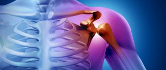
Lesions of the upper extremities lead to intrarotation-adduction contractures in the shoulder joints, extension contractures of the elbow joints, flexion contractures of the wrist joints against the background of ulnar deviation of the hand and flexion-adduction contracture of the first finger.
Limb contractures are caused by impaired tone, muscle degeneration, abnormal location of their attachment points, shortening of the bursal-ligamentous apparatus, depression of adjacent joint structures and slower growth of tubular bones.
Arthrogryposis Clinic - Department 10
The staff of the department specialize in the treatment of children with congenital and acquired deformities of the upper and lower extremities due to various diseases of the musculoskeletal system:
- Arthrogryposis;
- Osteogenesis imperfecta;
- Congenital malformations of the leg.
- Bone metabolism disorders;
- Flaccid paralysis of the upper limbs (consequences of obstetric paralysis, injuries of the brachial plexus and peripheral nerves, spinal cord tumors, neuroinfections);
The specialized department of arthrogryposis was organized on February 1, 2010. This was facilitated by the accumulated experience and results of scientific and practical developments of the institute’s staff over many years (Kazantseva N.D., Konyukhov M.P., Petrova E.V., Klychkova I.Yu., Lapkin Yu.A., etc.) , as well as a created database of children with arthrogryposis throughout Russia. To date, the department has accumulated the greatest experience in the world in the treatment of various forms of arthrogryposis.
In 2012, the G.I. Turner Institute was awarded the Vocation Prize for the development of a new method of treating children suffering from arthrogryposis.
In 2014, the second world symposium on arthrogryposis was held at the G.I. Turner Institute.
Employees of the department annually participate in international conferences and symposiums, and international scientific connections have been established.
The staff of the department are part of the international group for the study of arthrogryposis.
Under the guidance of Doctor of Medical Sciences Agranovich O.E. Doctors of Medical Sciences work in the department. Oreshkov A.B., Ph.D. Buklaev D.S., Ph.D. Petrova E.V., Ph.D. Trofimova S.I., Ph.D. Kochenova E.A., doctor Batkin S.F.
The team is working to introduce a neuro-orthopedic approach to the treatment of this pathology, developing and actively incorporating new treatment methods into clinical practice.
A system of early conservative treatment of children with arthrogryposis, starting from the first weeks of life, has been created, which has made it possible to reduce the severity of disability in patients with this pathology, significantly reduce the number of subsequent surgical interventions, significantly improve the ability for self-care and movement of these patients, and also reduce the frequency of postoperative relapses .
Treatment of clubfoot (the most common foot pathology in patients with arthrogryposis) is carried out using the Ponseti method. Methods for the conservative correction of flexion contractures of the knee and elbow joints have been developed; in addition, minimally invasive techniques such as temporary hemiepiphysiodesis are also actively used in the correction of various deformities of the limbs.
The arsenal of surgical interventions performed in patients on the upper and lower extremities includes hardware correction of contractures and deformities, tendon repairs, interventions on joints and bones using plates with angular stability and external fixation devices, various options for skin grafting, microsurgical transplantation of muscle flaps in order to restore the function of lost muscles and restore the ability to self-care.
In the complex treatment of patients with neuro-orthopedic pathology, the department successfully uses spinal cord stimulation techniques. Methods of brain stimulation are being developed to increase the effectiveness of conservative and surgical treatment of patients with arthorgryposis and flaccid paralysis of the upper limbs of other origins, through muscle retraining, as well as the development of new self-care skills.
The use of mechanotherapy (arthromotes for developing the joints of the upper and lower extremities), incl. robotic mechanotherapy (“Armeo”, “Lokomat”) allow a child to restore motor skills in a shorter time after surgery compared to traditional treatment methods.
The specialists of the department carry out consultations in the consultative and diagnostic department of the institute.
The result of treatment of contracture of the left wrist joint in a patient with arthrogryposis:
A – before surgery, B, C – 1 year after surgery.
Restoration of active movements in the right upper limb in a patient with arthrogryposis 2 years after transposition of various groups of donor muscles to the position of the missing ones:
A – patient before surgery, B-D – result of treatment.
The result of treatment of clubfoot in a patient with arthrogryposis:
A – before treatment, B-D – after treatment.
The result of surgical treatment of congenital flat-valgus foot deformity in a patient with arthrogryposis:
A, B – before treatment; V-D – 10 years after treatment.
Treatment of consequences of intrapartum brachial plexus injury
The incidence of birth injury to the brachial plexus is 0.12% (range 0.04 to 0.20%).
The main causes of this pathology are:
- Fetal shoulder dystocia (large fetus and narrow pelvis);
- Pathological position of the fetus during childbirth (occipital, pelvic, gluteal, tilting of the arm);
- Traction-rotation obstetric manipulations used during childbirth (manual, forceps, vacuum extraction).
All brachial plexus injuries (according to length) are divided into 4 groups:
Group I (or classic Erb's palsy) - level of damage C5-C6. Patients in this group make up 46% of all cases and have a good prognosis for restoration of limb function.
Group II – level of involvement C5-C7 (30%). Patients in this group have a worse prognosis for recovery compared to the first group.
Group III – total plexopathy, clinically represented by a dangling limb (20%).
Group IV - total plexopathy in combination with Horner's syndrome
The frequency of complete recovery of upper limb function is relatively high (up to 90%), however, in a number of patients, already at the age of 2-3 years, due to neurological deficits, disturbances in muscle balance and muscle function occur, which subsequently leads to deformation of bones and joints, as well as severe motor disorders. The most frequently observed anatomical and functional disorders are the shoulder and elbow joints.
If measures aimed at preventing the formation of contractures of the joints of the upper extremities in children with intrapartum brachial plexus injury are not observed, severe anatomical changes in the head of the humerus and scapula occur with age. The first changes in the shoulder joint are detected at the age of 3-5 months, pronounced - already at 2-3 years of life.
Formation of severe deformity of the right shoulder joint, shortening of the limb in a 16-year-old patient with Duchenne-Erb paresis.
Prevention of the formation of secondary deformities of the upper extremities in patients with intrapartum brachial plexus injury is:
- Adequate conservative treatment , including close cooperation between a neurologist, orthopedist and rehabilitation specialist.
- Neurosurgical treatment (according to indications).
- Botulinum therapy is aimed at restoring muscle balance, eliminating contractures in joints, and forming the correct pattern of movements.
- Orthopedic treatment aimed at restoring muscle balance, eliminating the vicious position of the limb, restoring the patient’s functionality, as well as preventing the formation of secondary deformities of the upper limbs.
The arthrogryposis department treats patients with flaccid paralysis of the upper limbs due to intrapartum brachial plexus injury, damage to peripheral nerves, tumors and injuries of the spinal cord, neuroinfections using modern methods of conservative and surgical treatment, which allows to quickly restore lost function and improve the patient’s ability to self-care.
The result of surgical treatment of a patient with consequences of left-sided Duchenne-Erb paresis:
A-D – before surgery; E-K 1 year after surgery.
Classification
There are mild, moderate and severe forms of arthrogryposis. Mild cases are characterized by rare cases of involvement of one or two limbs.
Arthrogryposis occurs most often in the classical (generalized) form and isolated, affecting the lower and/or upper extremities. A separate group of diseases is the distal form of arthrogryposis. Characteristic features include the presence of congenital multiple contractures and deformities of the hands, feet, as well as facial anomalies. The type of transmission of the disease in this form is hereditary. At least 9 varieties of distal arthrogryposis are known in the form of digitotal dysmorphism, Freeman-Sheldon syndrome, Gordon syndrome, trismus pseudocamptodactyly, pterygium syndrome and congenital arachnodactyly .
Contractures of the limbs in severe arthrogryposis
Joint contractures can be different - flexion and extension, adduction and abduction, and also in a combination of different forms. The condition of the limbs can be complicated by dislocations of the hips.
Is arthrogryposis completely curable?
The disease leads to contracture of the joints and, because of this, the inability to lead a normal lifestyle in the future. Contracture is a partial or complete restriction of mobility (for example, the inability to flex or straighten the hand). There is a chance to cure this disease completely or at least with minimal remaining complications only in its mild form. If several joints are involved in the process (usually the diagnosis is “distal arthrogryposis”) and the spine is not affected, then with the help of conservative and sometimes surgical methods it is possible to achieve lasting positive dynamics.
In situations where the pathology extends to multiple joints and the back, the main goal of treatment is to relieve the patient's pain and condition as much as possible. It is important to correct dislocations as much as possible and slightly increase muscle tone so that a person can move independently or at least work with his hands. A combination of surgical and therapeutic methods makes it possible in most cases to reduce the severity of the deformity and improve muscle sensitivity. After the course of primary treatment, such patients definitely need supportive physiotherapy and massage.
To prevent the joints from deforming again, special orthopedic braces and splints are used. Regardless of the severity of the disease, treatment and recovery of a child is a difficult process (mentally and physically) for parents.
Causes
The etiology of arthrogryposis has not been sufficiently elucidated to date; however, infectious agents (for example, the Zika virus ) presumably play a leading role in it, having a teratogenic effect on the embryo, thereby disrupting the normal development of the musculoskeletal system. However, scientists have suggested that the mechanisms of development of pathology can also be hereditary, neurogenic, myogenic, and even mechanical.
Risk factors during pregnancy
- bad habits of the mother - alcohol abuse, drug addiction and smoking;
- uncontrolled use of medications;
- exposure to radiation;
- severe oligohydramnios ;
- placental insufficiency;
- uterine anomalies (including bicornuity and underdevelopment);
- the presence of such severe somatic diseases in the decompensation phase as diabetes mellitus and systemic lupus erythematosus .
Symptoms
Arthrogryposis is usually characterized by local symptoms, including the following lesions and contractures of the limbs (impaired mobility):
- extension of the arms at the elbow joints;
- tightness of the arms to the body and club-handedness;
- tightness of the hands and fingers;
- violation of grasping functions;
- abduction of the lower extremities in the hip joints;
- extension of the knees;
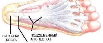
- deformity of the feet, usually deformed towards clubfoot;
- hip dislocations.
Symptoms of arthrogryposis
Characteristics of appearance in arthrogryposis
- the shape of the body is disproportionately elongated in relation to the deformed and underdeveloped limbs;
- narrow shoulders that lack muscle relief and have a beveled obtuse angle;
- the neck is wide with narrow shoulder girdles and massive folds - pterygia (pterygia);
- amniotic bands;
- skin syndactyly on the fingers;
- skin retractions over pathological joints;
- telangiectasia;
- hemangiomas of various localizations.
The onset of the disease is characterized by the appearance of fibrous and then developing bone contractures with the installation of the limbs in the form of an external turn and full arching, with deformation of the feet (equinovarus or planovalgus), hands, which are forced to be bent in the palms, while the fingers are bent. In some cases, dislocations and subluxations may be detected, which most often affect the hips and cause aplasia and hypoplasia of the patella. For this pathology, it is not uncommon to identify other developmental defects in the absence of systemic lesions of internal organs and preserved intelligence.
How to identify the disease?
The only type of prenatal diagnosis of the disease is ultrasound during pregnancy at different stages of fetal development. Some alarming signs help to suspect a terrible malformation. With a presumptive intrauterine diagnosis of “arthrogripposis,” the following changes are instrumentally noted:
- disturbance of fetal motor activity (its decrease or complete absence);
- thinning of the soft tissues of the extremities in volume;
- gross malformations of joints (for example, immobility or visual absence of feet and hands).
For screening purposes, expectant mothers need to undergo at least 3 mandatory ultrasounds: at 10-13 weeks, during about 5 months of pregnancy and after 30 weeks. The first 2 studies are especially important in diagnostic terms, since in the third trimester it is impossible to terminate a pregnancy even for medical reasons.
It is impossible to identify the pathology in any other way; it is not determined by donating blood or by touch during a routine examination by an obstetrician-gynecologist. After the birth of a child, X-rays and EMG are additionally used for diagnosis, although changes are visible externally to the naked eye.
Tests and diagnostics
During the examination, patients are found to have smoothing of the contours of the joints, muscle relief, and limited mobility of the affected joints, characteristic of arthrogryposis.
Morphological studies demonstrate a picture of severe muscle lesions and developed fibrous dystrophy.
An accurate diagnosis can only be made in the perinatal period due to its typical appearance. However, thanks to modern ultrasound, it is possible to detect soft tissue atrophy and deformation of the joints of the limbs. Also, indirect signs of pathology include low fetal mobility.
Classification of arthrogryposis
| Degree of disease | Bone apparatus | Muscles | |||||
| Joint pathology | X-ray features of bones | Morphological changes in bones | Force | Hypoplasia | Morphology | EMG | |
| Lightweight | Disorders of shape and limited mobility of hands and feet | Mild osteoporosis, fragility of long bones | Symptoms of bone and joint disorders | 1-2 points | Weakly expressed | Wasting and reduction of muscles | Signs of muscle and spinal cord damage |
| Medium-heavy | Violation of the shape and limitation of mobility of all joints of the limbs except the proximal parts | Moderate osteoporosis, fragility of long bones | Symptoms of bone and joint disorders | 2-4 points | Weak or moderately expressed | Wasting and reduction of muscles | Signs of muscle and spinal cord damage |
| Heavy | Violation of the shape and limitation of mobility of the spine and all joints of the arms and legs | Severe osteoporosis, fragility of long bones | Symptoms of bone and joint disorders | 1-2 points | Pronounced | Wasting and reduction of muscles | Signs of muscle and spinal cord damage |
Arthrogryposis in children
The disease in children begins with externally detectable disproportionate elongation of the body, deformation and underdevelopment of the limbs. The shoulders are sloping; they are narrow and lack muscle relief; the neck is wide with narrow shoulder girdles and massive folds in the shape of a pterygium. The changes are symmetrical, they do not progress after the birth of the child, but the deformities can recur with age, which may require prosthetic support for the patient.
The subsequent stage of the disease causes fibrous growths, which initiate bone contractures, immobilizing and placing the limbs more often in an external turn - up to full extension. Deformation of the feet and hands occurs, while the position of the palms and fingers is bent. In some cases, subluxations or dislocations occur that affect the hips against the background of aplasia and hypoplasia of the patella. In the most severe cases, lesions can affect not only the limbs, but also the spine and the muscles of the body. At the same time, other developmental defects are often detected; they are prone to respiratory diseases.
Children need social adaptation and training in specialized educational and rehabilitation centers provided for children with orthopedic pathologies.
Senya1985
How hard it is to realize that all this is not a dream...
1st pregnancy in 2006 - not developing, curettage at 12 weeks, then 5 years of infertility, 2012 after resection, my son was born (on May 3 we are 3 years old), in December the doctors said that I myself could not get pregnant, not the eggs are maturing, there is no ovulation, I need a lot of tests and treatment again, but on December 30 (before the new year) I take a test - and there is no limit to my surprise and happiness - we will be parents again! We did it despite all the doctors and their words! The pregnancy was going well, all tests were normal. At 6 weeks it anointed a little (but an ultrasound confirmed that it was smearing from the other horn - I have a bicornuate uterus, the baby is in the left, the right one is smearing), I suffered from heartburn and that’s it, everything else is normal. The belly grew and so did the belly. At week 18, I started to worry that I didn’t feel any movements, but G reassured me, saying that the baby can be calm, just wait. I was already getting ready to go on maternity leave again, on vacation, preparing and training the person who would come to take my place... 20 weeks.... Ultrasound.... I ran to him like crazy, ran and dreamed - who? boy? girl? It seemed to me that I was a butterfly, that wings had grown behind me and that I was not walking, but flying there. I came, lay down, the geneticist looked at me with an ultrasound and sent me to the toilet a little at a time, I went and walked for another 30 minutes until she called me. She came in again, lay down, she looked for a long time and again sent her out for a walk so that the child would turn around. As a result, I walked for more than an hour until she let go of this whole huge line and called me the last one. She looked silently and for a long time, and I looked with her (I had my monitor next to me), and almost shed a tear of happiness - I’m seeing my baby for the first time, God, how beautiful she is, what beautiful cheeks! I lay there and admired it until I noticed that she had been looking at me for 20 minutes! Then the manager came, they started whispering about something, but I didn’t hear what... And only when the manager sat down in her place and began to look at me, then I realized that something was wrong here, but I couldn’t even imagine I could have done that much! They sat me down on the table and gave me the verdict - the pregnancy must be terminated, the child has arthrogryposis...the hands and feet are deformed, the knees and elbows do not bend or straighten......There is a veil before the eyes and only tears of hopelessness....How is this possible? Why me? For what? It's just a bad dream...... I will not describe my condition, but God forbid anyone... On Tuesday (04/28/15) I go for amniocentosis (puncture and collection of amniotic fluid for examination), ultrasound of the baby’s heart, to the abortion commission, and on Wednesday I go to the hospital. That's all! Very hard. A child can only be born disabled and will never be able to walk or serve himself with his hands………It hurts……And today it became so difficult - the child began to hiccup…. and when you feel his movements and small movements, you understand that he is alive and he is yours….and you need to kill him…….
My son helps me not to go crazy and fall into depression. If I didn’t have it, I would 100% already be in a mental hospital. And in a week he will be 3 years old, and I will be in the hospital.....I want to wake up.....
Diet for arthrogryposis
Diet for sore joints
- Efficacy: therapeutic effect after 2-3 months
- Terms: 2-6 months
- Cost of products: 1700-1800 rubles. in Week
Children with arthrogryposis require the most balanced and rational diet, taking into account their needs for nutritional components and calorie content. At the same time, it is important that the diet is as rich in vitamins and varied as possible, and is also age-appropriate. The diet should be based on:
- porridge;
- vegetables and herbs;
- fruits;
- Fish and seafood;
- dietary meat;
- eggs;
- dairy products;
- natural vegetable oils, seeds and nuts.
Arthrogryposis: treatment should begin in the maternity hospital
For therapeutic actions to be effective, they are carried out from the very birth of the child. Before discharge, it is important to teach parents how to properly perform orthopedic exercises, which they can independently repeat daily at home. In the maternity hospital, children are usually prescribed the following types of treatment:
- massage;
- physiotherapeutic procedures (heat treatment);
- gymnastics;
- fixation of joints (using splints or plaster casts).
If the result of conservative treatment is not enough to recover or improve the child’s condition, surgical intervention is indicated. The sooner it is carried out, the higher its effectiveness. Deformities of the legs are operated on at the age of 3-4 months, and deformities of the arms at 6-8 months. Treatment at a later age requires more blue-eyed intervention in the bone apparatus and soft tissues, so orthopedists recommend not delaying this.