Injuries to the kneecap are a fairly serious pathology that requires timely and adequate treatment. The knees undergo enormous stress every day, because a huge weight presses on them while a person moves. If a crack occurs in the kneecap, the victim feels severe pain and begins to limp.
If a knee injury is left untreated, there is a risk that over time degenerative changes in the joint may occur, leading to impaired mobility, pathological pain and lameness. Complications such as inflammation of the joint often occur, so the patient needs to see a doctor immediately after the injury and begin proper treatment.
Anatomy
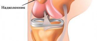
The knee joint is one of the largest joints in the human body; it is formed by three large bones: the femur, tibia and patella. The knee allows flexion and extension of the leg, which allows you to move normally and make fairly sharp and fast movements.
The joint is surrounded by large muscles and ligaments that are also involved in movement. Injuries to the kneecap may cause damage to ligaments and muscles, as well as cartilage and menisci. Such injuries are especially dangerous and, if not treated in a timely manner, lead to disability and inevitable surgical intervention.
Many patients believe that their knees are so strong that there is no need to worry about their integrity. Just remember the children who hurt their knees several times a year and the injuries heal without a trace. But do not forget that with age, the musculoskeletal system becomes less strong and takes longer to recover, so even if you fall from your own height, a fracture or crack of the kneecap is possible.
What are the dangers of meniscus damage?
A knee meniscus tear causes mechanical instability of the joint and requires immediate treatment. When moving, the knee joint may jam, causing a blockage of the joint. A torn meniscus in the knee damages the cartilage. The articular surfaces are deformed due to changes in the flexion/extension axis of the joint without a protective pad, the joint is deformed.
Damage to the knee meniscus, even after repair, can cause problems in the future. A person with such an injury is predisposed to developing arthrosis of the knee joint.
Causes
Cracks in the kneecap usually occur after an injury, such as falling onto a hard surface, especially from a height onto your knees. A crack can also occur when a pedestrian is hit, when the knees receive a strong blow, in which case a complete fracture is possible. It all depends on the force with which the impact occurred.
It's also worth noting that there are a number of people who are more susceptible to knee injuries:
- People leading an active lifestyle;
- Professional athletes;
- Workers in hazardous professions, for example, climbers, miners, etc.
- Aged people;
- Obese patients;
- Patients with diseases of bone tissue and joints;
- People who lead a passive lifestyle;
- Those who don't watch their diet.
In general, people whose bodies are weakened are more susceptible to injuries to the kneecap and other bones and joints. To reduce the risk of such an injury, you need to eat well, exercise, and strengthen your muscles and bones. In older people, taking calcium and vitamin D plays an important role; it is also necessary to wear comfortable, non-slippery shoes and avoid climbing to heights to reduce the risk of falling.
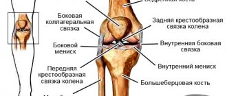
Anatomical features of the knee joint
The structure of the knee joint includes various segments, ranging from bones, muscles, menisci, ligaments, tendons to nerves and blood vessels.
The upper (femur) and lower bones (tibia and fibula) are connected by ligaments, tendons and muscles. Cartilage deserves special attention, as it performs several important functions: it minimizes frictional forces during movement and provides optimization under impact loads.
Cruciate ligaments are dense formations that secure the ends of bones. As for the menisci, they are responsible for distributing the weight of the human body, increasing the stability of the knee joint and performing other functions.
Damage to any component of the knee joint can lead to severe pain and requires immediate intervention from a specialist.
Professional athletes are at increased risk, but people who have nothing to do with professional sports also often experience knee injuries. Also, older people are at risk.
Symptoms
Cracks and fractures of the kneecap are accompanied by the following symptoms:
- Pain that appears immediately after an injury and does not go away after some time. So, with a soft tissue bruise, a person can walk normally, feeling aching pain, but with a crack, the pain is more pronounced. When a kneecap is fractured, the pain is severe; because of it, the patient cannot walk or straighten the leg. If you press on the knee when it is cracked, the pain will intensify.
- Redness and swelling appear, and over time, a hematoma may occur due to damage to the capillaries upon impact. Cracks and fractures are often accompanied by hemorrhage into the joint cavity, which is especially dangerous, since the injury can be complicated by inflammation.
- The patient protects and spares the leg, does not step on it and does not straighten it completely, so as not to feel pain.
It is worth noting that often the symptoms of a fissure are mild and more similar to a regular bruise: the swelling is small, the hematoma does not appear, so the patient does not see a doctor. You shouldn’t do this; lack of treatment can lead to chronic degenerative joint disease, which you will have to fight for the rest of your life.
Types of fractures
There are several types of patellar fracture:
- without offset . The parts of the broken bone fit tightly to each other and do not move to the sides. This type is considered the easiest, since it does not require reposition, and its treatment is relatively quick;
- with offset . In this type of injury, the bone is divided into two fragments, one or both of which are displaced relative to each other. Reposition is required to connect them.
- comminuted . In this case, the patella is divided into three or more smaller parts that are displaced and unstable (can move). The most difficult type to treat.
Open and closed fractures are also classified depending on the presence of skin disruption.
Symptoms of a fracture include severe pain immediately after the injury, swelling and deformation of the knee joint, and bruising or open bleeding. If these signs occur, you should immediately contact a clinic or call emergency help. The doctor will conduct an initial examination and X-ray examination, as a result of which he will determine the type of fracture and prescribe the appropriate treatment.
Important! With age, the risk of a patellar fracture, like other fractures, increases. This is because bones become more fragile over time. Therefore, older people should be as careful as possible.
First aid
In case of a knee injury, it is very important to immediately take the victim to the hospital. Since it is not known how badly the kneecap is injured, immobilization is necessary. If the leg is not immobilized, then displacement of fragments and other complications can occur.
In case of a knee injury, especially if the blow was strong or an elderly person was injured, an ambulance must be called. If it is not possible to call doctors, you need to immobilize the leg with a splint and transport the victim to a medical facility as soon as possible.
Diagnostics
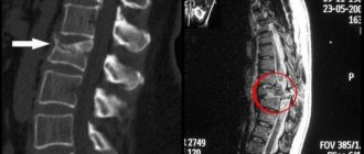
When a patient enters a medical facility, he is examined by a traumatologist, who can usually immediately suspect the presence of a crack, fracture, rupture of ligaments and other pathologies of the joint. To confirm the diagnosis and identify the location and extent of bone tissue damage, the patient is sent for radiography.
Sometimes radiography is not informative enough, but this happens extremely rarely. In this case, the doctor refers the patient to a CT scan. Computed tomography helps to view the joint from all sides and accurately identify the size and location of the fracture/crack. The patient also undergoes a puncture of the knee joint if hemorrhage occurs.
Diagnosis of post-traumatic arthritis of the joint
To clarify the diagnosis, a traumatologist or rheumatologist may prescribe:
- general and biochemical blood/urine analysis (as correct, sufficient to exclude similar diseases);
- immunological blood/synovial fluid test.
When diagnosing, it is important to establish the degree of damage and the presence of pathological processes in the joint. For this we use:
- radiography (detects accumulations of pus in the joint capsule, the presence of bone fragments, osteosclerotic changes);
- computed tomography (full assessment of the affected joint);
- MRI (indicates the presence of erosion, cartilage degeneration, bone defects);
- arthroscopy (for internal examination of joints and minor medical procedures);
- Ultrasound (to assess the condition of periarticular tissues).
Treatment
A crack in the knee is always treated conservatively, since with such an injury there are no bone fragments that need to be compared. If the patient consults a doctor in a timely manner, then the prognosis is positive. The crack heals quickly and rarely causes serious complications.
During treatment, the following measures are prescribed:
- The knee joint is immobilized therapeutically using a plaster splint or a special orthosis;
- Prescribe calcium and vitamin D for rapid healing;
- Painkillers or non-steroidal anti-inflammatory drugs are prescribed if the patient is bothered by pain.
- If hemorrhage occurs in the joint, a therapeutic puncture and antibiotics are prescribed.
During the treatment period, the patient must take care of his leg so that the kneecap heals faster. It is also necessary to monitor your diet, eat less salty, fatty and spicy foods and consume enough fresh vegetables, fruits, and dairy products. Good nutrition and normal metabolism will ensure rapid healing of the fracture. The patient will quickly return to normal life.
In cases where a fracture has occurred with displacement of the fragments, surgical treatment is most often prescribed. The doctor restores the kneecap by collecting the fragments and securing them (Libov's patella suture).
Various types of knee cracks and their treatment
The anatomical structure of the knee joint is quite complex. This may explain the variety of types of cracks.
Cracks localized in the condyles of bones
Knee Joint
The condyle is a cartilage-covered thickening on each of the three bones that make up the knee joint. Its rounded structure allows the knee to freely extend and bend, as well as perform rotational movements.
If an unnatural force is applied to the knee, a crack will occur.
Important: according to the practice of traumatologists, a crack in the knee is nothing more than a fracture, which is not accompanied by displacement of bone fragments and the formation of splintered fragments.
However, displacement of fragments, rupture (complete or partial) of ligaments, and disruption of the integrity of the meniscus can become concomitant pathological conditions.
Causes of condyle crack:
- targeted blow to the knee;
- falling with legs bent at the knee;
- falling from heights onto straight legs.
The external manifestations of the injury coincide with other injuries to the knee. Victims often note:
- pain that intensifies with any physical activity;
- joint swelling;
- signs of internal bruising (hemarthrosis).
Crack in the knee joint
An accurate diagnosis is only possible based on the results of an x-ray examination. Pictures are taken in several projections. This is done to exclude other injuries to the knee joint.
How to treat?
Treatment of a condyle fracture involves complete immobilization of the limbs until complete healing. To do this, a plaster cast is applied or a special design is used to prevent any activity in the knee.
If hemarthrosis is detected, complete removal of blood from the joint cavity is performed before immobilization. The period of wearing the brace is from 6 to 10 weeks, depending on the severity of the injury and the results of additional radiographic examination.
After removing the fixator, massage and therapeutic exercises are prescribed. Additionally, physiotherapeutic procedures are prescribed.
Crack in the patella
Patellar fracture is a common injury. It is diagnosed not only in athletes, but also in children, adolescents, and the elderly.
The patella is most often affected by a direct blow or fall on the knee due to the fact that it is the most unprotected part of the joint.
Damage can be of three types:
- horizontal - part of the bone can completely tear off and move out of its place;
Crack in the patella - vertical – divergence of parts of the patella is extremely rarely diagnosed;
- complex injury - numerous cracks emanating from the central part of the patella.
On palpation the patient complains of severe pain. Severe swelling and hematoma appear at the site of cracking. The condition is not accompanied by impaired mobility of the limb.
A fractured patella can be diagnosed solely based on the results of an X-ray examination.
How to treat?
Treatment must be immediate, otherwise the victim will develop increased mobility of the patella. This, in turn, increases the risk of re-injury.
Crack localized in the meniscus
An example of the most common type of traumatic knee fracture. The meniscus is a cartilage structure that bears most of the load on the joint. Unlike bone, the meniscus is porous and fragile, which is why it most often suffers from injury.
There are three types of traumatic meniscus injury:
- horizontal;
- vertical;
- longitudinal.
The most severe case is vertical damage due to the fact that it is often accompanied by lifting of the edges of the cartilage, their separation and, as a result, blocking of the entire joint.
Meniscal fracture
The causes of meniscus fractures can be either traumatic or non-traumatic. The condition is accompanied by the following clinical picture:
- pain that intensifies with movement;
- formation of edema;
- development of hemarthrosis if the red areas of the cartilaginous structure are affected.
Diagnosis of the condition by X-ray is not possible because the meniscus cannot be seen in such images due to its structure. Therefore, it is recommended to perform an MRI (preferably) or ultrasound (this method is less informative).
How to treat?
In most cases, cracks localized in the meniscus do not require surgical intervention. Temporary restriction of joint mobility by applying special splints is sufficient.
In complex cases of this type of crack in the knee, such as a violation of the integrity of the meniscus, treatment consists of plastic surgery of the cartilaginous formation. That is, sewing its edges together at the site of the crack. Extremely complex disorders require partial or even complete removal of the meniscus.
In order to restore the anatomically correct functioning of the knee joint after a meniscus fracture, great attention should be paid to rehabilitation. The prescribed measures help restore motor abilities, strengthen the joint, its ligamentous and muscular apparatus.
Rehabilitation
After the cast is removed, the patient is prescribed a course of rehabilitation, which is aimed at strengthening the muscles and gradually developing the knee. Do not give full load at once. This can cause pain, swelling and contracture (immobilization) of the joint. It is better to gradually increase the load, gradually moving from simple exercises to more complex ones. An indicator of the adequacy of the load is pain. When it appears, the load is reduced.
During the rehabilitation period, physical therapy is prescribed, which strengthens the muscles and develops the joint, and also improves nutrition and blood circulation in the tissues. Example exercises:
- You need to stand up straight and raise your straight leg above the floor, hold it in this position for a few seconds. Then lower your leg, rest a little and repeat the exercise with the second leg.
- You need to stand behind a chair and rest your hands on its back. Rise on your toes and hold for a few seconds, then relax and repeat 2-3 more times.
- You need to sit on the floor or on a flat sofa, straighten your legs. Bend over, trying to reach your toes with your hands. Don't stretch too much; you should feel pressure under your knee, but not pain.
- The gymnastics is completed with a massage around the knee joint, in the thigh and lower leg area, to improve blood circulation.
It is very important to do the exercises correctly and regularly. You can start practicing only after permission from a specialist. It is better to take the first classes in a physical therapy room. Later you can train at home. If knee pain occurs during exercise, then it is necessary to reduce the load. Also, after a fracture and crack of the kneecap, a patient is advised to attend physical therapy.
For a knee injury, the following physical procedures are prescribed:
- Magnetotherapy;
- Laser therapy;
- UHF;
- Electrophoresis;
- Ultrasound treatment;
- Paraffin and ozokerite, etc.
The physiotherapist decides which procedures to attend in a particular case. He prescribes the duration of the procedures and their number, and selects the device. The physiotherapist makes a decision based on the main diagnosis and concomitant diseases.
Symptoms of post-traumatic arthritis
The disease can occur in acute and chronic forms, and the intensity of symptoms will differ. PA can develop rapidly, within a couple of weeks after the injury, or it can drag on for years or decades before the first exacerbation.
Patients experience symptoms characteristic of all types of arthritis:
- pain and aches in the affected joint;
- local temperature increase;
- stiffness and reduction in the range of voluntary movements;
- unpleasant crunching sound when bending.
Pain after a joint injury may not go away for a long time. Often the disease is accompanied by swelling and the formation of edema. In the presence of severe inflammation, an increase in general body temperature, fever, chills, and leukocytosis is observed (determined by laboratory).
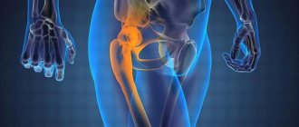
Most often, symptoms of post-traumatic arthritis are observed in the knee and hip joints, but the disease can begin in the elbow, ankle and even shoulder joints. Post-traumatic arthritis of other joints is less common - the interphalangeal joints of the fingers, the metatarsophalangeal joint of the big toes, and intervertebral joints.
Folk remedies
The use of traditional medicine recipes is only possible in the complex treatment of a cracked patella. You should not self-medicate, as there is a high risk of complications. It is very important to consult a doctor before using any product and make sure that you are not allergic to its components.
Remedies effective for fractures and cracks:
- To speed up bone tissue recovery, you need to consume enough foods with calcium. This substance is found in large quantities in cottage cheese, cheese, fermented milk and dairy products.
- Chamomile tea or tea made from mint and currant leaves has an excellent anti-inflammatory effect, soothes and normalizes sleep. It is better to drink it instead of regular tea, which contains caffeine, since caffeine interferes with the absorption of calcium.
- You can also replace tea and coffee with rosehip decoction; it contains a large amount of vitamins, strengthens the immune system and accelerates tissue healing after injuries.
- Dishes containing gelatin accelerate the healing of fractures: aspic, jellied, berry and fruit jelly with gelatin.