Fractures, bruises, and bone cracks often lead to degenerative processes in the joints. However, arthrosis of the elbow joint occurs not only due to injury. After 45 years, the body’s metabolism is often disrupted and a deficiency of vitamins and minerals develops. All this has a negative impact on the structure of the cartilage, the composition and amount of synovial fluid, first discomfort appears, and then pain. What are the prognosis for such a diagnosis and what treatment is most effective?
Arthrosis of the elbow joint is diagnosed infrequently and mainly in people over 45 years of age
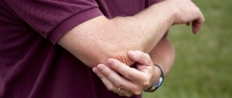
How do you know if you have arthrosis of the elbow joint?
Pathological changes in cartilage tissue make themselves felt as follows:
- when bending and straightening the arm, pain appears, which can be spontaneous, but over time it begins to radiate to the neck and is felt when pressing on the elbow;
- mobility in the elbow joint is limited, especially due to the growth of osteophytes;
- due to the friction of bone surfaces, a dry crunch appears (this sound should not be confused with normal physiological clicks during certain movements of the elbow);
- Some, mainly athletes, note muscle weakness, but this symptom is quite difficult to detect.
What are the most common elbow diseases?
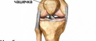
The elbow is a complex mechanism, which is represented by the connection of three bones (humerus, radius, ulna). Important blood vessels and nerve endings pass through it. Without realizing it, subjecting it to colossal stress in everyday life, people may increasingly encounter diseases of the elbow joint. Although the damage does not put the patient in mortal danger, it worsens the quality of life to a greater extent. Despite the periodic pain that bothers the elbow area, people attribute them to overwork, accidental bruises, the weather, and treat themselves by taking pills, applying compresses and using folk remedies. Unfortunately, in most cases, this leads to complications, loss of sensation and freedom of movement. Important! To determine the diagnosis, you need to consult a specialist.
Diseases of the elbow joints can be varied and the list of them is quite large:
- Various occupational injuries.
- Diseases caused by inflammation: tendonitis, epicondylitis, fasciitis, arthritis.
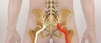
- Neurological diseases: nerve root lesions, carpal tunnel syndrome, neuritis, etc.
- Due to changes in tissues, both cartilage and bone: osteoarthritis, gout, chondrocalcinosis, osteochondrosis.
Who's at risk
Arthrosis of the elbow joint develops for many reasons:
- due to injuries;
- due to metabolic disorders - metabolic processes in the joints;
- against the background of a genetic predisposition or disease of the endocrine system;
- after intoxication of the body, an infection that has spread to the joint, hypothermia;
- as a complication of rheumatoid arthritis, etc.
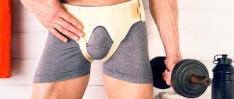
At risk are all representatives of the older age category, as well as those who seriously engage in weightlifting or expose the elbow joint to other mechanical stress. Women are also susceptible to the disease during periods of hormonal system disruptions, most often during menopause.
Arthrosis of the elbow joint is often diagnosed in tennis players
What it is?
A crack in a joint is an incomplete fracture , a violation of the overall integrity of the joint, which occurs when there is a strong mechanical impact on the joint, for example, with a severe bruise. Because the fracture is incomplete (that is, the bone does not break in two when damaged), this moment is not usually accompanied by a crunching sound.
The biggest danger in this situation is not paying attention to the pain and swelling in time, and thus contribute to the expansion of the crack, due to which the joint can literally begin to fall apart under the skin. Also, if the doctor’s recommendations are not followed, improper bone fusion may occur, which usually leads to complete or partial loss of limb mobility.
Stages of arthrosis of the elbow joint
Over time, the disease progresses:
- At the first stage, unexpressed pain is noted during exercise, but there are no external manifestations. Retraction of the arm back is somewhat limited, as is flexion-extension, but does not yet cause discomfort to the person. It is not difficult for a doctor to make a diagnosis for such symptoms, unlike patients who often attribute ailments to banal fatigue.
At the first stage of arthrosis, a slight narrowing of the joint space is noticeable in the image.
- At the second stage, noticeable pain appears, including at rest, as well as a dry crunching sound when moving. It becomes impossible to bend your arm at the elbow, as well as to move it back. Muscles begin to atrophy, joint tissues begin to deform, and leading a normal lifestyle is already quite difficult.
At the second stage of arthrosis, the image shows numerous osteophytes - bone growths
- At the third stage, the aching pain becomes constant. It is almost impossible to move your hand. To reduce pain, many people fix their hand in a certain position. The cartilage is completely destroyed, the joint space is absent in the picture, and the muscles are atrophied.
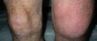
At the third stage of arthrosis, the damaged arm becomes smaller than the other
Crack in the elbow joint: symptoms, diagnosis and treatment
Joints are the moving joints of the ends of bones in the skeleton, which are located in those areas that are most often subject to flexion and extension.
They are a very important part of the musculoskeletal system. Their main function is to ensure smooth sliding of the ends of the bones relative to each other, without abrasion or deformation.
The joints are also responsible for maintaining the position of the body and moving it in space.
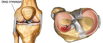
The elbow joint is where three bones meet: the humerus, ulna, and radius.
It is classified as "complex" because it consists of three simple joints enclosed in a common joint capsule, which provides mobility to the arm.
Damage to this joint is usually very dangerous and can lead to many complications, especially if you do not recognize the fracture by symptoms and do not promptly consult a doctor and begin treatment.
What it is?
A crack in a joint is an incomplete fracture , a violation of the overall integrity of the joint, which occurs when there is a strong mechanical impact on the joint, for example, with a severe bruise. Because the fracture is incomplete (that is, the bone does not break in two when damaged), this moment is not usually accompanied by a crunching sound.
The biggest danger in this situation is not paying attention to the pain and swelling in time, and thus contribute to the expansion of the crack, due to which the joint can literally begin to fall apart under the skin. Also, if the doctor’s recommendations are not followed, improper bone fusion may occur, which usually leads to complete or partial loss of limb mobility.
Causes of injury
There are usually not many of them. In addition to the severe bruise mentioned above, a crack can also appear when you fall on your elbow, get into an accident, play sports, and even in everyday life, for example, when the arm is heavily loaded.
The elbow joint, for all its strength, remains a very vulnerable place due to the lack of a muscular frame that could protect it from mechanical damage. Therefore, you should take care of your elbow and, if you fall, try to group your body in such a way as not to damage this particular joint.
Symptoms
Typically, victims experience the following symptoms with a crack in the elbow joint:
- Aching and throbbing pain at the site of the bruise that does not go away even when the hand is at rest.
- When you press on the sore area, there is a sharp sharp pain that radiates to the hand and shoulder.
- Stiffness of movements in the forearm area.
- Swelling of the skin around the joint injury.
- Rapid occurrence of hematoma.
- Discomfort when lifting objects.
- Complete or partial restriction of joint mobility.
- Partial or complete numbness of the forearm, hand and fingers.
Diagnostics
The diagnosis is made based on the nature of the incident in which the patient was injured. But an accurate diagnosis can only be made after radiography . Sometimes a fracture is confused with a severe bruise due to similar pain and hematoma with a tumor.
Treatment
First of all, the victim, depending on the severity of the damage, the size and position of the crack, is given a plaster cast or a fixing bandage to the site of the damage. The arm is bent at an angle of 90 degrees and fixed in this position. While healing is taking place, it is recommended to avoid putting excessive stress on the arm.
The victim is usually advised to adjust the diet by adding calcium-rich foods, such as sea fish, hard cheeses, milk, cottage cheese, kefir, sour cream, and beans.
Very often, vitamins and calcium are prescribed, which help accelerate the healing of cracks and strengthen bones. Use ointments to reduce swelling and blue discoloration only on the recommendation of a doctor.
As well as attending electrophoresis or magnetic therapy.
You can also begin to restore joint mobility with the help of therapeutic exercises , but in this case it is worth keeping in mind two very important factors. Firstly, therapeutic exercises must be fully agreed with the attending physician, down to the exercises performed. Secondly, you can start exercising only after applying a plaster cast or bandage.
Of course, the exercises should not include flexion and extension of the elbow joint. The best place to start is by clenching and unclenching your fingers and moving your wrist. You can also place your hand behind your head while lying down in the early stages of recovery.
Such exercises help to get rid of swelling faster and help avoid muscular dystrophy due to inactivity of the limb. During the healing process, you can gradually increase the complexity of the exercises and continue to avoid bending. Even after removing the cast or bandage, you should not bend or straighten your arm completely for some time to avoid re-fracture.
Surgery is rare for a fracture in the elbow joint. But sometimes the nature of the damage does not contribute to the natural healing of the bone. If the crack is severely widened, doctors use a special wire to fasten the damaged joints.
If the injury was so severe that the bone began to crumble, then it is also fastened with wire and the mobility of the arm is limited for a fairly long period of time.
Possible complications
The most unpleasant complication is complete or partial loss of limb mobility. This occurs when the joint does not heal properly or when the injury is ignored, leading to irreversible destruction of the joint.
Failure to comply with doctor’s instructions, for example, putting a lot of strain on the arms or frequently bending the injured part of the body, can also have an impact. As mentioned above, for some time after healing there is a danger of a second fracture, which can lead to complications.
It is important not only to take care of your joints, but also to promptly eliminate possible injuries in full accordance with medical recommendations.
Symptoms of deforming arthrosis of the elbow joint
An X-ray image allows you to distinguish ordinary arthrosis from deforming arthrosis, when pathological changes occur on the periphery of the humerus:
- at the first stage, osteophytes are noticeable not only in the joint area, but also in the area of the distal epiphysis, which does not happen with ordinary arthrosis;
- on the second, bone growths are distributed evenly around the elbow joint, without limiting mobility, but gradually leading to dysfunction;
- on the last one, the image shows signs of sclerosis of adjacent bone lobes.
About 50% of cases of arthrosis of the elbow joint are deforming arthrosis
Causes of injury
There are usually not many of them. In addition to the severe bruise mentioned above, a crack can also appear when you fall on your elbow, get into an accident, play sports, and even in everyday life, for example, when the arm is heavily loaded.
The elbow joint, for all its strength, remains a very vulnerable place due to the lack of a muscular frame that could protect it from mechanical damage. Therefore, you should take care of your elbow and, if you fall, try to group your body in such a way as not to damage this particular joint.
What physiotherapeutic methods are most often used in stages 1 and 2
- Electrophoresis with medications.
- Paraffin therapy to stimulate blood flow.
- Laser therapy to stop the growth of osteophytes and destroy existing ones.
- Mud compresses to eliminate atrophic changes in tissues.
- Acupuncture to reduce pain and restore muscle tone.
- Massage is relevant at the rehabilitation stage.
- Physiotherapy.
With the help of simple gymnastics, you can eliminate pain in the initial stages of arthrosis of the elbow joint. Contraindications to it are recent elbow injuries, elevated body temperature, influenza, ARVI, as well as a period of 3 months after surgery on the elbow joint.
Types of fractures
Elbow joint injuries are of the following types:
- Closed. Injury to the radius, its head, and neck is typical. This type of injury usually occurs due to too much stress while simultaneously placing pressure on the straight arm.
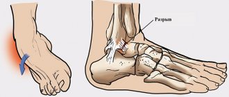
- Open. In addition to cracks, soft tissues are injured by bone fragments. In particularly difficult cases, the skin is torn, an open bleeding wound is formed, which leads to large blood losses.
- Fractures of the coronoid process. They develop due to a strong impact load on the bones of the elbow. This is the rarest form of injury and is characterized by additional dislocation and displacement in the forearm.
Open fracture
The fracture can be undisplaced or displaced, usually only one of the 3 bones that make up the elbow is injured.
Fractures are classified according to the place of formation:
- The radial head is the most common injury to the elbow joint. It occurs due to the lack of muscle protection in this structure.
- Radial necks.
- Transcondylar.
- Coronoid process, usually accompanied by additional injuries.
Depending on the location of the crack, fractures are:
- Intra-articular. Ligaments rupture, the integrity of the joint itself is disrupted, bones or fragments are displaced.
- Periarticular, which are not accompanied by displacement of bones and fragments.
Based on the number of damaged bones, fractures can be:
- multiple;
- single.
How Noltrex intra-articular injections work
Noltrex is an artificial endoprosthesis that can replace the missing synovial fluid in the elbow and any other joint. Once inside the joint space, the drug evenly covers the surfaces, creates a shock-absorbing effect and gradually restores the desired viscosity of the joint fluid. Therefore, cartilage and bones stop rubbing against each other, the joint space widens, and the pain goes away.
Unlike injections based on hyaluronic acid, Noltrex:
- It is biocompatible with body tissues, therefore does not cause rejection or allergic reactions;
- does not contain components of animal origin, therefore it is eliminated from the body much longer;
- has a minimum of contraindications, including only inflammation of the joint and in some cases diabetes.
The molecules of the polymer drug "Noltrex" are practically invisible to the body's immune cells, so the drug remains in the joint for a long time and has a prolonged effect. It is necessary to take a repeated course in the treatment of arthrosis of the elbow joint every 9-24 months, depending on the degree of the disease. During this period, the mobility of your elbow is completely restored, the pain goes away - you can enjoy life again!
Causes of injuries
The elbow is characterized by its susceptibility to injury because it lacks a dense framework of muscles. It is the muscles that reliably hold and protect other joints. Elbows are even more stressed in active children, who often find themselves in situations that lead to injury. You can get a cracked elbow due to a fall or blow. Usually the damage is internal.
Types of fractures
Elbow joint injuries are of the following types:
- Closed. Injury to the radius, its head, and neck is typical. This type of injury usually occurs due to too much stress while simultaneously placing pressure on the straight arm.
- Open. In addition to cracks, soft tissues are injured by bone fragments. In particularly difficult cases, the skin is torn, an open bleeding wound is formed, which leads to large blood losses.
- Fractures of the coronoid process. They develop due to a strong impact load on the bones of the elbow. This is the rarest form of injury and is characterized by additional dislocation and displacement in the forearm.
Open fracture
A fracture can be undisplaced or displaced, usually only one of the 3 bones that make up the elbow is injured.
Fractures are classified according to the place of formation:
- The radial head is the most common injury to the elbow joint. It occurs due to the lack of muscle protection in this structure.
- Radial necks.
- Transcondylar.
- Coronoid process, usually accompanied by additional injuries.
Depending on the location of the crack, fractures are:
- Intra-articular. Ligaments rupture, the integrity of the joint itself is disrupted, bones or fragments are displaced.
- Periarticular, which are not accompanied by displacement of bones and fragments.
Based on the number of damaged bones, fractures can be:
- multiple;
- single.
Symptoms
Characteristic manifestations of a crack:
- The pain is localized on the back of the elbow. In addition, the pain may radiate to other parts of the hand, even to the fingers. Palpation is always painful.
- When bones or fragments are displaced, a crunching sound is heard, the displacement is visually noticeable, and the joint takes on an uncharacteristic shape. The skin may begin to sag.
- If the injury affects the blood vessels, swelling and hematoma appear. In this case, the joint cavities are filled with blood.
- There is a limitation of mobility or its complete loss.
- When nerve fibers are injured, numbness in some areas of the hand is observed.
Pain from a fracture
Not all symptoms will necessarily appear at the same time. To confirm a fracture, the occurrence of several signs is sufficient, but pain is always present. A doctor, even without hardware diagnostics, can distinguish between a bruise and an articular fracture based on symptoms. In case of injury, you should immediately go to the traumatology department.
First aid
The tactics for implementing emergency care for an elbow injury correspond to the type of injury and severity:
- Immediately complete immobilization is carried out. It is better to use a splint; for this, the arm is bent at an angle of 90 degrees.
- For unbearable pain, analgesics are used.
Conservative treatment
Even a minor injury poses a threat to health, especially if there is a crack in the elbow or a displaced fracture. Treatment begins immediately after injury. Non-surgical options include:
- Limitation of mobility.
- Taking painkillers (Ibuprofen, Acetaminophen). They help relieve pain in the short term.
- Injectable corticosteroids for long-term pain relief.
- Medicines with gold salts or antimalarial drugs are also for long-term pain relief.
- Physiotherapeutic procedures.
Rehabilitation after treatment
At the rehabilitation stage, massage of the elbow joint and exercise therapy are required. They are used after the end of the immobilization period. The massage affects the shoulder and forearm, but not the area of injury. The implementation of folk recipes is not recommended.
Physiotherapy
The exercises involve performing flexion-extension complexes and rotations. In terms of time, it takes no more than 30 minutes to perform gymnastics three times a day.
The choice of specific therapy is based on many factors:
- age of the victim;
- severity of the fracture;
- the presence of concomitant pathologies;
- timely consultation with a doctor;
- proper emergency care;
- the patient’s diligence during treatment and rehabilitation.
If the recovery is favorable, a plaster cast is required for 3 weeks, and after 1–1.5 months the patient returns to normal life. In case of severe damage, recovery takes at least 3 months.
For elderly people with concomitant diabetes mellitus, plaster is placed for a long period - 4 months or even longer. Then it is replaced by a clamp to regulate the amount of arm activity.
Operative therapy
With an open type of fracture with displacement, surgery is necessary, otherwise the ability to flex the forearm is lost, and the chances of its recovery remain low.
The success of surgical intervention depends on the correctness of the surgeon’s manipulations, his ability to accurately compare bone fragments, and verify their anatomical fixation.
For a common injury to the elbow bone, therapy involves tightening the tissue with a medical wire loop. Additional fixation of bones using knitting needles is rarely needed.
To treat internal fractures with fragments, bone grafting is performed. It is difficult to tighten the tissue with a loop, since there is a high probability of shortening the surface of the joint. Instead, doctors use dynamic compression plates.
When a bone is crushed, it needs to be replaced with a special prosthesis. Implants are made of metal and plastic and are installed using bone cement.
Possible complications
A dangerous consequence of an elbow fracture and its ineffective treatment can be complete or partial loss of motor activity of the arm. The prerequisites for the development of such a complication are discomfort and significant pain, which persists even after completing the full course of therapy and rehabilitation. The problem can be prevented if you follow all the instructions of the traumatologist from the very beginning.
To prevent problems and loss of arm functionality after a crack in a child’s elbow joint, the patient should be under the close supervision of adults during the entire treatment. First of all, complete rest of the injury site is required throughout the entire treatment. It is forbidden to load the limb or make sudden movements. Negligence causes repeated fractures.
Immobilization with plaster
Rehabilitation after surgery
Rehabilitation should be aimed at restoring normal function of the injured limb. This will require:
- therapeutic massage course;
- physical therapy;
- physiotherapy procedures.
Exercise therapy
You should temporarily avoid sharp and frequent bending of your arms at the elbow. The main emphasis is on developing the wrist and fingers. The patient is required to place the injured arm behind the head while lying down, straining the forearm and shoulder. These exercises will speed up the elimination of edema by activating lymph flow in the tissues.
To normalize motor activity in a joint, its gradual development is necessary. After removing the plaster cast, you can perform measured movements of the limb.
During rehabilitation with the help of exercise therapy, full flexion and extension are prohibited - they can lead to a new fracture.
Massage
Massage
Massage is also allowed only after the plaster has been completely removed. The impact is required on the muscles of the shoulder girdle, back, only gentle manipulations. Regular procedures help relieve pain, strengthen the atrophied muscles of the affected limb, warm up the ligaments, and ultimately completely restore the previous activity of the arm.
Physiotherapy
It is most effective to alternate physiotherapy with gymnastics. The most effective procedures are: magnetic influence, UHF, electrophoresis, therapeutic mud therapy. During the rehabilitation stage, you will need to regularly visit a doctor to monitor your condition.
As a result, it is important to note that a patient with an elbow fracture after being discharged from the hospital department must first check with the doctor about further actions, find out about permission to lift weights, and about preventing deterioration and new cracks. Paying attention to your health will allow you to quickly return to a full life.
Symptoms
Characteristic manifestations of a crack:
- The pain is localized on the back of the elbow. In addition, the pain may radiate to other parts of the hand, even to the fingers. Palpation is always painful.
- When bones or fragments are displaced, a crunching sound is heard, the displacement is visually noticeable, and the joint takes on an uncharacteristic shape. The skin may begin to sag.
- If the injury affects the blood vessels, swelling and hematoma appear. In this case, the joint cavities are filled with blood.
- There is a limitation of mobility or its complete loss.
- When nerve fibers are injured, numbness in some areas of the hand is observed.
Fracture pain
Not all symptoms will necessarily appear at the same time. To confirm a fracture, the occurrence of several signs is sufficient, but pain is always present. A doctor, even without hardware diagnostics, can distinguish between a bruise and an articular fracture based on symptoms. In case of injury, you should immediately go to the traumatology department.
Conservative treatment
Even a minor injury poses a threat to health, especially if there is a crack in the elbow or a displaced fracture. Treatment begins immediately after injury. Non-surgical options include:
- Limitation of mobility.
- Taking painkillers (Ibuprofen, Acetaminophen). They help relieve pain in the short term.
- Injectable corticosteroids for long-term pain relief.
- Medicines with gold salts or antimalarial drugs are also for long-term pain relief.
- Physiotherapeutic procedures.