1.General information
Each joint, that is, a movable articulation of two bone structures, has a certain “degree of freedom” - and only within these limits are natural rotational, flexion-extension and other movements possible for a person.
If the head of one of the bones goes beyond the boundaries of the anatomical bed allocated to it by nature and cannot return on its own, this situation is called a dislocation or subluxation. The difference between one and the other can often be established only during instrumental examination (for example, x-ray): a complete dislocation is accompanied by destruction of the ligamentous apparatus; with subluxation, the ligaments can be stretched, but not torn. Another criterion is the presence or absence of contact of the articular surfaces: with subluxation they touch at least partially, with complete dislocation there is no contact.
Symptomatically, dislocation and subluxation are very similar - the joint turns red and swells, the limb is in an unnatural position, its mobility is sharply limited (with concomitant nerve damage, tactile sensitivity may also be impaired), the victim usually experiences severe pain.
It seems quite logical, from a mechanical and anatomical point of view, a trend long known to traumatologists: the more complex the joint, the more complex, diverse and dangerous its dislocations.
Shoulder dislocations lead in trauma statistics. Dislocations and subluxations of the forearm are in second place in terms of frequency of occurrence (20-25% of all recorded traumatic dislocations). According to some data, the gender and age structure of victims is dominated by males aged 10-30 years and females over 50 years old.
A dislocation (subluxation) of the forearm that has not been reduced in a closed manner for two weeks or more is considered old.
A must read! Help with treatment and hospitalization!
First aid
Before the victim, who is believed to have suffered a dislocated joint, gets to the trauma center, it is necessary to carry out pre-medical measures:
- Call an ambulance.
- Examine the hand, if there is no bleeding, try to position it motionless so that the hand is level with the heart.
- If there is damage to the skin, wash the wound with an antiseptic solution (hydrogen peroxide) and cover with a sterile cloth.
- Carefully apply a cold object to your elbow (cloth soaked in cold water, chilled foods) for 10-15 minutes. This measure will reduce the lumen of the blood vessels, which will reduce pain and reduce the likelihood of swelling spreading.
- Baralgin, Ketorol, Ketanov can be taken as painkillers.
When providing first aid, you should not move the patient’s arm or try to straighten the dislocation yourself. It should be borne in mind that the disease can be complicated by a fracture, which can be determined using hardware testing.
2. Reasons
The causes of (sub)luxation of the ulna and radius are as varied as the causes of dislocations in general. Absolute leadership belongs to traumatic mechanical impacts: a blow, a fall on a straight or half-bent arm, an attempt to hold an impossible load, etc. However, the topic of the article is not acute, but chronic dislocation of the forearm, and it is no coincidence that such an injury is identified as a special problem for traumatology. The fact is that with an old dislocation, intensive degenerative-dystrophic processes begin in the periarticular tissues, the so-called, within two to four weeks. Ossification: muscle and other elastic tissues are replaced by rapidly growing and ossifying scar tissue (in some cases, cartilage). Thus, a characteristic feature of chronic dislocation of the forearm is the impossibility (in the vast majority of cases) of its reduction using a closed method.
Visit our Traumatology and Orthopedics page
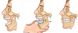
Incomplete dislocation
With this type of injury, the tooth can move vestibularly, orally, occlusally, towards adjacent teeth, or rotate around its axis. When a tooth is displaced in the vestibular or oral direction, the tooth appears shorter on the radiograph compared to the adjacent and uninjured teeth due to the fact that it is tilted. The greater the inclination of the tooth, the smaller the projection. The periodontal gap also changes. When the tooth displacement is insignificant, the periodontal fissure is widened only at the bottom of the alveolus. With a significant displacement, expansion occurs at the apex and at the lateral surfaces of the root (Fig. 1).
Sometimes incomplete dislocation to the vestibular side is combined with a fracture of the alveolar edge. On an x-ray, this looks like a violation of the continuity of the cortical plate limiting the tooth socket, and a strip of clearing appears in the cancellous bone (Fig. 2). When dislocated towards the occlusal surface, on the radiograph the injured tooth has the same length as the adjacent uninjured one. The root apex is located closer to the alveolar margin than that of the adjacent uninjured tooth.
The periodontal gap is widened on all root surfaces, especially in the area of the alveolar bottom. The cortical plate is not damaged in this type of dislocation, and therefore its continuity is not broken (Fig. 3).
When a tooth with an unformed apex dislocates, its growth zone appears elongated (Fig. 4).
Rice. 1. incomplete dislocation 21. On the RG-gram it looks shorter compared to the adjacent uninjured one 11. There is an expansion of the periodontal at the apex and at the lateral surfaces of the root.
Rice. 2. Dislocation 12 with a fracture of the alveolar edge.
Rice. 3. Incomplete dislocation of the central incisor in the occlusal direction.
Impacted dislocation
- The projected length of the injured tooth is the same as that of the adjacent uninjured one.
- There is no periodontal gap due to the impact of a wider part of the tooth into a narrower alveolus.
- The continuity of the cortical plate of the alveoli is disrupted.
Incomplete dislocation with significant displacement
- The tooth on Rg is projected shortened due to its oblique position.
- The periodontal gap is widened at the bottom of the alveolus and the lateral surfaces of the root.
Differential diagnosis of incomplete dislocation
- Incomplete dislocation with minimal displacement of the crown is differentiated from a bruise. In case of incomplete dislocation, the expansion of the periodontal fissure at one of the root surfaces will be determined. This is not observed with a bruise.
- Dislocation with significant displacement of the tooth in the oral or vestibular direction should be differentiated from impacted teeth, since the crown in the oral cavity may not be clinically determined due to swelling of the soft tissues.
- A differential diagnosis of tooth dislocation is carried out with rotation around the longitudinal axis in which the tooth rotates around the longitudinal axis.
- A slight expansion of the periodontal gap in incomplete dislocations should be differentiated from the physiological state of the periodontium during the period of completion of tooth root formation. It should be remembered that the periodontal fissure remains widened for a year after the completion of root development. In this case, knowledge of the timing of the completion of tooth root formation helps in diagnosis.
| Incomplete dislocation with tooth rotation around the longitudinal axis | Tortoanomaly of tooth position |
| Narrowing of the periodontal fissure around the root due to the fact that the wider part of it enters the narrow part of the alveolus | The periodontal gap has a uniform width throughout |
3. Symptoms and diagnosis
The clinical picture of chronic forearm dislocation is very diverse; it is determined, first of all, by the direction of the displacement. Up to 90% of such cases are posterior dislocations - with this option, the forearm appears unnaturally short, and the shoulder, on the contrary, appears disproportionately long. The opposite picture occurs when the forearm is dislocated anteriorly; in this case, severe tendon damage and muscle ruptures often occur. Displacements outward or inward, as well as divergent dislocation (divergence of the radius and ulna in any plane) are very rare and, as a rule, have the character of a combined fracture-dislocation.
The intensity of pain, limitation of mobility, severity of swelling and hematoma depend on the degree of displacement of the articular surfaces and damage to the ligamentous apparatus; in the most severe cases, ligaments and muscles are torn off, and conductive nerves and blood vessels are also damaged.
From a diagnostic point of view, dislocations and subluxations of the forearm are usually not particularly difficult. However, to clarify the situation, an X-ray examination is required, both before reduction (which often makes it possible to diagnose fractures, cracks or other complications), and after - to monitor the anatomical correctness of the joint.
One of the most informative techniques is an X-ray contrast study with the introduction of a contrast agent into the joint space.
About our clinic Chistye Prudy metro station Medintercom page!
Publications in the media
Frequency. Forearm dislocations rank 2nd (18–27%) among all dislocations. 90% of cases are dislocation of both forearm bones and anterior dislocation of the radius.
Cause: injury - falling on an outstretched arm while hyperextending the elbow joint.
Classification • Dislocation of both bones of the forearm (anterior, posterior, outward, inward, divergent dislocation) • Dislocation of the radius (anterior, posterior, outward) • Dislocation of the ulna.
Clinical picture. Posterior dislocation of the forearm is described • Pain • Forced position - semi-extended state • Active movements are impossible • Increase in the volume of the joint area • Symptom of spring fixation • Shortening of the forearm when viewed from the front • The olecranon protrudes posteriorly, is located above and posterior to the Huether line - the line connecting the epicondyles • The epiphysis of the humerus is palpated in the elbow bend.
Treatment
• Posterior dislocation of the forearm •• Anesthesia or local anesthesia •• Reduction. The patient lies on his back, the arm is abducted at the shoulder and bent at the elbow joint at an angle of 90°. They clasp the shoulder with both hands and press the olecranon with their thumbs. At the same time, the assistant pulls the forearm along its length and bends it at the elbow joint •• Immobilization with a plaster splint (on the back surface) for 1 week •• Exercise therapy from the first day, physiotherapy.
• Anterior dislocation of the forearm •• The assistant pulls the forearm along its length, slowly bending it at the elbow joint. The surgeon presses with his thumbs on the articular end of the humerus, while simultaneously moving the forearm proximally. After reduction, the forearm is extended and the limb is fixed with a plaster splint for 7–10 days in the supination position •• If conservative reduction is ineffective, surgical treatment is used •• If ossification occurs (after 2 weeks), phonophoresis with hydrocortisone, electrophoresis with lidase is prescribed, ossifications are removed, arthrodesis is used or elbow arthroplasty.
Features in children • 1st place among all dislocations in children • Fracture-dislocation is often observed in children - Galeazzi, Monteggia fractures, subluxation of the head of the radial bone • With external dislocation of the forearm bones, in 50% there is a separation of the apophysis of the internal epicondyle of the humerus • Reduction of uncomplicated dislocation is carried out without flexion or forearm extension.
Subluxation of the radial head. Causes: indirect injury, traction on the forearm, trying to hold the child’s hand when he falls, wearing clothes with narrow sleeves. The predominant age is 1–3 years; it is not observed in children over 5 years of age.
Pathomorphology: displacement of the head of the radius in relation to the capitate eminence of the humerus in the annular ligament and entrapment of the synovial fold of the joint. Clinical picture: the forearm is bent, pronated, the arm hangs along the body (pseudoparalysis); the child cannot localize the pain; on palpation - the greatest pain is in the area of the head of the radial bone; there is no active movement in the elbow joint, often movement is limited in the shoulder joint; passive movements: flexion and extension are painless, severe pain when rotating the forearm; X-ray examination reveals an increase in the distance between the head of the radius and the capitate eminence of the humerus. Treatment • Grab the wrist with one hand, and the elbow joint with the other hand (the surgeon’s first finger should be above the dislocated head of the radius). The forearm is pulled along the axis, then supinated and flexed to a right angle at the elbow joint with simultaneous pressure on the head of the radius. Reduction occurs during flexion with a characteristic click - the child calms down, active movements are restored. • After reduction, it is recommended to immobilize the arm with a scarf for 1-2 days. ICD-10. S53.0 Dislocation of the radial head
ICD-10 • S53.0 Dislocation of the radial head • S53.1 Dislocation of the elbow joint, unspecified
4.Treatment
In the acute period of dislocation or subluxation of the forearm, the method of choice is almost always closed reduction, the specific technique of which is determined by the clinical picture. This is especially true for pediatric and young patients, when the priority solution is minimal invasiveness and preservation of the integrity of the joint and periarticular tissues. This reduction is carried out either under local anesthesia or general anesthesia (depending on a number of individual factors).
Of course, the minimum condition for this is a timely visit to a doctor (attempts to straighten the joint on your own usually end in serious complications). The deadline is usually 21-28 days from the moment of dislocation - after which ossification makes conservative reduction impossible, i.e. It is no longer possible to do without surgery. However, in some cases, when an elderly or senile person with a chronic dislocation/subluxation of the forearm has no pain, and the functioning of the arm is preserved to a degree acceptable to the patient, surgery is also considered undesirable.
In all other cases, with chronic dislocation of the forearm, surgical intervention with arthroplasty is performed - reduction and restoration of the normal anatomy of the elbow joint. Hinge-distraction devices are used according to indications.
A common consequence of such a dislocation or subluxation is a significant decrease in strength in the injured limb.
Prevention
To prevent injuries, it is enough to follow safety rules on the road, in a work environment, and during sports training. To prevent forearm dislocation, it is important to wear comfortable shoes and engage in light sports to train the muscles and coordination. If you are involved in extreme sports, use protective equipment to soften the impact of a possible fall. If injury cannot be avoided, an immediate visit to the doctor is necessary for diagnosis and effective treatment.