Infectious diseases of the skeleton - this term sounds surprising to many. After all, infections are traditionally attributed to the respiratory organs and other body systems, but not to the bones. At the same time, there is such a category of diseases. But how to recognize it?
of the extrapulmonary tuberculosis research department of the Federal State Budgetary Institution "National Medical Research Center for Physical Research" of the Ministry of Health of Russia, Doctor of Medical Sciences, Honored Doctor of the Russian Federation, doctor of the highest qualification, told AiF.ru about what infectious diseases of the skeleton are, and also who should be especially wary of them. categories .
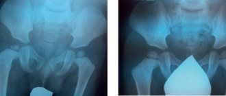
Uninvited bones. What is “second skeleton disease” and how is it diagnosed? Read more
The essence of the problem
“Infectious pathologies of the skeleton include inflammatory diseases of bacterial and viral origin, including osteomyelitis, purulent and viral arthritis. Such diseases are accompanied by local pain in the joint, segment of the spine, and increased body temperature,” says Evgeniy Peretsmanas. He also notes that in the advanced stage, a purulent abscess can form, destruction of the articular surfaces is noted, up to the loss of the joint and, accordingly, the motor function of the limb. Damage to the spine can lead to paralysis of the limbs and dysfunction of the pelvic organs. “With timely diagnosis and proper treatment, the prognosis for these diseases is relatively favorable, but otherwise even death is possible,” the specialist emphasizes.
These diseases are mainly dealt with by orthopedic traumatologists, neurosurgeons, and neurologists. The visit to the latter is due to the fact, the doctor notes, that it is they who first of all turn to patients with complaints of pain in the joints and spine. “Sometimes in such situations, diagnostic errors occur, radiation studies (X-ray, CT, MRI) are not always done on time, and infectious lesions of the skeleton can be effectively recognized and treated only on the basis of a visualized radiation picture,” says Evgeniy Peretsmanas.
Article on the topic
The child's bones hurt. What could it be?
Symptoms of bone and muscle diseases
Patients present the following complaints with diseases of the musculoskeletal system:
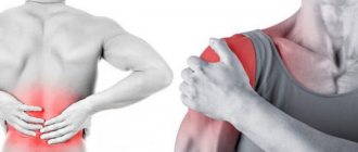
- Pain along the muscles or bones. May bother you in a certain position, at rest or after exercise. Starting pain is characteristic of osteoarthritis, morning stiffness is characteristic of rheumatoid arthritis.
- Deformation of the legs, arms, chest, flat feet, changes in the shape of small and large joints. Curvatures of the spine are possible in the lateral direction - scoliosis, in the anterior-posterior direction - kyphosis, lordosis.
- With muscle wasting, the limb looks thinner and its strength is reduced.
- Increased muscle fatigue, reduced range of motion.
- Swelling in the area of muscles, joints, at the site of injury.
- If the nerve that controls the muscle is damaged, the limb “does not obey” the commands of the brain, the person cannot perform the necessary movements or does it with difficulty. Sensory disturbances are also possible - “tingling”, “numbness”, “astringency”.
To exclude oncological diseases, an X-ray examination, CT scan of bones and joints, and MRI of soft tissues are performed. This allows you to differentiate diseases with similar clinical manifestations and determine the true size of the pathological focus. Electromyography helps distinguish nerve disease from muscle pathology. Sometimes, to clarify the diagnosis, doctors need to conduct a histological analysis of a piece of muscle tissue. Once the diagnosis is established, an individual treatment plan is drawn up.
How to treat the problem
Naturally, there should be no self-medication, because you can simply waste time and complicate your condition. In addition, it is worth understanding that there are a large number of infectious pathologies of the skeleton, therefore, various methods are used in their treatment. For example, due to the infectious component, antibacterial therapy is usually started at the initial stage of therapy. An analysis should first be carried out to determine the sensitivity of the pathogen.
If antibacterial therapy does not show the desired effect, then surgical treatment may be required as prescribed by a doctor, which is aimed at achieving several goals, says Evgeniy Peretsmanas. “Firstly, the infectious focus is removed, and then everything depends on the location of the lesion. In case of destruction of the joint after resection of the articular ends, arthrodesis is performed (fusion of the resected bones in a functionally advantageous position) or, if possible, endoprostheses are implanted to restore the function of the joint, but basically these operations are two-stage in nature with the installation of a temporary cement prosthesis at the first stage until the inflammatory process subsides,” - explains the doctor.
“It hurts my bones.” Why is rheumatism dangerous and why does it threaten young people? Read more
The doctor says that surgical treatment of the spine is particularly difficult. After all, first of all, it is necessary to remove the destructive infectious focus from the vertebral segments, and then the resected space is filled with either a bone autograft or an implant made of special bioinert materials. “In addition, to stabilize the affected part of the spine, posterior instrumental fixation is performed with special metal structures made of screws and rods. In case of complicated infectious spondylitis with compression of the spinal cord, these operations are complemented by decompressive interventions on the spinal canal,” explains the doctor.
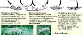
general information
Computed tomography (CT) is a cross-sectional imaging technique based on the X-ray principle.
A tomograph's X-ray tube is not static, unlike a conventional X-ray machine. Together with a digital detector, it rotates around the patient at high speed (up to 4 revolutions per second) inside the tomograph body, which is called a gantry. During scanning, the patient moves on a special table along the longitudinal axis of the body into the gantry aperture, so the X-ray beam “draws” a spiral trajectory on his body, hence the name spiral and multispiral computed tomography. From the data obtained by the detector, a three-dimensional model of the area under study is formed, and then sections in various planes are formed.
Diagnostics
To identify such pathologies, radiation diagnostic methods are primarily used: X-ray, CT and MRI. “If there is already suppuration with the formation of liquid pus, it can be detected using ultrasound. Various laboratory research methods are also used, including microbiological studies to determine microorganisms that are the direct causative agents of the disease,” says Evgeniy Peretsmanas.
Article on the topic
Fragile bones of the stronger sex. How a man can avoid osteoporosis People at risk should be especially wary of such problems. Today it includes people with diabetes, various chronic diseases, and HIV infection. “The cause of hematogenous introduction of infection into the osteoarticular system can be dental diseases, including carious processes in teeth, pathology of the ENT organs,” notes the specialist.
Of course, it is preferable not to wait until treatment is needed, but to engage in prevention. It should be understood that there is no specific prevention of infectious pathologies of the skeleton, the doctor notes. To maintain health, it is necessary to promptly and fully treat infectious diseases and sanitize foci of chronic infection. It is important to avoid injuries and receive qualified assistance in a timely manner in case of injuries (cuts, wounds, fractures).
Prevention of osteoporosis
In order to prevent the development of osteoporosis, it is important to assess the level of vitamin D in the blood, because we are mostly residents of northern latitudes. With a lack of sunlight, vitamin D deficiency develops, and without it, as is known, calcium is not absorbed, the deficiency of which leads to the progression of the disease.
Article on the topic
Deficiency A and D. About the vitamins that we lack The recommended dose of calcium after 50 years is 1000 mg per day. Organic, so-called “chelated” calcium, obtained from oyster shells, is best absorbed. It should be remembered that uncontrolled use of calcium supplements causes changes in arterial walls and increases the risk of developing myocardial infarction. Therefore, it is advisable to get calcium from food.
The leaders in calcium content are hard cheeses and sesame. Consume milk, cottage cheese, broccoli, cauliflower, salmon meat. But it is better to limit alcoholic and caffeinated drinks. And, of course, an important component of preventing osteoporosis is a healthy lifestyle and sufficient physical activity. After all, when moving, blood circulation improves and building material for bones is delivered in the proper quantity.
If you are over 50...
If you are over 50 years old, experience minor back pain, and are concerned about your health, you should be screened for osteoporosis. First, briefly: what is osteoporosis? Osteoporosis is a body condition in which bones lose calcium and become brittle and less dense. As a result, at the slightest injury they are easily deformed or broken. Why is this happening? The most common myth is that osteoporosis develops in people who get little calcium from their diet. In fact, you can eat calcium day and night and achieve nothing except disruption of salt metabolism and the possible formation of kidney stones. Osteoporosis is a disease in which calcium is poorly absorbed by the bones or is “washed out” of them, even if there is an excess of it in food. This could be due to several important reasons.
Gymnastics is the best helper!
Many methods are used to combat osteoporosis; they can be conditionally divided into two completely compatible and complementary groups: medications and a healthy lifestyle, which here means reasonable physical activity (gymnastics), dosed sun exposure and massage treatments. For example, with regular gymnastics 3-4 times a week for 30-40 minutes, you can achieve an increase in bone mass by 3-5% within the first months. In this case, of course, it is advisable that a specialist select a set of gymnastic exercises taking into account your characteristics and capabilities. Massage procedures enhance the effect of gymnastic exercises, allowing you to improve blood circulation and “pumping” of the back muscles: manual and vacuum massage are especially good in this sense. However, care must be taken and the procedures must be done very gently to avoid injury to the bones.
How is the CT scan procedure performed at Medscan in Moscow?
Before the examination, the x-ray technician carefully interviews the patient, collects an anamnesis of life and illness (information about previous diseases). Sometimes, before a contrast study, results of a test of renal excretory function (creatinine test) may be needed. If the patient has data from previous studies (CT, MRI, PET-CT, etc.), then their images are imported into the information system, and scans of extracts and analyzes are attached to the medical history.
The patient lies down on the tomograph table and the first “marking” low-dose scan (topogram) is taken, on which the rest of the study is marked; the table moves back and forth, and the patient may be asked to hold their breath through a speaker.
If a contrast study is required, after the topogram an injector (a special automatic syringe with a contrast agent and saline solution) is connected to the intravenous catheter.
Next, one or more CT scans are performed, and between them, a contrast agent is injected intravenously into the patient using an injector at high speed; The table moves forward or backward with each scan and instructions to hold your breath may be heard again. The patient can hear the tube and detector rotating inside the gantry, but the noise level is low and does not cause discomfort. The patient does not experience any sensations from the operation of the X-ray tube.
The x-ray technician controls the examination from the next room, which has a special window through which he can see the patient; The speaker and microphone built into the tomograph allow the laboratory assistant and the patient to be constantly in touch.
The entire CT procedure lasts from 3 minutes if the study is non-contrast, to 20 minutes if contrast is required.
The contrast agent is safe for the body and is excreted by the kidneys within 12 hours.
After the examination with contrast, we monitor the patient’s well-being for 20 minutes, during which time we prepare a digital medium on which all obtained images are recorded. After a 20-minute wait, the intravenous catheter is removed from the patient and the procedure is completed.
Then a team of radiologists comes into action, analyzing hundreds and thousands of images obtained during each study. The doctor carefully examines all the sections obtained, compares them with previous studies and analyzes what he saw, taking into account the anamnesis, medical history and treatment performed. The result of image analysis is a protocol (conclusion), which describes the identified changes and draws conclusions about their nature. It is ready within a few hours, maximum within a day.
This is not an easy job, but our centers employ real professionals who love their job; Our radiologists constantly improve their qualifications by participating in consultations, seminars, webinars and conferences, reading modern foreign literature in their specialty.
Calcium stealers
Products that impair the absorption of calcium or contribute to its “leaching” from the body: Sugar, honey, wheat flour products (pasta, white bread), coffee, tea. All refined foods and caffeine interfere with the absorption of calcium in the intestines. Factory-made meat products (natural meat does not affect the absorption of calcium). Excess salt. Displaces calcium from the body, which is lost along with urine. Just one extra teaspoon of salt per day can cause bone mass to decrease by 1.5 percent per year.
Key word - "calcium"
The strength of bones is affected not so much by the absolute amount of calcium in food, but by the ratio of calcium and phosphorus in the diet: the amount of calcium should be one and a half to two times the amount of phosphorus. In addition, for complete absorption of calcium, the diet must also contain magnesium, boron, copper, manganese, zinc, vitamins B6, C, K and folic acid, as well as certain proteins and fats that promote the absorption of vitamin D. A properly formulated diet containing all necessary components can seriously reduce the threat of osteoporosis.
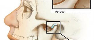
How common are joint diseases in older people?
A joint is the place where bones meet; it is a kind of cavity with articular fluid, which serves to reduce friction between the bones. They are the ones who help a person make all movements. While our joints are healthy, we do not notice their work. Only over the years, when faced with unbearable pain when walking, squatting or climbing stairs, do we begin to appreciate them. Joint diseases in older people are often irreversible and chronic.
The problem of joint diseases has always been relevant for humanity. Since ancient times, people have been looking for ways of salvation. Even now, despite the achievements of modern medicine, every third person in the world suffers from joint diseases. Each of us is at risk, regardless of gender and social status, age and habits. When we are young, we don’t think about serious illnesses.
- Recommended articles to read:
- Social services for older people
- Diseases of old age
- Valuable tips on how to choose a boarding house
Restriction of freedom of movement due to pain is very common. Unbiased statistics show scary figures: almost all elderly people over 75 years old have problems with joints. And at the age of 65 years, 70% of older people are already concerned about the symptoms of the disease.
It is obvious that joint diseases in older people occur against the background of age-related changes, namely: modifications of joints (lose their round shape), muscles (become flabby), tissues (elasticity decreases). In addition to natural aging, other reasons can intensify the disease or accelerate its exacerbation.
Stages of multiple myeloma
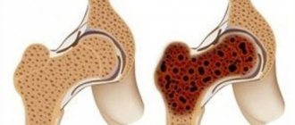
Stage 1 - characterized by mild anemia, calcium remains within normal values, M-protein is low, the number of bone lesions is no more than 5.
Stage 2. Moderate anemia (hemoglobin within 85-100 g/l), there is an increase in calcium (up to 3 mmol/l) and M-protein. There is also an increase in the number of osteolysis foci. For the second stage, their number should not exceed 20.
Stage 3 multiple myeloma is diagnosed when at least 1 of the following symptoms is detected:
- Hemoglobin is below 85 g/l, which corresponds to severe anemia.
- Exceeding the calcium value by more than 3 mmol/l.
- M-protein level is more than 70 g/l.
- The number of bone lesions is more than 30.
What should be on the table
The menu of a person predisposed to osteoporosis should include the following products: Milk and dairy products (preferably low-fat). They have an optimal ratio of calcium and phosphorus. In addition, fat makes it difficult to absorb calcium, so low-fat dairy products are preferable. Fresh vegetables and fruits, especially all types of cabbage (white cabbage, broccoli, cauliflower), carrots, turnips. In addition to calcium, they contain the entire “support group” of microelements necessary for the full absorption of calcium. Legumes, walnuts, pumpkin and sunflower seeds, and vegetable oils contain proteins and fats necessary to strengthen bone tissue and absorb vitamin D.