There are many rare pathologies, one of which is osteopetrosis (marble disease, congenital osteosclerosis, Albers-Schoenberg syndrome). This is a severe hereditary disease that is characterized by damage to the skeleton and bone marrow. Bone density increases, but at the same time they become more fragile, and the functioning of the hematopoietic system is disrupted.
Doctors distinguish 2 forms of the disease: early and late. Early osteopetrosis manifests itself in infants up to 12 months, it has a severe course and often ends in death. The late form is observed in adolescents, it manifests itself with less pronounced symptoms, and has a favorable prognosis. Treatment of the pathology is quite long and complex, but there is still a chance of cure.
General information
Osteopetrosis is a hereditary genetic disease that manifests itself in the form of diffuse compaction of the bones of the entire skeleton, increased fragility and reduced bone marrow hematopoiesis. This rare pathology was first described by the German surgeon Ernest Albers-Schönberg at the beginning of the twentieth century; it is also called Albers-Schönberg disease or congenital familial osteosclerosis .
In the classification of diseases, a code according to ICD-10 is assigned - Q78.2. The disease is rare, as it occurs in one person in 20 thousand individuals.
Causes
Osteopetrosis can be inherited in an autosomal dominant or recessive manner, and very rarely in an X-linked recessive pattern. The main defect in bone reabsorption is the insufficient production or dysfunction of cells called osteoclasts. These cells are responsible for bone resorption and help maintain bone health, which depends on the balance between bone resorption (osteoclasts) and bone formation (other specialized cells called osteoblasts). The human skeleton is completely regenerated every 10 years. In this context, osteoclasts are essential for bone turnover (replacing old bone with new bone), bone remodeling, and also repair of microcracks.
Human traits, including classical genetic diseases, are the product of the interaction of two genes for a given condition, one from the father and one from the mother
The adult type of osteopetrosis is inherited as an autosomal dominant genetic trait. Dominant genetic disorders occur when only one mutated copy of a gene is enough to cause a specific disease. The mutated copy of the gene may be inherited from either parent or may be the result of a mutational event occurring directly in the affected individual. The risk of passing an abnormal copy of the gene from the affected parent to the offspring is 50% in each pregnancy. The risk is the same for men and women.
Malignant childhood osteopetrosis is inherited as an autosomal recessive genetic trait. Recessive genetic disorders occur when a person inherits two abnormal copies of a gene, one from each parent. If a person receives one normal and one abnormal copy of a disease gene, they will be a carrier of the disease, but usually asymptomatic. The risk of two carrier parents passing on an abnormal copy of the same gene, and therefore having an affected child, is 25% in each pregnancy. The risk of having a carrier child, like the parents, is 50% with each pregnancy. The chance that a child will receive a normal copy of the gene from both parents is 25%. The risk is the same for men and women.
The X-linked form of osteopetrosis is recessive and extremely rare. X-linked recessive diseases are caused by an abnormal gene on the X chromosome and occur primarily in males. Women who have an abnormal copy of the gene present on one of their X chromosomes are carriers of the disease: they usually do not show symptoms because women have two X chromosomes. Males have one X chromosome inherited from their mother, and if a male inherits an X chromosome that contains an abnormal (pathological) gene, he will develop the disease. Women who are carriers of an X-linked recessive disorder have a 25% risk in each pregnancy of having a carrier daughter like themselves, a 25% chance of having a non-carrier daughter, a 25% risk of having an affected son, and a 25% chance of having an unaffected son. son.
Intermediate osteopetrosis can be inherited as an autosomal recessive or autosomal dominant genetic trait.
Pathogenesis
The basis of normal bone tissue development is the balance of formation of osteoblasts in bone tissue and resorption in osteoclasts. Osteoclasts by their nature are bone macrophages that have many lysosomes in their structure, upon release of which they release hydrolytic enzymes that cause resorption of the bone itself or calcified cartilage. The role of osteoblasts is to create and mineralize new bone tissue, and osteoclasts are to destroy old bone.
In osteopetrosis, regardless of the number of osteoclasts (their increase, decrease, or even normal), their function is impaired, and the amount of the enzyme carbonic anhydrase is insufficient. The mechanism of development of osteopetrosis is not fully understood, but enzyme deficiency provokes disruption of the proton pump in osteoclasts and, as a consequence, resorption processes, and an acidic environment is required for the transition of calcium hydroxyapatites to the microvasculature. Against the background of these disorders, the processes of bone formation, their compaction (osteosclerosis) and subsequently the formation of an excess amount ( hyperostosis ) are supported. Endosteal and enchondral ossification occurs with excessive formation of sclerotic tissue.
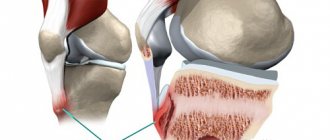
Functions and macro images of osteoblasts and osteoclasts
This mesenchymal dysplasia is accompanied by increased formation of a compact substance, the transformation of the spongy substance into a solid white mass resembling marble. As a result, the ability to retain large amounts of salts increases, which leads to deep-seated disturbances in the metabolism of phosphorus and calcium.
Fatal marble disease - osteopetrosis
The diagnosis of deadly marble sounds like a death sentence, because the proliferation of bone structures leads to the displacement of the bone marrow, which is responsible for hematopoiesis. Blood cells are constantly renewed and in order for mature functional leukocytes, erythrocytes, etc. to enter the bloodstream, hematopoietic functions must be fully exercised. Often with osteopetrosis, there are foci of extramarrow hematopoiesis, for example, in the spleen. Since the pathology develops slowly, they are a way to compensate for anemia , but at a certain point the body cannot cope and critical anemia occurs, decreased immunity , infection, the development of pneumonia and premature death.
It is immediately clear that osteopetrosis is a “deadly marble”, that it is a bone pathology: frequent fractures, deformities, growth retardation, pain in the legs and lower back. Sick children have noticeable facial changes - wider cheekbones, far-set eyes, a depressed nose, full lips.
Osteopetrosis in a child
Treatment methods
Treatment of “deadly marble” is an extremely complex process. Previously, doctors only fought symptoms with the help of iron-based drugs, blood transfusions (blood transfusions), vitamins, and glucocorticoids. As a result, the patient still faced disability and death.
Bone marrow transplantation is the only way to cure marble disease
Modern medicine makes it possible to get rid of even the malignant form of osteopetrosis, in which children died after 2–3 years. To do this, you need to undergo a bone marrow transplant. This is a complex procedure that has helped save the lives of many patients.
For the operation, you will need biological material from a close relative or a suitable donor. This is important because there is a risk that the child's body will reject the bone marrow. If the surgical intervention is successful, the development of the pathology stops. However, transplantation will not help eliminate complications that have already developed.
Important. A child with marble disease has a chance of recovery if a bone marrow transplant is performed before 6 months. Therefore, it is so important to diagnose the pathology as early as possible and begin to treat it.
The following medications will help supplement treatment:
- Vitamin D-based calcitriol stimulates bone resorption (destruction of bone tissue by osteoclasts) and stops further bone growth.
- Y-interferon activates the work of leukocytes, strengthens the immune system, reduces the volume of spongy bones, normalizes the level of hemoglobin and platelets. Interferon is recommended to be combined with calcitonins.
- Erythropoietin is prescribed for anemia.
- Glucocorticosteroids prevent bone hardening and anemia.
- Calcitonin (thyroid hormone) is used in large doses and helps improve the patient's condition.
To strengthen neuromuscular tissue and improve the patient’s general condition, it is recommended to supplement the complex with physiotherapy, diet, periodic examination by an orthopedist, exercise therapy, massage, and spa treatment.
Pathological fractures are treated with the help of reposition (comparing bone fragments), applying a plaster cast, and skeletal traction (gradually reducing the fragments using weights and special devices). If a femur is fractured in the neck area, endoprosthetics is performed (replacement of the hip joint with a prosthesis). If there is severe curvature of the legs, an osteotomy is prescribed, which eliminates the deformity. If a patient is diagnosed with osteomyelitis, then he is prescribed antibiotics, immobilization, the body is detoxified, the immune system is strengthened, and the abscesses are opened and drained.
Classification
There are several forms of the disease depending on the severity of the picture and the characteristics of inheritance:
- The malignant infantile early form (autosomal recessive) is typical for carriers of childhood, begins in intrauterine development, and most often leads to death.
- Autosomal dominant late benign form with thickening of the bones of the cranial vault.
- Classic pathology described by Albers-Schönberg, causing significant deformities of the spine and bone-in-bone changes in the pelvis. A chaotic accumulation of bone structures is observed, the bone marrow space is filled with layered conglomerates or lamellar bone tissues of an arcuate nature of gluing.
Types of pathologies
Doctors distinguish 2 forms of the disease:
- Malignant juvenile osteopetrosis. The pathology is detected in newborns up to 12 months; it has a severe course and often ends in death. The disease is manifested by a large head, wide-set eyes, squint, pale skin, and short stature. In this case, you cannot do without a bone marrow transplant operation, which must be carried out over 2–3 years.
- Autosomal dominant osteopetrosis. The disease is discovered in adolescents or adults, usually by chance during an x-ray. This is a milder, benign form of pathology. At first it has a gradual course, the only sign is frequent fractures. A little later, if left untreated, complications may appear in the form of deafness or paralysis of the facial nerve.
As you can see, the most dangerous is early marble disease, which requires quick radical action.
No ads 1
Symptoms
The first symptoms appear at an early age; children experience:
- causeless pain in the limbs;
- migraine;
- late first steps and first teeth;
- pallor;
- dizziness and tendency to faint ;
- weakened immunity ;
- weakened or lost vision, hearing;
- increased fatigue when walking;
- growth retardation;
- visually noticeable deformations of the skeleton and skull without deviations in physical development.
The disease is also called “marble” because with an increased amount of bone tissue and its calcification, bone fragility increases and elasticity is lost. Patients are at high risk of pathological fractures, most often of the hip bones. Frailty is the main manifestation of the disease in adults. Due to the preservation of the periosteum, their fusion is usually timely, but if the endosteum is not involved in the processes of osteogenesis and the bone marrow canal has succumbed to sclerosis, then it can be slowed down. In places of fractures, calluses form and osteomyelitis , which often becomes the cause of sepsis .
Excessive bone tissue and hyperostotic dysplasia leads to:
- deformations of the bones of the skull, chest, disorders of the spinal trunk;
- compression of nerves and, as a result, to peripheral paresis , paralysis and even blindness ;
- slow development of hypoplastic anemia and hemorrhagic diathesis .
Danger
Due to the increased fragility of bones, the patient often experiences fractures with minimal impact. Most often, the strong bones of the hip are damaged.
Doctors highlight other consequences of “deadly marble”:
- Slow jaw development, prolonged teething, dental caries, oral infections.
- Dropsy of the brain, increased blood pressure, blindness, deafness, facial paralysis, nystagmus.
- Frequent infectious diseases, for example, pneumonia, cystitis, sepsis.
- Anemia.
- Predisposition to bleeding.
- Deformation of different parts of the skeleton (skull, spine, limbs).
- Hepatosplenomegaly.
- Enlarged lymph nodes.
- Retarded physical and mental development.
In the absence of competent therapy, the risk of disability and death increases.
Tests and diagnostics
Processes of osteosclerosis can also be observed in other pathologies, so differential diagnosis with hypervitaminosis D, hypoparathyroidism, Paget's disease, melorheostosis, lymphogranulomatosis and metastases .
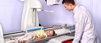
For diagnosis, it is necessary to conduct x-ray studies that reveal a sharp compaction of all the bones of the musculoskeletal system - the base of the skull, long bones, ribs, and spine. It is expressed in the form of absolute opacity for x-rays, the absence of the frontal sinuses and other air cavities, the medullary canal and the isolated cortical layer. X-rays of tubular bones may show transverse clearings resembling a marble pattern. With an unchanged external shape, the bones may have thickenings and roundings at the epiphyseal ends and club-shaped thickenings of the metaphyses of the bones.
X-ray of marble bones
Blood tests reveal an increased number of leukocytes and the presence of normoblasts (immature forms of blood cells), hypocalcemia . To study the dynamics of the disease, changes in bone density are monitored ( densiometry ).
Affected Populations
In the general population, 1 in 250,000 people are born with malignant childhood osteopetrosis. Higher rates were found in selected regions of Costa Rica, the Middle East, Sweden and Russia. Men and women suffer in equal numbers.
The adult type of osteopetrosis affects approximately 1 in 20,000 people. Men and women suffer in equal numbers.
The X-linked form of osteopetrosis predominantly affects men due to the way the mutation is inherited. Due to the rarity of cases, there are no population-based studies.
Marble disease in children
Sclerosis of the bone marrow canal and disruption of hematopoietic processes leads to the development of the following pathologies in children:
- hypochromic anemia ( anemia );
- compensatory hepatomegaly (scarcely enlarged liver);
- splenomegaly (pathologically enlarged spleen);
- lymphadenopathy (enlarged lymph nodes).
hydrocephalus may occur .
What it is
The diagnosis of osteopetrosis is made very rarely, in approximately one case in 300 thousand. However, in some Russian regions the disease is detected much more often. In particular, in Chuvashia this figure is 1 in 3,500 people, and in the Mari El Republic – 1 in 14,000. This difference in occurrence is due to the health characteristics of the indigenous population.
Marble disease is called because the patient’s bones, when cut, look like smooth marble, while healthy bone resembles a porous sponge. Because of this structure, bone tissue becomes too strong and at the same time fragile: it is very difficult to saw, but can be easily split.
Osteosclerotic changes develop mainly due to the endosteum - a thin connective tissue layer lining the inner surface of the bones and forming the medullary canal. In some areas, growth of the periosteum is possible - the periosteum, which forms segments of spongy tissue on the surface of the bones: normally there should be dense bone substance here.
The medullary canals of long tubular bones narrow or disappear altogether, being completely replaced by a dense mass. The most severe pathological changes are noticeable in the growth zones, especially at the top of the tibia and humerus, as well as in the lower part of the thigh and near the wrist - these areas literally “inflate” and take on the shape of a flask.
The spongy tissue of flat bones - the skull, shoulder blades, vertebrae, collarbones - changes to dense bone substance, which can lead to enlargement and deformation of the head. Due to the narrowing of the openings through which the optic and auditory nerves exit, progressive deterioration of visual and auditory function may occur.
The cortical layer of the bones of the skull can be rebuilt unevenly and form roughness on the inner surface of the bones. This sometimes provokes hemorrhages into the epidural space between the skull and the dura mater. In some cases, blood may accumulate in the area between the arachnoid and dura mater (subdural hemorrhage).
The exact mechanism by which marble disease develops has not been established. The number of osteoclasts may remain normal or even increased, but their function is in any case impaired
With marble disease, weight increases and the structure of the bone substance is disrupted. Bone cells are randomly arranged and form typical streamlined layered structures that are found in zones of enchondral growth.
When there are many such structures, the bone loses its natural strength and can no longer bear the usual loads.
Diet for osteopetrosis
Diet on vegetables and fruits
- Efficiency: 3-4 kg in 7 days
- Lead time: 3-7 days
- Cost of products: 1200-1300 rubles. in Week
Nutrition must be nutritious and include in full all the necessary nutrients and nutrients. The diet should be rich in vitamins , so the menu includes a lot of fresh vegetable and fruit salads and juices with berries.
To prevent increased processes of calcification and bone formation, it is necessary to limit the consumption of foods containing calcium.
Forecast
Since a pathogenetic treatment strategy has not been developed, it is not possible to normalize the processes of bone formation, gradual disability occurs, one can only try to cope with the consequences and complications of this process. However, timely treatment of anemia and bone deformities makes it possible to create favorable living conditions.
To prevent pathology in the child, parents should undergo prenatal diagnosis after the 8th week of pregnancy, especially if there were people in the family tree suffering from this disease.