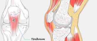
Tendinitis (ICD10 code No. M70-M79) is a disease in which an inflammatory-degenerative process develops in the tendons. A pathological process can develop in the muscle tendons that provide movement in the knee, hip, shoulder, and elbow joints. There is tendonitis in the wrists and feet. Rheumatologists at the Yusupov Hospital diagnose the disease using the latest equipment from leading global manufacturers.
Which doctor treats tendinitis? At the Yusupov Hospital, tendonitis is treated by a rheumatologist. With the development of degenerative changes in the tendon, the need for surgical intervention arises. The operations are performed by orthopedic traumatologists. Rehabilitators implement a rehabilitation treatment program. Psychologists help the patient cope with the problem, avoid depression and quickly return to normal life.
In most cases, treatment for tendinitis is performed on an outpatient basis. Patients with severe pathological processes undergo inpatient treatment in the therapy clinic. The level of comfort in the rooms meets European standards. Doctors take an individual approach to the choice of medications, physiotherapeutic procedures, type of massage and gymnastic exercises.
Severe cases of tendinitis are discussed at a meeting of the Expert Council with the participation of doctors and candidates of medical sciences, doctors of the highest category. Leading specialists in the field of rheumatology and orthopedics collectively determine patient management tactics. If conservative therapy is ineffective, the patient is offered surgical treatment. Orthopedists are fluent in the tactics of modern surgical interventions on tendons.
Knee tendonitis
Tendinitis of the tendons of the knee joint (ICD code 10 No. M76) is a consequence of an injury. Post-traumatic tendonitis develops in athletes, older people and people involved in heavy physical labor. The provoking factors of knee tendinitis are constant jumping on hard surfaces, an ill-conceived training regimen, prolonged use of antibiotics, wearing uncomfortable shoes, injuries to the knee joint, flat feet, valgus position of the feet, poor posture and pathological changes in the spine. The cause of secondary tendinitis can be rheumatic diseases, metabolic disorders, or previous infectious diseases. Over time, tendons can undergo degenerative changes and develop tendinitis ossificans.
The following symptoms are characteristic of tendinitis of the tendons of the muscles that provide movement in the knee joint:
- During active movements that are performed with the participation of the affected tendon, while similar passive movements are painless;
- Pain on palpation along the affected tendon;
- Redness, increased body temperature over the tendon area;
- Crepitus (crunching) when performing movements in the knee joint.
Doctors at the Yusupov Hospital make a diagnosis of tendinitis based on the patient’s complaints, life and illness history, characteristic clinical manifestations of the disease and instrumental research data. Changes in blood and urine tests are detected only with secondary symptomatic tendonitis. In patients suffering from rheumatic diseases, anti-citrullinated antibodies and rheumatoid factor are detected in the blood. Metabolic disorders increase the level of creatinine and uric acid. Ultrasound examination and magnetic resonance imaging make it possible to determine the localization of the pathological process. Changes on the x-ray will be present in the case of bone pathology.
Diagnostics
Research methods • Laboratory studies: changes are observed only with concomitant rheumatic pathology • X-ray examination •• Possible calcium deposits in the tendons •• Heel spurs - with tendonitis and tendobursitis of the Achilles tendon or plantaris tendon •• With tendonitis of the patellar ligament, signs of aseptic necrosis of the tuberosity are possible tibia (Osgood-Schlatter disease) • Special studies •• Echography of the tendon - contraction of the tendon, changes in its structure. It is necessary to ensure that the ultrasound wave does not cross the tendon along the oblique diameter. •• CT/MRI are informative for identifying tendon ruptures, but are not very informative in diagnosing stenosing tenosynovitis.
Differential diagnosis • Tendon avulsion • Bursitis (often combined with tendonitis should be remembered) • Infectious tenosynovitis (usually on the arm; pain on palpation and swelling are located along the tendon sheath, and not at the point of attachment to the bone).
Shoulder tendonitis
Inflammatory-dystrophic diseases of the shoulder joint are based on neurodystrophic reflex disorders, which are closely related to cervical osteochondrosis. Tendinitis of the shoulder joint (ICD code 10 - M75) develops under the influence of direct or indirect trauma to the arm, shoulder against the background of osteochondrosis of the cervical and upper thoracic spine. The disease develops in the presence of the following factors:
- Occupational hazards;
- Microtrauma of the tendons of the muscles of the shoulder girdle, which are associated with monotonous and frequently repeated movements of the arm;
- Excessive loads on the upper limb;
- Hand inactivity for a long time.
Tendinitis of the supraspinatus muscle is based on microcirculatory disorders with the subsequent development of a focus of tendon necrosis. This causes reactive inflammation and swelling of the tendon segment near the location of the lesion. Due to the thickened segment of the affected tendon, it is difficult to move the arm to the side and upward. The patient experiences pain, most pronounced in the lateral part of the shoulder below the edge of the acromion process. The intensity of the pain increases at night. Sometimes pain does not interfere with daily activities, but it limits self-care.
Using palpation, the doctor determines pain in the anterior outer area of the shoulder under the edge of the acromion process of the scapula. The pain intensifies when the shoulder is moved to the side. For supraspinatus tendonitis, doctors identify a positive Dowborn's sign. It manifests itself as maximum pain under the edge of the acromion process on the outside of the left shoulder at the 15 o'clock level and the right one at the 9 o'clock level during active abduction of the arms to the sides and raising them upward. The pain disappears or weakens with further upward movement of the arm. When you lower your arm, severe pain appears again. It can cause a real arm block. Active movements of the affected upper limb become impossible.
Coracobrachialis tendinitis is characterized by anterior tenderness at the level of the coracoid process of the scapula. The arm is slightly abducted, extension is limited. When performing passive rotational movements, the patient does not feel pain. Pain appears during active movements of the affected upper limb.
A sign of tendovaginitis of the long head of the biceps muscle is Ergason's symptom: passive rotational movement of the forearm, bent at the elbow joint at an angle of 90°, outward, with resistance from the patient, causes pain in the front of the shoulder along the projection of the tendon of the long head of the biceps muscle. With tendonitis of the infraspinatus and teres minor muscles, pain occurs with external rotation of the shoulder. In order to determine the exact cause of shoulder pain, rheumatologists prescribe radiography of the shoulder joint, ultrasound and magnetic resonance imaging to the patient. With the help of clinical and biochemical studies, the inflammatory and autoimmune nature of tendonitis is excluded.
Shoulder tendinitis
Shoulder tendonitis is damage and inflammation of the ligaments of the shoulder joint.
The most common injuries:
- Supraspinatus or infraspinatus tendinitis is an injury to the supraspinatus tendon.
- Tendinitis of the teres minor (major) muscle is an injury (tear) to the tendon of the teres minor (major) muscle.
- Biceps tendinitis - damage to the tendons of the small or large head of the biceps.
- Rotator cuff tendinitis. The rotator cuff is made up of ligaments and tendons of four muscles: supraspinatus, infraspinatus, teres minor and subscapularis. And accordingly, the term “rotator cuff tendinitis” refers to damage to one or more tendons that form the cuff itself. The cuff itself performs the function of stabilizing the shoulder.
Calcific tendinitis of the shoulder
Calcific tendinitis of the shoulder is a disease of the ligamentous apparatus of the shoulder caused by the accumulation of calcium salts in the ligaments and tendons of the shoulder. Such a cluster can be point-like or quite massive.
Calcium salts are clearly visible on an x-ray.
Most often, calcium salts are deposited on the tendons that form the rotator cuff (supraspinatus, infraspinatus, teres and subscapularis).
What is the root cause of the disease - the deposition of calcium salts or tearing of the ligaments - is not yet known with certainty. Everything is very interconnected: a tear in the ligaments can cause the formation of calcifications and, conversely, accumulated calcifications can damage the ligaments.
But it has been reliably clinically proven that physiotherapy, and mainly shock wave therapy, is the main conservative method of treating this disease. And that is why shock wave therapy is often called biosurgery, since in the recent past such diseases were treated mainly by surgical methods.
Tendinitis of the knee joint (patella)
Knee tendinitis
Knee tendonitis
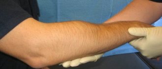
Elbow tendonitis
Inflammation of the elbow tendon is called tendonitis. In ICD 10, the disease is assigned code M70.9 (soft tissue diseases associated with stress, overload and pressure). Tendinitis of the lateral tendon of the elbow is often called “tennis elbow,” and tendinitis of the tendons located on the inner surface of the elbow is often called “golfer’s elbow.” At the initial stage, symptoms are mild or absent altogether. A person feels slight discomfort in the hand, which is attributed to overload or fatigue.
As the inflammatory process intensifies, the intensity of clinical manifestations also increases. The pain is usually clearly localized and does not radiate to the forearm or wrist. Painful sensations first occur when flexing, extending the joint, or turning the hand outward or inward. The intensity of the pain syndrome with strong tension in the skeletal muscles, since their contraction leads to tension in the tendon. In the absence of medical intervention, the patient begins to feel pain at rest. It becomes aching and constant, preventing you from sleeping peacefully and doing your daily work efficiently.
There is a restriction of movements in the elbow joint, swelling of the periarticular tissues. Pathological exudate interferes with the smooth sliding of the tendon. A person deliberately spares his hand, since flexion, extension, and rotation cause him severe pain.
Sometimes, when palpating an inflamed tendon, a rheumatologist discovers characteristic nodules. Their formation indicates the traumatic nature of tendinitis. Fibrous tissues grow and become denser over time. Their dimensions can exceed several millimeters. When you press on the nodules, they move slightly due to the tension of the muscle fibers. The seals are elastic, elastic, but can harden during the crystallization of calcium salts. Sometimes fibrous nodes sometimes resolve, but only if there are no calcium deposits in them.
When listening with a stethoscope, you can hear the specific sounds that a swollen, inflamed tendon makes during movement. They are almost indistinguishable at the initial stage of the disease. The noise is more accentuated when fibrous nodes form.
The inflammatory process from the tendon quickly spreads to the subcutaneous tissue and epidermis. Due to the rush of blood to the surface of the skin, it appears reddened. This is clearly visible when comparing diseased and healthy elbow joints.
Ultrasound diagnostics is the most informative for traumatic tendonitis, as it allows you to detect fiber ruptures. X-rays allow you to assess the condition of bone and cartilage tissues. Magnetic resonance imaging helps to determine the degree of damage to tendon tissue, the number and location of fibrous nodules, and calcifications.
Achilles tendonitis
Achilles tendonitis (ICD 10 code in adults – M76.6) develops for the following reasons:
- Excessive load on the Achilles tendon (overstrain of the calf muscle, frequent running uphill or downhill, a sharp increase in the amount of physical activity);
- Using uncomfortable running shoes;
- Frequently wearing high-heeled shoes and changing heels to flat soles every night.
The patient experiences pain along the Achilles tendon, usually closer to the heel. Swelling and local redness of the skin appear in the tendon area. The pain intensifies when standing on your toes and jumping on your toes. Pain may occur within the first few minutes of walking in the morning. Mobility of the ankle joint is limited.
Short description
Tendinitis is an inflammation of tendon tissue, usually observed at the point of attachment to the bone or in the area of the muscle-tendon junction; usually combined with inflammation of the tendon bursa or tendon sheath.
Code according to the international classification of diseases ICD-10:
- M65 Synovitis and tenosynovitis
- M75 Shoulder lesions
- M76 Enthesopathies of the lower limb, excluding foot
- M77 Other enthesopathies
Tendinitis of the patellar tendon and thigh muscles
Chronic patellar tendinitis is known as “jumper's knee.” The quadriceps muscles are subject to excessive stress during many sports. Microtraumas systematically occur in them. Tendinitis develops more often in athletes who take anabolic steroids.
At the initial stage of the disease, touching the tendon causes pain. With more serious injuries, swelling of the tendon may occur. Over time, a degenerative process develops and the tendon may rupture. In the patellar ligament, degeneration occurs at the inferior pole of the patella or below, at the site of attachment to the tibial tuberosity.
At the onset of the disease, the patient feels pain only after physical activity. It is gradually felt during exercise, and over time does not leave the person even during rest. During the examination, rheumatologists determine the location of the pain by asking the patient to straighten the leg at the knee and applying pressure to the affected tendon. The diagnosis is confirmed using ultrasound. X-rays and magnetic resonance imaging can help identify calcifications within the tendon.
Quadriceps tendonitis occurs for the same reason as inflammation of the patellar tendon. The disease is rarely detected in young athletes. Due to the development of degenerative processes in older patients, rupture of the quadriceps tendon may occur due to tendonitis.
1.What is tendonitis and its causes?
Tendonitis is inflammation or irritation of tendons, the connective tissue fibers that attach bones to muscles.
Causes of tendinitis
Tendonitis most often occurs due to minor but repeated stress to the tendon or from a sudden serious injury. There are many activities that can cause tendonitis.
– gardening, working with a rake, carpentry, drawing, playing tennis, golf, skiing, throwing some objects.
Incorrect postures during all these activities and poor stretching before sports increase the likelihood of tendinitis.
Other risk factors for tendinitis include:
- Problems with the Achilles tendon, including complete or partial ruptures of the Achilles tendon;
- Bone and joint problems (eg, leg length differences, joint arthritis);
- The influence of other diseases - rheumatoid arthritis, gout, psoriatic arthritis, thyroid diseases. This group also includes an unusual reaction of the body to medications;
- Excessive stress after a long period of rest. Tendons can become inflamed if, for example, you exercise intensely on weekends and avoid exercise on other days;
- Sometimes the cause of tendonitis is infection. For example, due to a dog bite on the hand or finger.
Tendonitis can occur in any person, but more often this disease occurs in people over 40. The fact is that with age, the tendons connecting bones and muscles become less elastic and are easier to damage.
A must read! Help with treatment and hospitalization!
Treatment of tendinitis
At an early stage of the disease, to relieve the inflammatory process, doctors carry out conservative treatment of tendinitis:
- Peace;
- Cold;
- Non-steroidal anti-inflammatory drugs;
- Injections of glucocorticoids into periarticular tissues;
- Physiotherapeutic procedures (ultrasound, laser or magnetic therapy, UHF, phonophoresis of drugs).
At the Yusupov Hospital, patients suffering from tendinitis receive massage of the affected area. Rehabilitators individually compose a set of gymnastic exercises depending on the location, severity of the pathological process and the phase of the inflammatory process. If, despite complex therapy, treatment is ineffective, the disease progresses, a degenerative process develops in the tendons or stenosing tendinitis develops, patients are offered surgical intervention. After surgery, patients restore impaired functions in a rehabilitation clinic. The rehabilitation therapy program is compiled and implemented by a multidisciplinary team of specialists: rheumatologists, orthopedists, physiotherapists, and rehabilitation specialists.
When the first signs of tendinitis appear, call the Yusupov Hospital at any time of the day, regardless of the day of the week. Contact center specialists will make an appointment for you with a doctor. After an examination and an accurate diagnosis, the doctor will prescribe a comprehensive treatment aimed at relieving pain and inflammation and preventing the degenerative process.
Treatment
Treatment • Management •• In the acute phase - rest, immobilization ••• Shoulder sling or splints for the upper extremities ••• Braces, cane and/or crutches for the lower extremities ••• Plasters placed tightly on the forearm slightly distal to the elbow joint - for epicondylitis •• Exercise therapy • Drug therapy •• NSAIDs ••• Piroxicam 10 mg/day ••• Indomethacin 25 or 50 mg 3 times a day ••• Ibuprofen 1800–2400 mg/day ••• Ointments with NSAIDs, for example ibuprofen, 3 times a day •• GK (injection into painful areas) ••• 40 mg of methylprednisolone with 4-6 ml of 1-2% r-ralidocaine ••• 1-20 mg of hydrocortisone with the same volume of 1-2% r - ra procaine. It is necessary to avoid insertion into the tendon sheath; in case of medial epicondylitis, the proximity of the ulnar nerve should be taken into account. After periarticular injections, despite a significant reduction in pain intensity, it is recommended to exclude physical activity due to the risk of tendon rupture • Surgical treatment - dissection of tendon aponeuroses, is used in the absence of the effect of conservative treatment, in the presence of signs of stenosing tendonitis, in Osgood-Schlatter disease.
Complication is tendon rupture.
The prognosis is favorable.
ICD-10 • M65 . 2 Calcific tendonitis • M75 . 2 Biceps tendinitis • M75 . 3 Calcific tendinitis of the shoulder • M76 . 0 Gluteal tendinitis • M76 . 1 Psoas tendinitis • M76 . 5 Patellar tendinitis • M76 . 6 Heel [Achilles] tendinitis • M76 . 7 Fibular tendinitis • M77 . 9 Enthesopathy, unspecified