Foot anserine bursitis, also known as anserine bursitis, is an inflammatory disease of the bursa of the joint tendon of the gracilis, sartorius, and semitendinosus muscles. We can find it on the inside of the middle part of the knee joint, 5 cm below the projection of the medial part of the joint space between the pes anserine tendons.
Anatomy
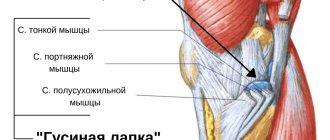
Anatomy of a crow's foot
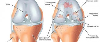
The anserine bursa, or crow's foot bursa, is a sac filled with fluid. It secretes synovial fluid to reduce friction between tissues and also serves as a shock absorber for bones, ligaments and muscles. Inflammation of the bursa does not occur suddenly, but develops over a long period of time. Bursitis can also occur in the joints of the shoulder, knee, hip, elbow and thumb.
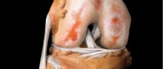
The pes anserine is the insertion site of the combined tendon of the sartorius, gracilis, and semitendinosus muscles on the superomedial portion of the tibia. The three pes anserine tendons are located superficial to the medial collateral ligament (MCL) of the knee. The sartorius and gracilis muscles are hip adductors (that is, they bring the thigh toward the median axis of the body). The semitendinosus muscle is part of the hamstring group and is located on the back of the thigh. Together, these three muscles are the flexors and pronators of the knee.
Forms and types of disease
The form of the disease “crow's foot” of the knee joint is determined by the anatomy of the localization of the affected bursa - the anserine bursa.
The disease is divided into two types - deep and superficial
Such bursitis is classified according to the factors of its development into four types:
- Aseptic, in which inflammation is caused by excessive mechanical stress or friction. Causes excessive production of synovial fluid and its accumulation in the cavity of the articular bursa. This type of disease has the most favorable prognosis.
- Infectious (purulent), caused by the invasion of virulent flora into the anserine bursa by blood flow or by an open wound. Without proper treatment it leads to sepsis. Requires treatment in a medical facility under the supervision of a physician.
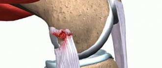
- Post-traumatic, developed through injury to tendons, ligaments, articular surfaces, muscle fibers of varying severity.
- Caused by the appearance of osteophytes or the accumulation of calcium salts. It is a complication of chronic arthrosis.
Epidemiology
Foot anserine bursitis usually occurs when the gracilis, sartorius, and semitendinosus muscles contract regularly during knee flexion and hip adduction. This increases friction between the fabrics and puts more pressure on the bag. Bursitis can also be caused by trauma, for example, a direct blow to the bursa area. Contusion of this area leads to an increase in the secretion of synovial fluid in the mucous membrane of the bursa. For these reasons, the bursa becomes swollen, weak or painful. This mechanism underlies osteoarthritis of the knee joint.
Reports show that anserine bursitis is more common in middle-aged and overweight women. An explanation for this may be the fact that this category of women has a wider pelvis. This leads to an angular displacement of the knee joint in the frontal plane, which leads to an increase in pressure in the “crow's foot” area due to the valgus position of the knee.
We can say that inflammation of the bursa is more likely a consequence of earlier complications than the original pathology.
Diagnosis of anserine bursitis
Table: types of bags
Numerous observations and studies have proven that the main provoking factor for the development of pes anserine bursitis is deforming osteoarthritis. Among other catalysts, experts call:
- diabetes;
- arthritic process in the knee;
- rupture (partial or complete) of the meniscus;
- flat feet;
- excess body weight;
- serious and regular injuries to the tendons and knee joint;
- strong inward turning of the foot.
Orthopedists and traumatologists outline the risk group in which bursitis most often develops:
- professional runners;
- prolonged stay in a standing position, combined with stress on muscles and ligaments (for example, running);
- people who professionally load their legs;
- congenital hypersensitivity of the hamstrings.
The “crow's foot” on the leg carries shock-absorbing and stabilizing functional loads. Prevents joint instability and prevents excessive rotational movements of the joint. Direct injury to the anserine bursa is difficult.
The causes of the development of the disease are previous diseases of various etiologies and localizations:
- infectious, purulent arthritis;
- DOA (deforming osteoarthritis);
- consequences of knee joint injuries;
- dysfunction of the tendon-ligamentous apparatus;
- autoimmune diseases (lupus erythematosus, rheumatoid arthritis);
- endocrine diseases (diabetes);
- excessive body weight;
- acquired or congenital flatfoot;
- pathological disorders of the development of the muscular and bone structure of the skeleton;
- penetration of pathogenic flora from a nearby inflammation site by hematogenous route.
The presence of risk factors in everyday life may contribute to the appearance of inflammation of the anserine bursa:
- Professional affiliation - the work of workers, construction workers, agricultural workers, associated with prolonged kneeling positions.
- Belonging to professional sports. The disease affects weightlifters, wrestlers, weightlifters, football players, and skiers. There is a direct correlation between the appearance of bursitis and excessive physical activity.
- Hereditary predisposition.
- Congenital pathologies and developmental anomalies of the musculoskeletal skeleton and tendon apparatus.
- History of autoimmune disorders.
- Hormonal imbalance. In women during menopause, as well as during long-term treatment with steroid hormones.
- Sudden and prolonged hypothermia of the lower extremities or the body as a whole.
Risk factors include: professional affiliation, congenital pathologies.
To clarify the diagnosis, laboratory and instrumental studies are prescribed:
- physical examination - stress test;
- blood test with leukocyte formula;
- test for rheumatic factor;
- blood test for uric acid levels;
- X-ray of the knee joint;
- Ultrasound of periarticular tissues;
- MRI;
- puncture and culture of synovial fluid.
Diagnosis at the initial stage of the disease allows you to reduce the duration and intensity of treatment and speed up the recovery process.
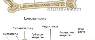
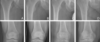
X-ray of the knee joint
Clinical picture
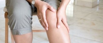
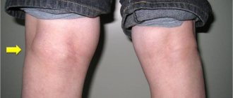
Anserine bursitis causes pain in the inside of the knee joint (most often when running or walking up stairs). Spontaneous pain may also occur in the anteromedial part of the knee when going up or down stairs. There will be swelling around the bursa, and the area itself will be painful on palpation.
Other clinical symptoms may include:
- decreased muscle strength;
- gait disturbances;
- decreased function;
- limitation of range of motion in the joint;
- postural dysfunctions/biomechanical disorders of the lower extremities.
Factors that aggravate symptoms include movements such as knee flexion and internal rotation, as well as external rotation and hip adduction. Spinning, kicking, squatting, and moving quickly from side to side will cause further irritation in the crow's foot area.
Symptoms
The disease has a characteristic clinical picture, which includes the following symptomatic manifestations:
- pain in the inner part of the knee joint, which increases when climbing up a hill or up steps;
- swelling localized below the knee fossa;
- increased sensitivity of the knee;
- sensation of a foreign body inside the joint;
- hyperemia of the skin at the site of inflammation;
- deterioration of general condition;
- decreased range of motion or limited joint mobility;
- the development of muscle atrophy caused by sparing the diseased joint.
Manifestations of the disease depend on the reasons that caused its appearance. A differentiated diagnosis is established taking into account diseases with a similar clinical picture.
Symptoms vary from the causes of the disease
Differential diagnosis
Foot anserine bursitis is often confused with other causes of knee pain. These include:
- A stress fracture of the tibia on the medial side will cause pain in the crow's foot area.
- Patellofemoral pain syndrome.
- Medial meniscus injury and osteoarthritis: Pain and tenderness will be found in the medial aspect of the knee joint, while anserine bursitis causes pain below the projection of the medial aspect of the knee joint space. With increased stress on the medial collateral ligaments, with or without instability, their damage can be diagnosed.
- Knee pain due to L3-L4 radiculopathy is associated with lumbar pain, but pain does not occur with finger pressure on the crow's foot area.
- Panniculitis occurs in obese people and causes painful inflammation of the subcutaneous fat.
- Semimembranosus tendonitis is a common injury among runners.
- Mediopatellar fold syndrome, which can cause pain and tenderness on the medial aspect of the knee.
- Extra-articular cystic lesions: synovial cyst, ganglion cyst, parameniscal cyst, pigmented villonodular synovitis, synovial sarcoma.
The specialist should also keep in mind the following problems that have similar symptoms:
- atypical cysts of the medial meniscus of the knee joint;
- periarticular bone cysts;
- semimembranosus bursitis;
- bursitis of the medial collateral ligament.
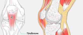
Knee tendonitis: ICD 10 code
In the International Classification of Diseases, 10th revision, tendinitis of the knee joint is associated with a variety of diseases that give rise to the pathological process. Pathologies characterized by inflammatory and degenerative-dystrophic processes, with the exception of foot pathologies, have code M76. This category includes tendinitis of the patellar region, which is assigned the code M76.5.
The use of the International Classification of Diseases is a prerequisite for making a diagnosis. Knee tendinitis, symptoms and treatment, photos of which are described and demonstrated by rheumatologists at the Yusupov Hospital Therapy Clinic, can be treated conservatively and surgically.
Diagnostics
Lateral radiographs of the patient's knee help rule out stress fracture, arthritis, and osteochondritis dissecans. MRI is necessary when it is necessary to clarify whether there are other damage to the medial surface of the knee joint. Also, MRI will allow you to do without arthroscopy. The MRI image must be compared with the data obtained as a result of the physical examination. Sinusography (radiography of the sinus after administration of a contrast agent) is the best assistant for making a diagnosis if other imaging methods, including MRI and CT, are not feasible for some reason. Injections of lidocaine/corticosteroids into the bursa area are possible, which will help assess the contribution of this pathology to the overall structure of the pain syndrome of the knee joint.
Rating scales
Lower Extremity Functional Scale (LEFS)
Physical examination
The pes anserine bursa can be palpated at a point slightly above the tibial tuberosity and 3-4 cm medially (about two finger widths).
Palpation of the crow's foot bursa
Examination of hamstrings should be carried out in the supine position. Flex the patient's hip 90° and then extend the knee as far as possible. The degree of knee extension will be an indicator of the length and tone of the hamstrings. If the patient's knee straightens completely, the hamstring is not constrained.
Estimating the length of hamstrings
If bursitis occurs due to sports activities, symptoms can be reproduced by resisting internal rotation and flexion of the knee. In the chronic version in older people, bending or straightening the knee usually does not cause pain.
First aid
When you are concerned about pes anserine bursitis, you need to know the rules of first aid:
- Complete immobilization (ensuring immobility) of the knee joint. The maximum period if it is not possible to go to a medical institution is no more than 10 days.
- Cold compress. It is necessary to apply ice packs and apply cooling ointments.
- The use of pharmaceutical or self-made compression products (elastic bandages, tight bandages from improvised means, compression stockings, knee pads). Allows you to reduce pain.
- Ensuring that the limb is elevated to reduce swelling.
Treatment
The initial stage of treatment for anserine bursitis should include relative rest of the affected limb and the use of NSAIDs. Additional agents, including corticosteroid injections (for example, methylprednisolone injections), are prescribed individually. Intrabursal injections of local anesthetics and corticosteroids are second-line therapy. Surgical treatment is indicated when conservative methods are unsuccessful. Several studies suggest that a small incision and drainage of the swollen bursa may help improve symptoms of the disease. The bursa may be removed if the chronic infection does not respond to antibiotics. After surgery, if the bursa has been removed, follow the rehabilitation and recovery tips listed below in the Physical Therapy section. If bursitis is accompanied by infectious inflammation and standard antibiotic therapy does not work, surgical decompression of the bursa is possible.
Physical therapy
Physical therapy is the cornerstone of treatment for pes anserine bursitis. Rest is initially indicated to reduce pain. The patient should refrain from walking on stairs, climbing, or other activities that promote inflammation (Level of Evidence: IIa). Nonsteroidal anti-inflammatory drugs (NSAIDs) can also help relieve pain. During the inflammatory phase of the disease, you need to limit movements in the joint and alternately apply ice. Ice massage for 15 minutes every 4-7 hours will reduce inflammation. To reduce swelling and prevent it, it is possible to use a bandage.
In order not to miss anything interesting, subscribe to our Telegram channel.
Be careful not to increase the friction of the bag. Teach the patient general strengthening physical exercises (level of evidence: I). These include leg stretching exercises: stretching the hamstrings, calf muscles (standing), quadriceps femoris (standing), hip adductors, as well as sliding the heels on the floor, isometric tension of the quadriceps femoris and hamstrings. The next step could be closed-chain exercises, such as single-leg squats, regular squats, and leg presses. Raising your legs with resistance (such as using resistance bands) is also recommended.
Closed kinetic chain exercises are also recommended as a measure to prevent the development of collateral instability of the knee joint. This pathology, in turn, is considered one of the risk factors for pes anserine bursitis (level of evidence IIIb).
In anserine bursitis, ultrasound has been documented to be effective in reducing inflammation (Level II Evidence).
Some patients are given an injection consisting of a solution of an anesthetic and a steroid. The physical therapist then gives the patient a hamstring stretching program and, at the same time, a quadriceps strengthening program consisting of closed kinetic chain exercises. These exercises should be performed several times a day. These measures will reduce pain within 6-8 weeks.
Kinesio taping is more effective in reducing pain and swelling than physical therapy or naproxen (Evidence Level Ib).