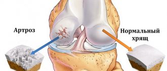
Arthrosis of the knee joint is a degenerative-dystrophic pathology that leads to deformation and destruction of articular cartilage. Gradually the limb loses mobility. According to statistics, almost every third person on the planet suffers from arthrosis, and this number is not decreasing. Elderly people, especially those who are overweight, are at risk. After 65 years, arthrosis is diagnosed in 70-85% of cases of treatment for knee pain.
A rheumatologist helps maintain a patient’s quality of life with joint pathology.
Causes of arthrosis
- Destruction of the joint due to natural wear and tear (aging of the body).
- Hormonal disorders (menopause, endocrine diseases).
- Congenital defects of the musculoskeletal system.
- Injuries, surgery on the knee joint.
- Professional sports.
- Monotonous physical work with increased load on the knee joints.
- Excess body weight.
- Genetic predisposition.
- Autoimmune diseases.
Treatment of gonarthrosis using UVT
Extracorporeal shock wave therapy has been used in the West since the 1990s, and in Russia since the beginning of this century. During this period, the shock wave therapy method has significantly evolved and improved; numerous medical studies have been conducted that have proven its effectiveness and safety.
In addition, the shock wave destroys osteophytes - intra-articular bone growths, and promotes the removal of slag compounds and salts from the joint. The UVT method produces analgesic, anti-inflammatory and anti-edematous effects, leading to significant relief in the area of the knee joints. The nutritional system of the joint tissues is restored, thanks to which their gradual restoration begins.
Only one course of shockwave therapy out of several procedures can “push back” the disease from the second stage to the first, and three such courses, carried out at intervals over 2-4 years, can return the condition of the joints to the initial stage of gonarthrosis or to complete recovery.
Symptoms of the disease
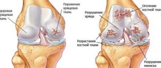
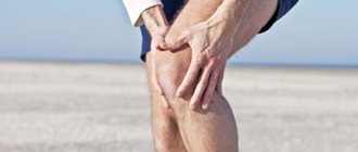
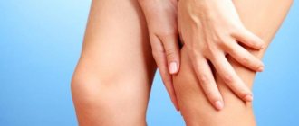
Deforming arthrosis of the knee joint (gonarthrosis) develops slowly and is chronic. In the early stages, the disease does not cause pain: the person feels only discomfort and stiffness in the lower limb. Gradually, motor restrictions increase. Without adequate treatment, the knee becomes noticeably deformed. Motor functions are so impaired that it is difficult for a person to walk, sit down, or stand up. Deforming arthrosis progresses to the point of disability of the patient. To preserve the joint, you must consult a doctor when the first symptoms of pathology appear.
Depending on the severity, there are three degrees of arthrosis of the knee joint:
- 1st degree. Clinical manifestations of the disease are mild. Most patients do not pay attention to the symptoms and continue to lead their usual lifestyle. With grade 1 arthrosis, a person may feel discomfort in the knee after standing for a long time, intense walking, or physical activity. On an x-ray, a narrowing of the joint space is visible, osteophytes growing inside the joint are visible. If arthrosis is accidentally detected in the first stage, for example, during a medical examination, its development can be significantly slowed down and even stopped.
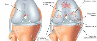
- 2nd degree. Pain with arthrosis of the knee joint becomes intense and difficult to ignore. The leg is especially bothersome early in the morning or in the evening. During the day, at rest, aching pain persists. Degenerative processes in the joint are reflected in the gait: the person begins to limp. When moving, a crunching sound is heard in the knee. Arthrosis of the 2nd degree can be complicated by a “joint mouse” - this is a condition in which a particle of bone or destroyed cartilage enters the synovial cavity. A foreign body causes severe pain that interferes with the movement of the limb. On examination, deformity of the knee is obvious. Possible inflammation and swelling. The x-ray shows a narrowed joint space and osteophytes, thickening of the bone.
- 3rd degree. A severe form of the disease that develops without treatment. Arthrosis of the 3rd degree is the cause of permanent disability. The pain in the knee is very severe, mobility is limited, the person cannot walk independently, every step is painful. The leg becomes deformed and begins to crunch heavily. On an x-ray, the doctor determines degeneration of cartilage tissue, destruction of ligaments, menisci, and proliferation of connective tissue.
HOW TO GET TREATMENT FOR JOINT DISEASES AT RCMC
In order to take advantage of the rehabilitation capabilities of the Center, you must have a referral from your attending physician for physiotherapeutic treatment of joints.
If you do not have a referral, you can make an appointment with a rehabilitation specialist at the Center, who, after studying the patient’s medical history, will prescribe the necessary procedures. To do this you need:
- Call the Contact Center to make an appointment with a rehabilitation specialist
- At the reception desk, enter into an agreement for the provision of paid services.
- Pay the bill at the RCMC cash desk or through ERIP
- Come to your appointment at the appointed time.
Treatment of arthrosis of the knee joint
Therapy includes a set of procedures, medication, and recommendations for lifestyle changes. It is important not to try to treat arthrosis on your own. Often, patients in the first stages of the disease use painkillers and consult a doctor when the joint is already destroyed. The earlier treatment is started, the more effective it is.
Drug treatment
The doctor prescribes medications to relieve inflammation, swelling, reduce pain, activate metabolic processes and tissue regeneration. Medicines are selected individually.
The following medications can be taken:
- Nonsteroidal anti-inflammatory drugs (NSAIDs) in the form of tablets, ointments, injections. The drugs relieve pain and swelling well, and improve the patient’s well-being.
- Glucocorticosteroids in the form of injections directly into the knee joint. Injections are indicated for severe cases of the disease, when the limb is practically immobilized.
- Painkiller blockades. They help cope with symptoms and alleviate the course of the disease.
- Chondroprotectors. The drugs help restore cartilage tissue and slow down the destruction of the joint.
Conservative treatment
Shock wave therapy
The method is non-invasive, helps remove salt deposits and improve the trophism of connective tissue. Physiotherapy improves blood circulation and has a beneficial effect on the elasticity of ligaments. Shock wave therapy is carried out in courses of 4-10 procedures.
Plasmolifting (PRP therapy)
The patient's own platelet-rich plasma is injected into the joint. A course of plasma lifting accelerates tissue regeneration.
Phonophoresis
The method combines the effects of ultrasound and medicinal ointments. Physiotherapy products, as a rule, have a complex composition and are prepared in a pharmacy according to a prescription. Ultrasound increases the penetrating ability of the active substance.
Massage
The procedure is contraindicated in the acute stage of arthrosis. When the inflammation is relieved and the pain is reduced, you can begin a massage course. Lymphatic drainage technique helps prevent the accumulation of synovial fluid. Massage also improves blood circulation in the knee and relieves muscle spasm. The procedure is most effective after performing special exercises for arthrosis of the knee joint.
Taking a bath
You can conduct the course at home as prescribed by a doctor or as part of a spa treatment. For arthrosis, radon, turpentine, and hydrogen sulfide baths are indicated. The procedures have a beneficial effect not only on the knees, but also on the hip and ankle joints.
Hirudotherapy
Medical leeches are placed around the deformed joint. The saliva of these creatures contains active substances that promote the restoration of cartilage. Hirudotherapy is usually prescribed for grade 1 and 2 arthrosis to relieve swelling and reduce pain.
Physiotherapy
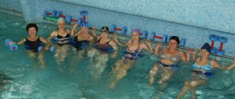
Gymnastics for arthrosis of the knee joint is an essential part of complex treatment. Special exercises help maintain muscle tone in the sore limb and prevent congestion. They begin to do gymnastics in the morning, without getting out of bed. Then during the day, perform another 3-4 sets of exercises for several minutes. It is useful to supplement therapeutic exercises for arthrosis of the knee joint with swimming.
Surgery
Surgery is indicated for grade 2 and 3 arthrosis:
- Puncture. Using a syringe, the accumulated fluid is pumped out of the joint cavity. Internal pressure decreases, swelling and inflammation decrease, and mobility improves. The procedure is carried out on an outpatient basis, at the surgeon's appointment.
- Arthroscopy. The method is used for rehabilitation of the knee joint. Arthroscopy is performed through small punctures, so the operation is quite easy to tolerate and the recovery period is short.
- Corrective osteotomy. A classic method of treating deforming arthrosis, which consists of correcting the deformed anatomical axis of the lower limb, followed by fixation of the wedge-shaped resection of the bone with a titanium plate. After osteotomy, the patient requires rehabilitation for several months.
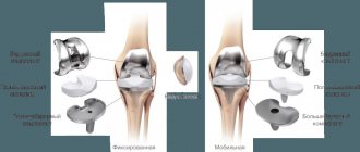
- Endoprosthetics. Installation of an artificial joint is performed in extreme cases of deforming arthrosis of the knee joint and allows the knee to return to its previous range of motion without pain. After total endoprosthetics, the patient requires long-term (about 2-3 months) rehabilitation.
The relevance of physiotherapy for injuries and fractures of the limbs
The need for this article arose due to the fact that patients with various injuries and their consequences often seek rehabilitation help at a relatively late date.
The rehabilitation process is delayed and often has little success. However, with a timely and comprehensive approach to solving the issues of treating such patients, it is possible not only to improve the patient’s quality of life, but also to completely restore the functions of the injured limbs of the victims.
For fractures without bone displacement, outpatient conservative treatment is usually prescribed. The principles of fracture treatment are simple, with restoration of bone integrity being the most important. The patient is given a fixing bandage, usually a plaster cast. This allows you to reduce pain and ensure limb immobility. For fractures with complications, for severe fractures with bone fragments, with displacement, surgical intervention is performed. In the most severe cases, fixation with metal knitting needles is used.
The patient is forced to limit his movements for a long time and, unfortunately, his rehabilitation treatment during this period. The deepest misconception is the fact that the recovery period begins only from the moment the plaster splint and all fixing devices are removed. That only from this moment you can strengthen muscles, develop movements in joints, and restore the supporting abilities of injured limbs.
It is important to understand that the treatment of fractures has two main tasks:
- complete anatomical restoration of a bone fracture (reduction and immobilizing bandage)
- full functional restoration
Thus, simultaneously with the precise reposition of bone fragments, properly carried out rehabilitation with physiotherapeutic means plays a decisive role in the patient’s cure.
It should be carried out simultaneously with surgical and orthopedic measures and carried out until complete recovery. This means that constant close contact between the surgeon and a physiotherapy and rehabilitation specialist is necessary.
General physiotherapeutic factors
There are general principles for the use of therapeutic physical factors in a specific patient during rehabilitation treatment:
- Physiotherapeutic factors must correspond to the patient’s current condition. It is necessary to adjust the parameters of physical factors throughout the entire treatment period. The dynamics of the disease requires changes in localization, area and time of exposure, intensity and frequency of the physical factor, and the appointment of additional therapeutic physical factors in the treatment complex
- Individual approach. When prescribing physiotherapy, it is necessary to take into account the patient’s age, gender and constitutional characteristics. The presence of concomitant acute and chronic diseases, contraindications for the use of a specific physical factor, general and local reactivity of the body, sensitivity threshold, autonomic background of the nervous system, psycho-emotional state of the patient.
- The absence of a pronounced therapeutic effect after the first procedures is not a reason to cancel or replace one physical factor with another.
- A significant therapeutic effect of physical factors, as a rule, occurs as a result of a course of treatment. Its duration can be 5-10-15 procedures daily or every other day.
- When undergoing a course of treatment, there is a so-called aftereffect period of the physical factor. In some cases we are talking about several weeks, and in others – up to 6-12 months. An unfinished course cannot guarantee a full aftereffect period. This should be kept in mind by patients who independently interrupt treatment after receiving initial visible improvement.
- Treatment can be combined, if two physical factors or a physical factor are used simultaneously with medications, and sequential, in which one factor follows the other, either directly or at some interval. It is also necessary to take into account that the previous and subsequent procedures should not negatively affect the body's responses to each of these procedures separately.
The main objectives of physiotherapy for injuries and fractures
The main objectives of physiotherapy (after reduction of fragments and immobilization of the limbs) in the treatment of fractures are:
- have an analgesic effect
- eliminate swelling and improve blood circulation
- combating wound infection in open fractures, preventing the development of osteomyelitis
- relieve muscle tension
- accelerate wound healing and callus formation
- prevention of the development of muscle atrophy and joint contractures
- accelerate the recovery of limb function as a whole
Physiotherapeutic methods effectively complement the surgical and medicinal components of the complex treatment process, avoid unwanted side effects and achieve lasting positive treatment results. But the key link in a successful diagnostic and treatment process is the doctor.
Therefore, procedures should be performed only after prior consultation with a specialist - for maximum effectiveness of therapy and patient safety.
Be healthy!
Head of the physiotherapy department Evgenia Alekseevna Tsymbalova