Magnetic resonance imaging shows pathological changes in internal structures without invasive manipulation. The method is more informative in relation to loose tissues containing a significant amount of liquid. Hydrogen atoms in water molecules respond to a directed electromagnetic pulse, providing a stable signal. Trabecular (from the Latin trabeculae - plates of spongy substance) edema is clearly visible on MRI of the spine due to the accumulation of fluid in the bone marrow tissue.
Lumbar spondylodiscitis with hyperhydration (increased fluid volume) of the spongy substance
The tomograph sensors record the signal generated by the resonance of charged atoms. Using complex algorithms, information is converted into a series of monochrome images and transmitted to a computer monitor. Layer-by-layer scanning is carried out in increments of 1 mm, which allows you to visualize the slightest changes in the substance in the area of interest.
In some cases, contrast-enhanced MRI is performed. The method involves intravenous injection of a “coloring” solution based on gadolinium chelates. The drug does not cause allergies and is eliminated from the body naturally.
What is a hematoma?
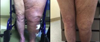
A hematoma occurs when a blow ruptures a blood vessel close to the surface of the skin. In this case, a small amount of blood enters the tissue under the skin. The area of the blow appears initially red and then turns blue or purple, green and yellow-brown, and when the bruise heals, the skin returns to its normal color.
Bruising can occur not only under the skin, but also deep in tissues, organs and bones. Deep bruises do not have visible signs of bleeding; the bruise can cause severe pain.
In 1988, scientists discovered a condition they called bone marrow edema. It was found that people with hip and knee pain had changes in bone marrow density on magnetic resonance imaging (MRI) that were not seen on X-rays.
Bone marrow edema is now called "bone bruise" to reflect the traumatic nature of the condition. A bone bruise is also sometimes called a bone contusion or hematoma.
Changes in bone marrow bones can be caused by the following reasons:
Blood congestion leads to stagnation of blood flow and causes severe inflammation.
Fluid inside the bone. When muscle injuries occur, fluid accumulates in the muscles and causes swelling. The bones are unable to swell; instead, the fluid in the bones creates pressure, which leads to pain.
Reactive hyperemia - This occurs when blood flow increases after a temporary stop.
Fracture: There may be a small crack in the layer of bone just below the cartilage in the joint.
Trabeculae are septa that support fibrous tissue in bone. A complete bone fracture means that all the bone trabeculae in that particular area of the bone are damaged, causing a rupture. Bone hemorrhage is often described as a pre-fracture stage. In this case, only some of the trabeculae are disrupted.
Results and discussion
In the article by G.V. Dyachkova et al. [8] presents the results of ultrasound and MRI4 of intra-articular injuries of the knee joint for topical (exact localization and extent) diagnosis. The authors found RCM of varying severity before treatment (immediately after injury) in 7 patients and within a year after surgery in 5. Changes in the structure of the medial and lateral menisci in the form of heterogeneity, destruction, partial or complete tears were identified by MRI in 8 patients with closed intra-articular fractures of the upper third of the ankle joint; changes in the structure of the ligamentous apparatus in the form of heterogeneity, swelling, thickening, partial or complete rupture of the ligaments - in 9 people. According to the authors, it was the MRI results that made it possible to obtain the necessary diagnostic information about all structural formations of damaged joints: bones, cartilage, menisci, determine the nature and extent of damage to bone structures, cartilage, and also study the condition of the bone marrow (Fig. 1). The authors established a direct correlation between the size of the RCM and the duration of the injury. The significance of this work for forensic doctors is the visualization and confirmation of the fact of injury, indisputable evidence of its volume and specific (topical) localization, as well as the duration of injury to the victim under the conditions of a specific mechanism (impact, impact with twisting elements, stretching, etc.) in the specified circumstances (traffic accident, etc.).
Rice. 1. MRI of the left knee joint of patient P., 42 years old.
The dynamics of MRI imaging after knee joint injuries were monitored by G.V. Dyachkova et al. [9]. The authors noted that after injury, MRI can reveal a number of significant changes in the mineralized and non-mineralized components of the distal femur and proximal aspect of the femur. In particular, the authors found RCM of varying severity, manifested by an increase in signal intensity on T2 images and on the same tomograms with fat suppression, before treatment (fact of injury) and after removal of the Ilizarov apparatus in all examined patients 1 year after the end of treatment (prescription injuries). The best visualization of RCM was observed on T2-weighted images (Fig. 2). In addition, the authors found that consolidated fractures of the knee joint differ in hypointense signals on T1- and T2-weighted images from areas of bone marrow sclerosis, planes of former fractures, post-traumatic bone cavities, proliferation of scar tissue, and heterogeneous periosteal callus. These signs were visualized on MRI up to 4 years or more after the end of treatment, even during primary healing of the fracture. The work noted that with the phenomena of post-traumatic arthrosis of the knee joint, not all patients showed such classic symptoms of injury as subchondral sclerosis of the articular surfaces (especially the femoral condyles) and marginal bone growths.
Rice. 2. MRI of the left knee joint of patient B., 51 years old.
In the work of I.R. Kuzina et al. [10] showed that the use of MRI in clinical practice has significantly expanded the ability to detect not only “fresh” but also old fractures of the knee joint. RCM is determined on T1-WI as a hypointense signal, and on T2-WI as a hyperintense signal. The swelling was localized in the area of the former fracture in 7 patients with an injury duration of 1 to 1.5 months, and its size depended on the duration of the injury: the more time passed since the injury, the smaller the zone of hyperintense signal from RCM on T2-VI was. The authors note that in case of old fractures, the signal characteristic of the RCM does not differ from that in case of “fresh” injury and did not depend on the duration of the injury.
After 2 years I.R. Kuzina et al. [11] continued similar studies. They examined 88 patients with fresh (up to 4 days) and stale (up to 28 days) trauma5 (see table). The authors noted that MRI detected fractures 4.6 times more often6 than radiography ( p
<0.00001). We studied MRI symptoms of 144 knee joint fractures in three planes. On T1-WI and T2-WI, the signal characteristics of damage to bones, intra- and extra-articular soft tissue components, effusion in the joint cavity and synovial bursae, etc. were assessed. RCM on T1-WI was detected in the epimetaphyses of the femur, LBD and MCL, as well as in the patella in in the form of hypointense local foci or diffuse signal changes. On T2-weighted images, the signal from the RCM region was hyperintense. In case of “fresh” fractures, RCM was found in all examined patients. As the duration of treatment increased, edema was observed less frequently, and at a later date it was detected only in 56.1% of cases. The authors noted that such bone marrow changes were detected in all bones forming the knee joint in all fractures, regardless of the mechanism of injury. The swelling was localized in the area of the fracture, as well as in other parts of the same bone at some distance or in another bone, occupying a small area of the bone (local OCM) or had a diffuse nature. The MR signal from the RCM was round, oval, radiant, or shapeless in shape. According to the authors, only on T2-weighted images was it possible to differentiate the MR signal caused by RCM from diffuse bone marrow sclerosis, which on T1-weighted images gave the same hypointense signal as edema.
Changes identified using MRI in fresh and non-fresh fractures of the bones of the knee joint (according to I.R. Kuzina et al. [11])
Thus, the significance of these publications for forensic doctors is to confirm the fact of injury, reliably determine the volume (area) damage to bone structures (joint) and, most importantly, the age of the identified injuries with possible differential diagnosis of the circumstances (mechanism) of causing the injury.
Similar data were obtained by foreign specialists in the field of radiation diagnostics. In the works of M. Schmid et al. [12] and D. Weishaupt et al. [13] provide MRI data indicating the presence of RCM in foot injuries and HS. The authors noted that RCM on MRI (Fig. 3) represents areas of hypo- or hyperintense signal (depending on the conditions and recording mode), which morphologically corresponds to the presence of fluid, hemorrhage, fibrosis or tissue necrosis. Depending on the extent of the process in the bones of the joint, a distinction is made between local (in one bone) and multifocal RCM, in which several bones of the same joint are involved in the pathological process.
Rice. 3. MRI (sagittal plane, fat-suppressed T1-weighted image) of the right ankle joint of a 63-year-old man with diffuse RCM of the talus (arrows) in the form of an increased signal intensity [12].
Multifocal bone marrow edema
According to N. Shabshin et al. [14], the most common causes of multifocal RCM in children, affecting several bones of the joint joint and foot, are unusual increased load on the joint, changes in the usual biomechanics of movements, bruises or fractures, immobilization of the joint, complex regional pain syndrome, heart attacks , osteoarthritis, inflammatory arthritis, neuroarthropathy and transient osteoporosis. Differential diagnosis of these nosological forms is based mainly on clinical signs in combination with MRI data: identification of fracture lines, osteophytes, erosions, double line sign and local skin changes in the area of the joint in the form of edema, hyperemia, smoothness of contours. The authors note that the initially “asymptomatic” development of the process can further evolve and with symptomatic “abuse” of excessive physical activity and stress reactions against the background of increasing pain and expanding the boundaries of multifocal RCM (Fig. 4), the formation of a hidden (“fatigue”) fracture is possible . In case of cessation or complete abolition of physical activity, which helps reduce pain, gradual resorption of RCM is observed in the affected bones and joints over several months.
Rice. 4. MRI of the right ankle joint of a 12-year-old girl (sagittal STIR image with short T1 inversion time and signal suppression from adipose tissue). Multifocal RCM is visualized in the bones of the GJ [14].
According to I. Elias et al. [15] multifocal RCM due to immobilization
in its form it can be spotty and localized subcortically or subchondrally with the duration of stabilization or cessation of the process within 18 weeks. An appropriate clinical history (prolonged immobilization and immobility) and the absence of a number of symptoms can help in the differential diagnosis of the process from transient osteoporosis and complex pain syndrome. The latter may also have macroscopic distinctive signs: swelling of the skin in the joint area and its thickening. According to the authors, a wide range of degenerative and inflammatory diseases such as osteoarthritis, rheumatoid arthritis and seronegative spondyloarthritis can contribute to the development of periarticular multifocal subchondral RCM in several bones of the AJ. Tomographic correlations, as well as the characteristic features of osteophytes in osteoarthritis, periligamentous soft tissue swelling, synovitis, and marginal erosions in rheumatoid arthritis, can help in the diagnosis of such conditions.
Research by D. Weishaupt et al. [13] showed that heart attacks
several bones of the GSS, but they, as a rule, differ from OCM by a characteristic tortuous “geographical” pattern and a double line sign.
D. Chatha et al. [16] and M. Ahmadi et al. [17] observed a variant of multifocal periarticular RCM predominantly in the midfoot in patients with diabetes mellitus
in the acute phase of the onset of neuroarthropathy. If MRI is performed before the onset of relevant bone changes, clinical presentation and medical history may be needed to distinguish them from the onset of inflammatory arthritis.
Local bone marrow edema
Focal (local) RCM, occurring in isolation in one bone, is often post-traumatic in nature, and according to the mechanism of formation, it occurs either as a result of an avulsion fracture (indirect impact) or from a direct blow (bruise) to the area of the joint. In addition to injuries, diseases (pathological conditions) such as osteoarthritis, inflammatory arthritis, and impingement syndrome can lead to local RCM. The MRI diagnosis of this type of RCM is helped by a medical history (injury) and the location of the edema in relation to adjacent bones, joints, capsules, ligaments, tendons and fascia.
RCM of the distal LBC
The occurrence of RCM in the LBD, especially in its distal section, is often caused by injuries and complications such as hidden (“fatigue”) fractures, ruptures of the retinaculum of the foot flexor muscles, distal tibiofibular syndesmosis and deltoid ligaments. In addition, in this area of the LBD, reactive RCM associated with dysfunction of the posterior tibial tendon and secondary periarticular RCM in arthropathy may occur.
Diffuse options
the distribution of RCM in the LCL may reflect physical stress in the indicated area or a hidden fracture in the area of the anterior and/or anteromedial surface of the LCL, while
osteoarthritis
more often forms
a central location
of RCM.
According to Z. Rosenberg et al. [18] and W. Morrison et al. [19], local
Anterior internal and anterior external localization of RCM can be caused by avulsion of the anterior deltoid or anterior tibiofibular ligament, respectively.
An important pattern, from a forensic point of view, is the presence of a weaker signal from RCM on MRI images for avulsion injuries
, in contrast to
direct impact
injuries. The location of the RCM in the posteromedial segment of the distal LCT is most often caused by dysfunction of the posterior tibial tendon due to its contusions, while the detected osteochondral damage at the site of its insertion is often associated with the opposite RCM of the talus. The authors noted that similar bone marrow changes were caused by avulsions of the posterior deltoid ligament, posterior tibiofibular ligament, and retinaculum of the foot flexors.
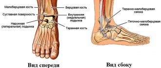
RCM of the distal part of the cervical spine
The MBC is a small bone; therefore, various injuries and pathological processes can lead to the formation of diffuse RCM. The emergence of focal zones of RCM in the MBC, as well as in the LBC, and their difference from each other are due to adjacent (neighboring) bone and soft tissue structures (attachments of ligaments and tendons). The proximity (in shape) of the distal part of the ICD to the spiral often creates an artificial increase in the signal on MRI images. In this regard, suppression of the signal from the adjacent subcutaneous fat base helps to eliminate this artifact and determine the true boundaries of RCM.
In the work of Ph. Robinson [20] showed that the localization of RCM in the area of the apex of the disc may be associated with an avulsion fracture, rupture of the calcaneofibular ligament, or calcaneofibular impingement. The distribution of typical localization of RCM in the distal parts of the LBC and MBC and their relationship with the most common causes of edema are schematically presented in Fig. 5. The authors noted that RCM in the medial part of the disc joint is often associated with ligament avulsion through a stretching (traction) mechanism. In this area, inflammatory changes in the form of osteoarthritis can also lead to edema in combination with obligatory similar changes on the opposite side (OCM of the talus).
Rice. 5. Schematic illustration illustrating the various causes of RCM in the distal tibia of the GJ (axial section).
Research by X. Wang et al. [21] and D. Weishaupt et al. [13] demonstrated that the location of the RCM in the lateral and posterolateral sector of the spinal cord may be the result of injury to the superior peroneal retinaculum or a consequence of damage to the peroneal tendons.
Thus, MRI diagnostics of RCM in cases of injuries or diseases of bones and joints is a routine study in the work of specialists in radiological diagnostic methods. The most common causes of OCM are injuries of different nature and mechanism of formation (local impacts, sprains, dislocations, twisting, etc.), as well as a number of pathological conditions (diseases) not directly related to traumatic influences (arthritis, arthrosis, osteoporosis and etc.).
Classification of bone hematoma
There are three types of bone hematoma:
- Subperiosteal hematoma - blood accumulates under the fibrous membrane that covers the surface of the bone;
- Interosseous hematoma , which involves bleeding and swelling in the central cavity of the bone where the red and yellow bone marrow is located;
- Subchondral lesion - bleeding and swelling occurs between the cartilage and bone below.
The type of bone bruise depends on where the injury occurs.
Subperiosteal hematoma mainly occurs after a traumatic blow to the bone. It often occurs in the lower parts of the body.
Interosseous hematoma occurs with regular injuries. This type of bruise is mainly found in football players, basketball players and runners.
Subchondral damage is caused by compressive force. This type of bone bruise is more common in football and basketball.
Jumping on hard surfaces can cause all three types of bone hematoma.
Spinal cord
Trabecular edema of the spinal cord on x-ray Swelling
of the spinal cord can occur as a result of new or old injuries and diseases of the spine, tumors, and circulatory disorders. Local and general swelling are divided. The first is observed, for example, with a pinpoint injury or dislocation of the spine or vertebrae, so swelling (or redness) occurs only in the place where the damage occurred. General swelling of the spinal cord occurs in patients with severe forms of diseases (osteochondrosis, spondylosis, dysplastic processes), as well as household injuries (falling from a height, heavy lifting, accidents), in which swelling of the entire spinal region often occurs. Damage to the bone marrow of the spine can manifest itself in different ways, depending on the damaged area: cervical region - impaired function of the arms and legs, thoracic region - torso and lower limbs, lumbar - impaired function of the lower torso. Symptoms:
- The sensitivity of the limbs decreases (legs and arms are taken away);
- Fatigue, dizziness, nausea;
- Acute pain when walking in the area of shock;
- Decreased emptying of urine and rectum (constipation, incontinence).
- Spasm of capillaries and blood vessels.
Symptoms of bone marrow edema
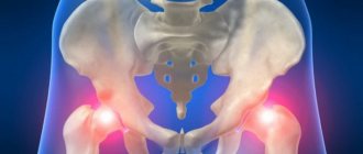
Bone marrow edema is often seen in the knee, wrist, heel bone, ankle and hip. They often occur with sports injuries, falls, car accidents, or impacts. Bone hematomas can also be caused by medical conditions such as arthritis.
A person may be at risk for a bone hematoma if they:
- Takes part in sporting events;
- Has heavy physical work;
- Does not wear protective equipment for sports or work;
- If he has osteoarthritis.
Causes
Trabecular edema occurs more often in people prone to constant injury, for example, athletes, those who lead an active lifestyle, as well as elderly people in whom swelling occurs after injury or with chronic joint pathology. Doctors identify the following factors for the development of the disease:
- Edema manifests itself as a result of fractures, operations, inflammation and violations of the integrity of the capillaries of bone tissue.
- Accumulation of internal fluid of the bone, as well as collagen fibers (at the site of damage, a tumor of varying degrees appears).
- Bone cells (osteoblasts, osteocytes, osteoclasts) swell. This is a more serious disorder than the accumulation of fluid in the alveoli (pneumonia).
Stopping movement and accumulation of bone fluid in the trabeculae may indicate a bruise or sprain, for example, of ligaments or vertebrae. It can be observed in patients with knee arthritis and spinal osteochondrosis. In this regard, excess fluid inside the bone marrow leads to disruption of hematopoietic processes. Trabecular edema occurs most often with pathologies that are localized in the very center of the bone marrow lesion or near it. This disease develops due to a violation of the composition and integrity of cartilage (tendons, ligaments).
Treatment of bone marrow edema
A bone hematoma can be treated with rest, ice, and pain medications.
The doctor may suggest:
- Rest the injured limb;
- Reducing swelling by elevating the injured limb above the level of the heart;
- Applying ice several times a day;
- Medicines to reduce pain and inflammation;
- Wearing a brace to limit movement.
It is important to avoid constant or strong pressure or heavy weight on the affected area. If the bone or joint does not get enough rest, the healing process may be slower and more damage may occur.
It is necessary to maintain a diet rich in calcium, vitamin D and protein. Smoking can delay bone healing.
Most bone bruises heal within a few months. In rare cases, in the absence of blood supply, avascular necrosis of the bone can occur. If the bone dies, the damage is irreversible.
While bone bruises cannot always be prevented, eating a balanced diet, regular exercise, stopping smoking, limiting alcohol consumption and using protective equipment when playing sports will help keep your bones healthy.
Why and who gets trabecular edema. Risk group.
The main “candidates” for inclusion in the risk group are people leading an active lifestyle. However, at the same time, this includes older people who have recently undergone surgery, since surgery with reduced immunity may well provoke changes in the structure of bone tissue.
The main reasons for the occurrence of trabecular edema are:
- fractures of various types;
- spondylosis;
- arthritis;
- osteocondritis of the spine.
If you belong to a risk group, do not under any circumstances diagnose this pathology yourself and do not begin to treat it. This should only be done by a professional specialist.