Gonarthrosis or arthrosis of the knee is not a disease of an inflammatory nature. This pathology is a progressive degenerative-dystrophic disorder in the functioning and structure of intra-articular cartilage tissue. The disease can progress. It has 4 stages of development. When stage 2 arthrosis of the knee joint occurs, the patient is still capable of independent movement, although there is a significant decrease in his activity. Let's look at the main causes of this disease and effective treatment methods.
Types of gonarthrosis 2 degrees
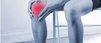
As joint activity decreases, the patient becomes irritable. The occurrence of severe painful sensations leads to a deterioration in the quality of life. The risk group includes people who are professionally involved in sports or ballet. The disease also develops in people whose work involves constant heavy lifting and prolonged standing. Office employees also suffer from gonarthrosis, which is associated with prolonged sitting.
Arthrosis of the knee joint stage 2 can be primary or secondary. Often, patients are diagnosed with a secondary form of pathology. In this case, the disease develops against the background of:
- previous knee injury;
- advanced forms of arthritis and some other pathological processes occurring in the body
- as a consequence of the progression of diseases of the endocrine system (diabetes, problems with the thyroid gland, etc.).
The primary form of knee arthrosis is characterized by the causeless appearance of a degenerative-dystrophic process in the cartilage tissue of the knee joint. This condition is observed in rare cases.
The joints are affected symmetrically, there is a complete absence of joint space. In turn, both primary and secondary forms of grade 2 gonarthrosis can affect not only one knee, but both at once (both right and left). If the cartilage tissue of only one knee joint is affected, orthopedists call this disease unilateral. If the knees of both lower extremities are affected simultaneously, this is bilateral gonarthrosis.
Bilateral knee arthrosis of the 2nd degree is much more difficult for patients to endure, since serious deformation of the articular tissue is observed simultaneously in both legs. Based on etiological characteristics, this type of pathology is most often classified as idiopathic (primary) type. The same applies to the unilateral form of gonarthrosis; it is almost always secondary.
Regardless of the type of arthrosis of the knee joint, stage 2, conservative methods are used at the initial stage of treatment. If the desired result after the therapy is not available, surgical intervention is resorted to.
Non-surgical treatment has a positive effect only if stage 1-2 knee arthrosis is diagnosed. In some cases, at this stage, surgical intervention may be required.
It should be noted that the use of conservative methods cannot cure gonarthrosis completely (any arthrosis is an incurable disease). Non-surgical therapy is:
- competent prevention of subsequent destruction of cartilage tissue with properly selected medications and physical preparations;
- symptomatic relief from painful symptoms due to the use of painkillers;
- a means of maintaining muscle and ligamentous tissue by performing special gymnastic exercises. This helps to some extent to activate motor function.
The use of conservative technologies is advisable at an early stage of the disease. If the middle phase of arthrosis of the knee joint is diagnosed, such techniques become less effective, although in some cases they help improve the patient’s condition.
If the last phase of gonarthrosis is identified, non-surgical treatment methods become useless. The use of alternative medicine for therapeutic purposes in severe stage 2-3 gonarthrosis is ineffective. Without seeking specialized help, the disease will progress. In this case, destructive processes appear inside the knee cartilage.
If you do not consult a doctor in a timely manner and do not undergo appropriate treatment, this may even cause disability.
A person may be assigned a disabled group in the following cases:
- grade 2 arthrosis of the knee joint was diagnosed;
- the disease is accompanied by persistent and pronounced functional disorders;
- the disease is characterized by a rapidly progressing degenerative and dystrophic process in the cartilage tissue of the joint.
When the pathology reaches the last phase, the patient is in any case recognized as disabled. No gentle therapeutic techniques are used in such a situation. Treatment in this case consists of removing non-viable articular tissue, after which an artificial joint is installed.
With the help of endoprosthetics at an advanced stage of gonarthrosis, it is possible to restore lost locomotor-support functionality for a long time (for a period of 15 to 30 years).
Clinical picture at the middle stage
Pathological disorders of moderate severity are more intense than at an early onset. Painful sensations intensify and become more frequent; they can no longer go unnoticed. In addition to morning pain and discomfort after a long stay in a maximally immobilized state, pain syndrome naturally appears after walking and physical activity, and much more often begins to disturb for no apparent reason. When going up and down stairs, a person experiences difficulties, since the flexion/extension of the joint is already seriously limited.
Steps can be an insurmountable challenge.
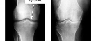
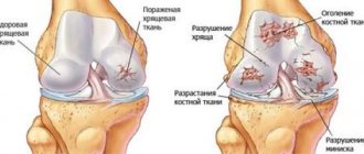
As for the results of the X-ray examination, they usually contain the following content:
- in direct and lateral projections, a significant narrowing of the joint space is clearly visible (2-3 times the required norm);
- a change in the density of articular cartilage is detected;
- there are large osteophytes at the ends of the articulating bones;
- there is pronounced subchondral osteosclerosis of the surfaces;
- the first deformations of the bones and the curvature of their axis are visualized.
Due to pathological transformations and acute pain, a person protects the problematic leg in every possible way, trying to transfer the main part of the load to the healthy limb. Due to the forced gentle regime, muscle structures weaken, lose their functional tone, decrease in volume, in other words, they gradually begin to atrophy. Muscle atrophy further complicates motor problems: along with inadequate range of motion during exercise, lameness appears. Due to the destruction of cartilage, non-physiological sounds are felt in the knee at the moment of movement - crunching, clicking. The contracture is moderately expressed.
Symptoms and diagnosis
For stage 2 arthrosis of the knee joint, the clinical picture is more pronounced, although the symptoms are not yet critical. This degree of the disease is accompanied by the following symptoms:
- Regular painful manifestations in the knee area, but at this stage they do not yet become permanent. The pain starts to bother me in the morning, not every day. In addition, after about 15 minutes the discomfort goes away. At this stage, prolonged walking or intense physical activity is accompanied by the appearance of an unpleasant pain syndrome.
- Painful manifestations that often occur against the background of increased loads on the damaged area. To prevent them, the patient tries to reduce the load on the knee joint as much as possible. This is not recommended. Such actions can lead to atrophy of the muscle tissue surrounding the joint.
- A dull aching pain syndrome that occurs in damaged cartilage tissue of the knee joint under the influence of reduced atmospheric pressure. In medical practice, this phenomenon is called weather dependence, and such a condition does not have an exact definition. According to some doctors, when the pressure in the atmosphere decreases, this leads to an increase in the pressure of the intra-articular fluid. This condition leads to painful sensations.
- Crunching in the knees, characteristic of stage 2 gonarthrosis. In addition, as the pathology progresses, it intensifies.
Also, with stage 2 arthrosis of the knee joint, another characteristic sign is observed, but only a specialist can determine it on an x-ray. The space in the knee joint tissue narrows, and this process is accompanied by the formation of bone growths - osteophytes. On X-ray film they are visualized as tubercles.
At this stage of pathology progression, a certain deformation of the cartilage tissue in the joint occurs. For this reason, in some cases there is not complete, but partial flexion and extension of the injured limb. This is fraught with the appearance of pain.
Diagnostic measures for identifying stage 2 knee arthrosis consist of interviewing the patient, collecting anamnestic data, and also conducting some examinations. A differential diagnosis is also required, since the symptoms of the disease are similar to some diseases of the knee of inflammatory etiology.
Surgical treatment of gonarthrosis
As a rule, from the 2nd stage, if conservative therapy is ineffective, and up to the 4th stage of development of gonarthrosis, surgical treatment is indicated. The most effective method today that ensures full restoration of knee joint mobility and elimination of pain is endoprosthetics. This is a high-tech operation that consists of replacing a worn-out joint with an endoprosthesis artificially created from biocompatible materials that can last 15-30 years.
Depending on the severity of the situation, the patient may be offered partial (unicondylar) and total endoprostheses. In the first case, only the damaged area of one of the condyles is replaced with an artificial prosthesis; in the second, the entire articular surface is replaced. Total arthroplasty is preferable, although it has a higher cost. But this treatment option allows you to count on the maximum service life of the endoprosthesis, while a partial one can work up to 7 years and can only be used in the presence of degenerative-dystrophic changes in the hyaline cartilage of only one of the condyles, provided that the functionality of the articular ligaments is preserved. Next, replacement of the implant is required, but with each subsequent operation its size increases, and the success of the intervention decreases in direct proportion. Although initially, partial knee replacement is associated with significantly less blood loss and requires easier and faster rehabilitation.
To install the endoprosthesis, the surgeon makes a 12-14 cm long incision on the front side of the knee, bypassing the patella. After this, the joint is carefully released from the soft tissues and the patella is shifted, which provides access to the knee joint. The surgeon sequentially loosens the tension of the ligaments and soft tissues, and then removes the deformed fragments. The resulting cut edges are polished and adjusted to the parameters required for installation of the endoprosthesis.
The lower part of the femur is then replaced with a titanium prosthesis, a flat plate is fixed to the upper part of the tibia, followed by a polyethylene liner and a trial prosthesis. The surgeon checks the functionality of the knee joint, corrects defects if necessary and, using the chosen method (using special cement or a tight fit method), fixes the permanent endoprosthesis, suturing the wound, installing drainage and applying a bandage and splint.
Endoprosthetics today has no analogues in terms of efficiency and long-lasting results. The operation is performed under general anesthesia or epidural anesthesia and takes on average 1.5–3 hours. After its successful completion, discharge is carried out on 5-7 days, but all patients require rehabilitation, which lasts about 3-4 months. After its completion, the patient returns to a full life without pain and limitations in performance.
Thus, gonarthrosis is a serious disease that can lead to loss of the ability to self-care and the development of severe complications from the spine and other parts of the musculoskeletal system. Therefore, it is important to pay attention to the slightest changes in the knee joints and immediately make an appointment with an orthopedist for diagnosis and treatment. Otherwise, it will be possible to restore motor functions and get rid of pain only through surgery. But, fortunately, modern surgical techniques make it possible to achieve a positive outcome in more than 95% of cases.
Carrying out an external inspection
Initially, the patient should be examined by an orthopedist. During a visual examination, the doctor determines:
- roughened contours of bone tissue;
- deformed joint;
- curved limb axis.
When moving transversely, the kneecap crunches and a click may be heard. During a palpation examination, the doctor can detect zones in and around the gap of the knee joint that, when pressed, respond with the appearance of pain.
Functional tests are required to assess motor activity and muscle strength.
Stage 2 arthrosis of the knee is often accompanied by synovitis (an inflammatory process in the synovial membranes). This condition is characterized by the occurrence of swelling in combination with hyperemia of the skin. There is a smoothing of the contours of the knee, the formation of a rounded seal. If you press on it, you can feel the fluid moving in the middle of the joint. This phenomenon is called fluctuation.
Symptoms of the disease
Symptoms of the disease are determined by the degree of development of pathological processes.
The development of pathology is gradual. Grade 1 knee joint gonarthrosis is characterized by symptoms such as:
- superficial pain when performing movements;
- stiffness, a tightening sensation in the popliteal region;
- “starting pain” that occurs when taking the first steps after a long static stay (standing/sitting/lying).
Grade 2 knee joint gonarthrosis has symptoms such as:
- increased intensity of pain;
- localization of pain in the anterior internal articular surface;
- decreased motor function;
- visually noticeable deformation, expansion of the knee.
Grade 3 knee joint gonarthrosis is accompanied by the following symptoms:
- constant pain of varying intensity;
- loss of mobility;
- increased volume of the knee, noticeable x-shaped or o-shaped deformity;
- unsteady gait.
Clinical diagnosis
To assess the condition of the patient’s body, the doctor prescribes general clinical urine and blood tests. If synovitis occurs in the body in a latent form (as evidenced by the detection of leukocytosis and an increased level of ESR), this indicates the development of inflammation in the joint tissue.
In some cases, serological and biochemical studies may be required. This will help differentiate systemic and infectious diseases. Intra-articular fluid and cartilage tissue can also be collected to carry out a morphological study. This makes it possible to detect degenerative-dystrophic disorders in the articular tissue of the knee.
X-ray examination
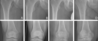
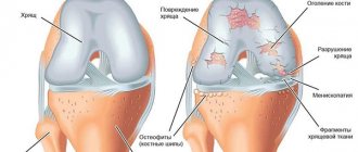
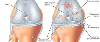
X-ray diagnostics is the most informative when diagnosing stage 2 knee arthrosis. On the resulting x-ray you can see:
- how the joint space has narrowed;
- see multiple bone growths (osteophytes);
- see deformation of the bone tissue of the joint in the form of the appearance of cystic formations or signs characteristic of subchondral osteosclerosis.
Thanks to X-ray examination, it is most often possible to determine the cause of the problem, for example, incorrect fusion of ligament and tendon tissue.
Causes
Most often, the primary form of knee arthrosis is detected in older people, mostly women. The consequence of the disease is the natural wear and tear of the cartilage in the knee joint. In young and middle-aged people, the cause of the disease often lies in excess load on the joint.
The development of secondary gonarthrosis occurs as a concomitant disease with the following health problems:
- Bekhterev's disease, reactive, psoriatic, chronic sluggish arthritis of an infectious nature;
- systemic diseases - systemic lupus, scleroderma, rheumatoid arthritis;
- endocrine, metabolic pathologies - gout, diabetes mellitus, hyperthyroidism, hypothyroidism;
- injuries suffered in the past - fracture of a tubular bone, rupture of the meniscus, ligament or tendon tissue, intra-articular fracture;
- disorders in the knee joint of a congenital and acquired nature.
A predisposing factor to the formation of arthrosis of the knee joint is the presence of excess weight, which is often accompanied by varicose veins. The development of this condition is facilitated by a sedentary lifestyle, smoking, and a lack of essential vitamins and minerals in the body.
Types and forms of the disease
There are:
- Primary arthrosis develops as an independent disease and is associated with age-related changes and heredity.
- Secondary gonarthrosis occurs against the background of various diseases or injuries, for example post-traumatic osteoarthritis.
Depending on the main causes and pathogenesis, the following types of osteoarthritis are distinguished:
- Ischemic, associated with problems with blood supply to tissues and vascular disorders;
- Post-infectious, caused by infections, viruses, autoimmune processes that cause damage to the articular surfaces;
- Idiopathic, the causes of the occurrence and development of the pathology remain unclear;
- Metabolic is associated with metabolic disorders - gout, hemochromatosis, chondrocalcinosis;
- Involutionary, as a result of age-related processes of destruction of the body;
- Post-traumatic arthrosis and previous injuries cause the development of degenerative changes in the joint;
- Dishormonal - due to hormonal imbalances, changes occur in the musculoskeletal system, primarily in the skeletal system of the body.
When to see a doctor?
It is important to consult a doctor if the first signs characteristic of knee arthrosis are detected:
- prolonged and intense pain that increases with movement;
- periodic crunching in the joint area;
- restrictions in physical activity;
- noticeable knee deformity;
- swelling
If these symptoms occur, it is imperative to visit an orthopedic traumatologist for an examination and to undergo appropriate diagnostic procedures and prescribe therapy. You can also contact a rheumatologist or surgeon with similar complaints.
If there are no doctors of this specialization in the nearest clinic, it is recommended to contact your local physician - he will be able to advise where specialized medical care of this type is provided.
Prevention of deforming gonarthrosis
Gonarthrosis belongs to the group of diseases that are better and easier to prevent than to treat expensively and for a long time.
The following can be recommended as preventive measures:
- When engaging in dancing and sports, try to avoid serious injuries (torn ligaments, fractures, severe bruises of the patella);
- Maintain regular physical activity, as movement is the mechanical basis for cartilage nutrition;
- Make up your diet taking into account the nutritional needs of bone and cartilage tissue;
- Monitor your body weight;
- If you are injured or experience discomfort in your knee joints, immediately seek qualified medical help;
- Starting from the age of 35, undergo preventive courses of treatment with chondroprotectors;
- Drink enough water daily.
Proper nutrition for gonarthrosis of the knee joint 2 degrees
If articular pathology is detected, the patient will have to follow some rules regarding his own diet:
- Eat jelly, jelly, jellied fish and other dishes containing gelatin.
- Eliminate regular table salt from your diet. As a replacement, you should use sea or iodized salt when cooking.
- Be sure to diversify your menu with fresh or boiled vegetables, fresh herbs, eggs, legumes, cereals, and bran.
- It is healthy to eat low-fat dairy products, marine fish, boiled beef in small quantities, chicken and rabbit.
- It is recommended to diversify your diet with herbal infusions, still mineral water, green tea, vegetable and fruit juices.
- Refractory fats of animal origin will have to be replaced with vegetable oils.
You will have to give up foods that contribute to the deterioration of the condition of joint tissue and the body as a whole:
- too salty and spicy dishes;
- smoked meats
- sweet sparkling water;
- fatty meat and fish products;
- lard, canned food, semi-finished products;
- buns;
- instant food products;
- mixtures containing preservatives and dyes.
During the day, it is necessary to drink clean water in an amount of about 2 liters, since a lack of fluid can negatively affect the condition of cartilage.
Restoring cartilage tissue requires an integrated approach to treatment. One of the components of therapy is a properly formulated diet. It is also important to reduce weight to a certain level in accordance with the patient’s age and height. Due to excessive stress on the affected joint tissue, healing is slowed down.
Lifestyle, useful tips
If, when visiting a doctor, you are diagnosed with stage 2 gonarthrosis, you will have to follow these recommendations:
- Maintain a positive mood. Although knee arthrosis cannot be cured, you should not despair. The degenerative-dystrophic process in the joint can be slowed down by using intra-articular injections (liquid endoprosthesis) with continued active lifestyle.
- Move as much as possible, starting the day with light physical activity on your legs, which can be done right in bed. Throughout the day you need to regularly load your joints. If you lead a sedentary lifestyle, you need to get up and do a little warm-up. Prolonged stay in a stationary position is harmful to joint tissue.
- Drink plenty of water. To maintain joints and metabolism in normal condition, the daily intake of clean water (in addition to tea, juices, coffee, soup) is 1 liter.
- Control body weight. This is necessary to reduce the load on the affected joint. To this end, you need to switch to healthy food, excluding from the menu chips and sweets, semi-finished products, factory-made chicken, sausage products and store-bought pates, yoghurts. These products can be replaced with vegetables, beans, rose hips, sauerkraut, cottage cheese, and dried fruit compote.
- Take chondroprotective drugs. Experts recommend regular treatment with chondroprotectors, and it is better to give preference to injectable agents.
- Take calcium-containing medications. A course of vitamin D is also sometimes recommended. To select the correct remedy, you should consult your doctor.
- Do not put too much stress on your knees when playing sports, as this causes the cartilage to wear out faster. This leads to the progression of the pathology. You need to properly combine activity and rest.
- Workout. For gonarthrosis, it is better to choose swimming, skiing or cycling, since these sports are low-traumatic. Swimming helps to activate blood circulation, pump up the periarticular muscles, and restore tone.
Drug treatment of arthrosis
The main role in the fight against the manifestations of gonarthrosis is played by non-steroidal anti-inflammatory drugs and chondroprotectors.
- Non-steroidal – relieve inflammation and eliminate pain. However, if you take them for a very long time, the symptoms of the disease are “masked” and it becomes impossible to prescribe adequate treatment. In addition, according to recent studies, drugs of this particular group negatively affect the synthesis of proteoglycans, which is why the affected cartilage is destroyed even faster.
- Chondroprotectors – nourish and restore the functioning of cartilage tissue. At the third stage, these medications can no longer help - they are not able to restore completely destroyed cartilage or return the knees to their original shape.
The positive effect of treatment with chondroprotectors occurs only after 6-18 months
- Medicinal ointments and creams only alleviate the patient’s condition.
- Compresses have an anti-inflammatory and analgesic effect, but, like ointments, they are only an adjuvant in the treatment of osteoarthritis and arthrosis.
Gymnastics
A set of gymnastic exercises is selected by the attending physician in each specific case. During exercise, movements should be smooth and slow, no need to jump or squat. The optimal time to do exercises is in the morning. Each exercise should be done 10 times, 20 minutes should be allocated for gymnastics for training in a sitting and lying position.
The purpose of gymnastic exercises for gonarthrosis is to prevent the destruction of cartilage tissue, slow down the process of decreased mobility, activate blood circulation and relax muscle spasms.
How does the disease develop?
Under the influence of external and internal factors, the blood supply and nutrition of joint tissues is disrupted, degenerative dystrophic damage to articular cartilage develops, with subsequent participation in the inflammatory process of the underlying bone structures of the femur, tibia and patella. The gradual destruction of cartilaginous surfaces leads to loss of function of the knee joint, limits movement and disrupts the patient’s quality of life.
Forecast of the incidence of gonarthrosis among the population for the next 20 years, according to the World Health Organization (WHO)
Gymnastics Evdokimenko
The gymnastic complex of Dr. Pavel Evdokimenko was designed to be performed by patients during remission. When the pathology begins to worsen, performing these exercises is prohibited.
Dr. Evdokimenko has developed many different exercises, but one session will require a maximum of 10 of them. You need to train every day for half an hour.
Let's consider several exercises from the Evdokimenko gymnastic complex:
- Lie on your back, raise your lower limb 10 cm from the floor surface and fix it in this position for a couple of minutes.
- We sit on a chair, keep our back straight, straighten and lift first one and then the other leg in turn. We hold them in this position for 40-50 minutes.
- We stand near the chair, turn to face it and lean on the back. We rise on our toes and stay in this position for 3-5 seconds.
Gymnastics Popova
Gymnastic exercises were developed by the famous chiropractor and traumatologist Peter Popov. The training is perfect for patients suffering from left- or right-sided arthrosis of the knee joints, as well as bilateral gonarthrosis.
Let's look at some exercises from Popov's gymnastic complex:
- We sit on a chair and imitate walking, raising and lowering our heels.
- We lie down on the floor and turn to our side. Raise your leg up and lower it. Then we turn over to the other side and repeat the exercise with the other leg.
- We stand near the chair. Leaning against the back, we move one leg back a little and rotate the foot. Then we change the leg and perform the exercise with the second leg.
Before you begin performing such a gymnastic complex, you need to warm up the damaged joints by rubbing them with your palms. Any movements when performing such exercises should be performed measuredly.
How to treat gonarthrosis?
The method of treating gonarthrosis differs little from the methods of treating arthrosis of other joints.
Step 1 – relieve inflammation
For this purpose the following are traditionally used:
- NSAIDs are nonsteroidal anti-inflammatory drugs that are prescribed intramuscularly or intravenously. Medicines in the form of injections provide a longer-lasting and stronger pain-relieving effect. These include drugs such as diclofenac, olfen, diclac, ibuprofen, indomethacin, ketoprofen.
- NSAIDs COX-2 are the most effective and gentle compared to NSAIDs COX-1. They can be used long-term, for several months. These are meloxicam, celecoxib, nimesulide and etoricoxib.
- Hormonal drugs. This group of medications is used for intra-articular injections in the presence of synovitis of the knee joint (inflammation of the synovial membrane). The goal of therapy is to relieve inflammation and pain as quickly as possible. The disadvantage of use is the damaging effect on cartilage tissue, a large number of contraindications and side effects. The most commonly used synthetic hormones for gonarthrosis are: hydrocortisone, Kenalog, Diprospan;
- Antienzyme drugs. They neutralize the synthesis of certain enzymes and prevent further joint degeneration. The most well-known drugs in this group are: contrical, ovomine, gordox. For gonarthrosis they are administered intra-articularly.
Step 2. We provide an anabolic and anti-catabolic effect
For this purpose, medications are used that replace the substances necessary for the synthesis of cartilage, providing a highly specific protective effect on the cartilage tissue. They are also called chondroprotectors. Such preparations contain substances that are part of the cartilage matrix. These medications are natural, well accepted by the body and actively stimulate collagen synthesis.
The drugs justifiably used for arthrosis of the knee joint include structum, DONA, alflutop, rumalon, mucosat. All of them are slow-acting medications that need to be taken over long periods. Some of them are available in the form of injection solutions. This form of application is the most effective.
Step 3. Apply, warm, rub
To do this, you can use various kinds of gels, ointments and creams. For the most part, they are warming and anti-inflammatory. The purpose of their use is to activate local blood circulation and relieve inflammation. The most well-known drugs in this group: apizartron, finalgon, dolobene, feloran, fastum gel, nikoflex.
Step 4. Improve blood circulation
Vasodilators are used to reduce intravascular muscle tone. Such medications allow you to increase internal blood flow and improve the trophism of tissues located around the joint. For gonarthrosis, Cavinton, Trental and Actovegin are recommended. To strengthen the vascular walls, upsavit or ascorutin are used.
Step 5. Removing excess tone
Antispasmodics such as mydocalm, sirdalud, tizalud and drotaverine (no-shpa) can remove excess muscle tension in the damaged segment. It often occurs as a compensatory reaction of the body.
Step 6. Inject synovial fluid prostheses
The most progressive method of treating gonarthrosis in recent years has been the inclusion of hyaluronic acid-based drugs in the treatment protocol. It is a natural component of articular cartilage and synovial fluid. Therefore, its introduction into the knee joint does not cause inflammation, rejection or other negative reactions.
At the same time, the use of drugs such as otrovisk, sinocorm or hyalual can soften movements and relieve pain caused by friction of articular surfaces. For gonarthrosis, the most recommended drug in this group is fermatron.
The sequence of treatment is determined by the doctor according to current protocols. In this case, anti-inflammatory therapy, a course of chondroprotectors and physiotherapy can be prescribed simultaneously. Hyaluronic acid preparations are allowed to be injected into the joint only when the inflammation is completely relieved. Otherwise, instead of a therapeutic effect, you can, on the contrary, aggravate the course of the disease.