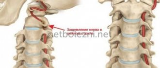
Lumbar radiculopathy (radicular syndrome) is a neurological condition caused by compression of one of the L1-S1 roots, which is characterized by low back pain radiating to the leg. Compression of the root can be manifested not only by pain (sometimes of a shooting nature), but also by impaired sensitivity, numbness, paresthesia or muscle weakness. Radiculopathy (radicular syndrome) can occur in any part of the spine, but it most often occurs in the lumbar region. Lumbosacral radiculopathy occurs in approximately 3-5% of the population, in both men and women, but, as a rule, the syndrome occurs in men at the age of 40 years, and in women the syndrome develops between the ages of 50 and 60 years. Treatment of radicular syndrome of the lumbosacral spine can be carried out using both conservative methods and surgical techniques.
Causes
Any morphological formations or pathological processes that lead to compression effects on the nerve root can cause radicular syndrome.
The main causes of lumbar radiculopathy are:
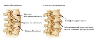
- A disc herniation or bulge can put pressure on the nerve root and lead to inflammation in the root area.
- A degenerative disease of the joints of the spine that results in the formation of bone spurs on the facet joints, which can lead to a narrowing of the intervertebral space, which will put compression on the nerve roots.
- Trauma or muscle spasm can put pressure on the root and cause symptoms in the area of innervation.
- Degenerative disc disease, which leads to wear and tear of the intervertebral disc structure, and a decrease in the height of the discs, which can lead to a decrease in the free space in the intervertebral foramen and compression of the root as it exits the spinal column.
- Spinal stenosis
- Tumors
- Infections or systemic diseases
In patients under 50 years of age, the most common cause of radicular syndrome in the lumbar spine is a herniated disc. After 50 years of age, radicular pain is often caused by degenerative changes in the spine (stenosis of the intervertebral foramen).
Risk factors for developing lumbar radiculopathy:
- age (45-64 years)
- smoking
- mental stress
- Strenuous physical activity (frequent heavy lifting)
- Driving or vibration exposure
What to do if a nerve is pinched in the back
If there is a suspicion of pinched spinal nerves, first aid must be provided. The person must be carefully placed on a flat, hard surface and given a painkiller tablet (for example, Ibuprofen, Ketoprofen).
Next, you need to call a doctor at home or take the victim to the hospital yourself, positioning him in the car so as to minimize his mobility, and therefore the possibility of damage to pinched nerves.
Drug therapy
In the hospital, after the necessary diagnostic procedures to treat the pinching, the specialist will prescribe the necessary drug therapy to the patient. It usually includes non-steroidal anti-inflammatory drugs in the form of tablets, injections, ointments or gels (Diclofenac, Voltaren, the same above-mentioned Ketoprofen or Ibuprofen). They have an effective analgesic and anti-inflammatory effect.
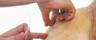
If after using the above drugs for several days the pain does not subside, a specialist may prescribe the use of novocaine blockade. To do this, the area of the back located above the damaged spinal nerves is injected intramuscularly with Novocain. The blockade allows you to get rid of pain and improve the patient’s well-being.
Exercise therapy and massage
Did you know that...
Next fact
In parallel with drug treatment for pinching, the patient is prescribed daily exercise therapy , as well as a visit to the massage room. Physical therapy must be done under the supervision of an experienced specialist who will monitor all your movements and help prevent unnecessary actions that can lead to additional complications in the event of pinched spinal nerves.
Most often exercise therapy includes the following exercises:
- Starting position: lying on the floor. Both legs need to be bent at the knee joints, with the soles of the feet pressed to the floor. Stretch your arms along your body. As you inhale, lift your back off the floor, trying to lift it up, while your shoulder blades should not come off the floor. As you exhale, your back returns to the floor. Perform 10-15 repetitions.
- Lying with your back on the floor, bend both lower limbs at the knees and pull them towards the chest as much as possible. In this position, roll on the floor so that your shoulder blades and pelvis touch the floor alternately. Do 10-15 rolls.
- Starting position – standing on all fours. As you inhale, bend your lower back like a cat, then, as you exhale smoothly, straighten your back, and then, on the contrary, bend in the opposite direction, as if rounding your back. Repeat about 15 times.
Therapeutic exercise and manual therapy are the main part of the complex treatment of a pinched nerve. Regular exercise therapy is especially necessary during the recovery period after surgical treatment of pinched spinal nerves. They will help restore the strength of the spinal column and restore its flexibility. Regular swimming is also an ideal option to speed up spinal recovery.
Physiotherapeutic procedures and manual therapy may also be prescribed to treat pinched spinal nerves . Massage must be performed by a qualified chiropractor. All his actions should be aimed at working not with the spine, but with the muscular corset that supports it.
The massage is first performed from bottom to top, and then vice versa from top to bottom. The therapist begins to warm up the muscles with light stroking movements in a circular direction. Then his movements should become rubbing, and then the specialist directly moves on to kneading the muscle corset. The massage session should end with stroking movements of the specialist.
Note! During the massage, pressing and patting movements are not allowed! This can lead to complications with the spine! Throughout the entire massage procedure, the patient should lie on his stomach on a special massage table, with his arms freely placed on both sides of the body.
Video: “Exercise for piriformis muscle spasm”
Symptoms
Symptoms resulting from radicular syndrome (radiculopathy) are localized in the area of innervation of a particular root.
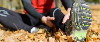
- Back pain radiating to the buttock, leg and extending down behind the knee, into the foot - the intensity of the pain depends on the root and the degree of compression.
- Disruption of normal reflexes in the lower limb.
- Numbness or paresthesia (tingling) may occur from the lower back to the foot, depending on the area of innervation of the affected nerve root.
- Muscle weakness can occur in any muscle innervated by a pinched nerve root. Prolonged pressure on a nerve root can cause atrophy or loss of function of a specific muscle.
- Pain and local tenderness are localized at the level of the damaged root.
- Muscle spasm and postural changes in response to root compression.
- Pain increases with exercise and decreases with rest
- Loss of the ability to make certain movements of the body: inability to straighten back, bend towards the localization of compression, or stand for a long time.
- If the compression is significant, activities such as sitting, standing and walking may be difficult.
- Change in normal lordosis of the lumbar spine.
- Development of stenosis-like symptoms.
- Stiffness in the joints after a period of rest.
Patterns of pain
- L1 - back, front and inner surface of the thigh.
- L2 - back, front and inner surface of the thigh.
- L3 - back and front, and the inner surface of the thigh with a downward extension.
- L4 - back and front of the thigh, to the inner surface of the leg, into the foot and big toe.
- L5 – Along the posterolateral part of the thigh, the front part of the lower leg, the top of the foot and the middle toe
- S1 S2 – Buttock, back of the thigh and lower leg.
The onset of symptoms in patients with lumbosacral radiculopathy (radicular syndrome) is often sudden and includes low back pain.
Sitting, coughing or sneezing can aggravate the pain, which radiates from the buttock down the back of the leg, ankle or foot.
You need to be vigilant for certain symptoms (red flags). These red flags may indicate a more serious condition that requires further evaluation and treatment (eg, tumor, infection). The presence of fever, weight loss, or chills requires careful evaluation.
The patient's age is also a factor when looking for other possible causes of the patient's symptoms. People under 20 years of age and over 50 years of age are at increased risk for more serious causes of pain (eg, tumors, infections).
Treatment of the lower back in the network of clinics “Hello!”
A good choice in order to quickly receive a complete and effective diagnosis and treatment for symptoms of a pinched nerve in the back is to contact any clinic in the “Hello!” network.
We employ the best specialists in the field of treatment of diseases of the musculoskeletal system with many years of practical experience in medicine.
Our clinics have everything you need for a complete, accurate diagnosis. Therefore, our doctors will be able to quickly determine what caused the disease and select a treatment regimen that meets the needs of each individual patient.
In order to make an appointment at any clinic in our network, you can call the phone number indicated on the page or request a call back through the online form on the website.
Diagnostics
The primary diagnosis of radicular syndrome of the lumbosacral spine is made based on the symptoms of the medical history and physical examination (including a thorough examination of the neurological status). A thorough analysis of motor, sensory and reflex functions allows us to determine the level of damage to the nerve root.
If the patient reports typical unilateral radiating leg pain and there are one or more positive neurological test results, then a diagnosis of radiculopathy is very likely.
However, there are a number of conditions that may present with similar symptoms. Differential diagnosis must be carried out with the following conditions:
- Pseudoradicular syndrome
- Traumatic disc injuries in the thoracic spine
- Damage to discs in the lumbosacral region
- Spinal stenosis
- Cauda equina
- Spinal tumors
- Spinal infections
- Inflammatory/metabolic causes - diabetes, ankylosing spondylitis, Paget's disease, arachnoiditis, sarcoidosis
- Trochanteric bursitis
- Intraspinal synovial cysts
To make a clinically reliable diagnosis, as a rule, instrumental diagnostic methods are required:
- X-rays – can detect the presence of joint degeneration, fractures, bone defects, arthritis, tumors or infections.
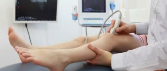
- MRI is a valuable technique for visualizing morphological changes in soft tissues, including discs, spinal cord and nerve roots.
- CT (MSCT) provides complete information about the morphology of the bone structures of the spine and visualization of spinal structures in cross section.
- EMG (ENMG) Electrodiagnostic (neurophysiological) studies are necessary to exclude other causes of sensory and motor disorders, such as peripheral neuropathy and motor neuron disease
Conservative treatment:
- Rest: avoid activities that cause pain (bending, lifting, twisting, turning or bending backwards. Rest is necessary for acute pain syndrome
- Drug treatment: anti-inflammatory, painkillers, muscle relaxants.
- Physiotherapy. For acute pain syndrome, the use of procedures such as cryotherapy or chivamat is effective. Physiotherapy can reduce pain and inflammation of the spinal structures. After the acute period has stopped, physiotherapy is carried out in courses (ultrasound, electrical stimulation, cold laser, etc.).
- Corseting. The use of a corset is possible in case of acute pain syndrome to reduce the load on the nerve roots, facet joints, and lumbar muscles. But the duration of wearing a corset should be short, since prolonged fixation can lead to muscle atrophy.
- Epidural steroid injections or facet joint injections are used to reduce inflammation and control pain in severe radicular syndrome.
- Manual therapy. Manipulations can improve the mobility of the motor segments of the lumbar spine and relieve excess muscle tension. Using mobilization techniques also helps modulate pain.
- Acupuncture. This method is widely used in the treatment of radicular syndrome in the lumbosacral spine and helps both reduce symptoms in the acute period and is included in the rehabilitation complex.
- Exercise therapy. Exercise includes stretching and strengthening exercises. The exercise program allows you to restore joint mobility, increase range of motion and strengthen your back and abdominal muscles. A good muscle corset allows you to support, stabilize and reduce tension on the spinal joints, discs and reduce the compression effect on the spine. The volume and intensity of exercise should be increased gradually to avoid relapse of symptoms.
- In order to achieve stable remission and restore full functionality of the spine and motor activity, it is necessary for the patient, after completing the course of treatment, to continue independent exercises aimed at stabilizing the spine. The exercise program must be individual.
Physiotherapy
When acute symptoms are relieved, the patient can gradually move on to a number of physical exercises:
- Body tilts.
- Walk with your knees high.
- Twisting the body from a side lying position.
- Bend the legs at the knees and bring them to the chest from a supine position.
- Rolling on your back, clasping your knees with your hands (“Roller”).
- Swing your legs.
You need to perform therapeutic exercises every day, for at least 10 minutes. Exercise activates blood circulation, improves nutrition and oxygen supply to muscle tissue and joints.
Surgery
Surgical methods for the treatment of radicular syndrome in the lumbosacral spine are necessary in cases where there is resistance to conservative treatment or there are symptoms indicating severe compression of the root such as:
- Increased radicular pain
- Signs of increased root irritation
- Muscle weakness and atrophy
- Incontinence or bowel and bladder dysfunction
As symptoms worsen, surgery may be indicated to relieve compression and remove degenerative tissue that is affecting the root. Surgical treatments for radicular syndrome in the lumbosacral spine will depend on which structure is causing the compression. Typically, these treatments involve some way to decompress the spine or stabilize the spine.
Some surgical procedures used to treat lumbar radiculopathy are:
- Fixation of vertebrae (spinal fusion - anterior and posterior)
- Lumbar laminectomy
- Lumbar microdiscectomy
- Laminotomy
- Transforaminal lumbar
intercorporeal fusion - Cage implantation
- Correction of deformity
Forecast
In most cases, it is possible to treat radicular syndrome in the lumbosacral spine conservatively (without surgical intervention) and restore ability to work. The duration of treatment may vary from 4 to 12 weeks depending on the severity of symptoms. Patients should continue to perform exercises at home to improve their posture, stretching, strengthening, and stabilization. These exercises are necessary to treat the condition causing radicular syndrome.
Prevention
The development of radicular syndrome in the lumbosacral spine can be prevented. To reduce the likelihood of developing this condition you should:
- Practice good posture while sitting and standing, including while driving.
- Use proper body mechanics when lifting, pushing, pulling, or performing any activity that places additional stress on the spine.
- Maintain a healthy weight. This will reduce the load on the spine.
- No smoking.
- Discuss your profession with a physical therapy doctor, who can analyze work movements and suggest measures to reduce the risk of injury.
- Muscles should be strong and elastic. It is necessary to consistently maintain a sufficient level of physical activity.
Surgeon's help
Surgery is prescribed if the treatment does not give the expected effect for two months or longer. Indications for surgical intervention may include:
- ineffectiveness of conservative methods;
- intervertebral hernia;
- violation of the integrity of the nerve.
To avoid recurrent problems after completion of therapy, patients must strictly follow the recommendations of the attending physician and go through all the necessary stages of the rehabilitation course.
Avoiding a recurrence of an unpleasant situation with the lower back will help by eliminating increased physical stress on the back and protecting the spine from injury. A course in a specialized sanatorium will also benefit.