Muscle fatigue
In our technological age, many people are gradually forming a new “bad habit”. Spending most of the day in front of a computer screen or holding mobile gadgets in our hands, we may not even notice that we are sitting in a completely uncomfortable and unnatural position. It especially affects the muscles of the shoulder girdle, neck, back and right arm, which is constantly on the computer mouse of office workers1. Staying in one position for a long time or using stereotyped movements can lead to overstrain of the muscles involved, which we feel as soreness1.
Exercise stress
“Body aches” after physical activity can occur both in untrained people and in professional athletes8. And the cause of discomfort is the accumulation of under-oxidized metabolic products in muscle cells; in particular, painful sensations are caused by an excessive amount of lactic acid (lactate)2. Less commonly, the cause is microtrauma, but this can only occur if the training rules are violated8.
Muscle soreness due to overwork does not occur immediately, but after a few hours or within 1-2 days after training or unusual physical activity and disappears within a week8.
Causes of hip pain
Traumatic injuries
A hip bruise occurs when a blow or fall occurs and is manifested by moderate local pain.
At first the pain is sharp, then dull, aching, intensifying with palpation and movement. Due to the large array of soft tissues, severe swelling often develops. Bruising is possible. The function of the limb is usually slightly limited, support is preserved. A hematoma also forms after blows and falls; its manifestations resemble a bruise. It differs from a bruise in the bursting or pressing nature of the pain, the formation of dense local edema, in the area of which, after some time, an area of fluctuation appears. With deep-lying hematomas, the swelling is nonspecific, fluctuation may be absent. Hematoma can be suspected by a significant increase in swelling in the first days or its persistence for a long time.
Damage to the quadriceps tendon is observed in athletes, but less often develops in everyday life. It is characterized by acute pain along the front surface of the thigh, just above the knee joint, sometimes by a cracking sound at the time of injury. Subsequently, the pain subsides, but remains quite intense. In the area of damage, pain, swelling of soft tissues, and a defect at the site of the rupture are detected. With a complete rupture, support and independent straightening of the lower leg are impossible; with an incomplete rupture, it is difficult.
The cause of a diaphyseal femoral fracture is high-energy impacts: falls from a height, road and industrial injuries. The damage is accompanied by unbearable explosive pain. Subsequently, the pain is very intense and intensifies when attempting to move, palpate, or passively shift the limb. The thigh is swollen and deformed. The leg is shortened. Crepitation and pathological mobility are detected. The general condition is disturbed, traumatic shock is possible.
A pathological fracture of the femoral diaphysis can be diagnosed with bone cysts, tumors, and some hereditary and infectious diseases. It has mild symptoms. The pain is minor or moderate. Displacement, pathological mobility and bone crunch are absent, which is why the injury is often mistaken for a bruise. Distinctive features of a pathological fracture are the long-term persistence of symptoms and the presence of previous pain during exercise or at rest.
Inflammatory diseases
Myositis of the thigh muscles occurs after unusual physical activity: prolonged walking or running, the first workouts after a long break. It is characterized by aching pain in the projection of the affected muscle or muscle group. There is increased pain on palpation and muscle tension. When palpated, a diffuse thickening of the muscle is determined; slight swelling and slight local hyperthermia are possible.
Quadriceps tendonitis is usually found in active people over 40 years of age and manifests itself as vague, low-intensity nagging pain in the lower parts of the front of the thigh. The pain intensifies when the leg is extended. With biceps tendonitis, which is typical for runners, the pain is localized deep in the buttock, spreads down the back of the thigh, and intensifies during running, especially when accelerating. Subsequently, the pain becomes long-lasting, constant, and occurs after minor exertion, at rest, and at night.
Pain on the outer surface of the thigh is sometimes accompanied by aseptic arthritis of the hip joint of various etiologies (reactive, allergic, post-traumatic). The pain is aching, dull, non-localized, complemented by soreness in the groin and buttock, limitation of movements, and difficulty walking.
Hip pain
Bone infections
Periostitis of the femur develops infrequently, as a rule, it is the result of injuries (bruises, fractures) or purulent lesions (deep infected wounds, abscesses, phlegmon, arthritis). In the first case, the process is aseptic in nature, accompanied by moderate pain, aggravated by palpation. Minor swelling is detected. The general condition is not disturbed.
Sometimes serous post-traumatic periostitis is observed, characterized by significant accumulation of fluid under the periosteum. The pathology is manifested by moderate arching pain, swelling, and deformation. With purulent inflammation of the periosteum, an acute onset with intense twitching, throbbing pain is observed. Local hyperemia, significant swelling of the segment, increased body temperature, chills, and fever are detected.
The femur is one of the most common locations of hematogenous osteomyelitis. Bone inflammation develops in childhood, often due to minor trauma or after an acute infection. It occurs with a sharp increase in temperature, fever, chills, and delirium. A day after the onset of hyperthermia, intense localized deep pain occurs in the thigh. The pain increases, becomes unbearable, forcing the patient to freeze in bed.
There is a local form of hematogenous osteomyelitis, in which the pain syndrome is less pronounced and the general condition is slightly disturbed. In some cases, a severe hurricane course of the disease is observed with a predominance of general symptoms. Post-traumatic and postoperative osteomyelitis are also manifested by pain, swelling, hyperemia, and disturbances in general condition, but the pain appears against the background of suppuration of an open fracture or postoperative wound, progresses more slowly, and does not reach the level of unbearable.
Infections of blood vessels and soft tissues
Pain in the thigh is observed with purulent lesions of soft tissue structures. At first it has a bursting or pressing character. The intensity of the pain syndrome quickly increases, the pain becomes throbbing, tearing, jerking, sharply limits movement, and disrupts night sleep. Swelling and increased local temperature are detected. The severity of general manifestations (hyperthermia, fever, symptoms of intoxication) depends on the extent of the purulent process. The cause of pain is:
- Furuncle.
A limited focus of inflammation, which is a convex, sharply painful formation, covered with bluish-purple skin. In the center of the formation there is a black core surrounded by pus. The general condition is usually not affected. - Carbuncle.
It consists of several closely spaced small foci merging into one infiltrate with a diameter of up to several centimeters. Severe hyperthermia and fever are observed. - Abscess.
A limited purulent focus, the diameter of which can vary from 2-3 to 10 or more centimeters. Accompanied by weakness, weakness, fever to febrile levels. - Phlegmon.
Widespread purulent inflammation without clear boundaries. It is characterized by intense pain throughout the thigh and severe intoxication.
From foci in soft tissues, the infection can spread through the lymphatic vessels. The manifestation of reticular lymphangitis is indicated by increased pain, deterioration of the general condition, and the appearance of a marble pattern around the primary inflammatory focus. With stem lymphangitis, a longitudinal strip appears on the thigh, along which pain on palpation, swelling, hyperemia, and cord-like tissue compaction are determined. When deep vessels are affected, stripes on the limb are not detected; rapidly increasing arching pain and lymphedema of the underlying sections are noted.
With phlebitis and thrombophlebitis of the superficial veins of the thigh, developing against the background of infectious lesions or varicose veins, rapidly progressing pain is detected along the vein. The pain intensifies when walking and is usually localized in the lower third of the segment. Phlebitis and thrombophlebitis of the deep veins are manifested by constant increasing pain, swelling, general hyperthermia, milky white coloration of the skin of the limb.
A special form of venous damage is postpartum thrombophlebitis, which occurs in the first weeks after childbirth. Intense pain on the inner side of the thigh is observed with inflammation of the great saphenous vein, combined with swelling and deterioration of the general condition. When the deep femoral veins are involved, unbearable sharp pain along the anterior inner surface of the thigh, significant swelling, pallor of the skin of the limb, and fever are noted.
Oncological diseases
Benign tumors of the femur are characterized by slow growth and a long, often multi-year course. Osteomas and chondromas manifest themselves as minor, transient, unclear pain; with chondromas in children, deformities and shortening of the limb are possible. With osteoid osteomas, the pain is intense and sharp. The growth of neoplasia is accompanied by increased pain and the appearance of palpable bone density formations.
The most common malignant tumors of the hip include osteosarcoma, chondrosarcoma, and Ewing's sarcoma. Chondrosarcomas are usually localized in the upper part of the bone, osteogenic sarcomas in the upper or lower part, and Ewing sarcomas in the lower part. The pain with such neoplasms is initially dull, unclear, quickly intensifies, becomes unbearable, and is combined with the appearance of a venous pattern, deformation, weight loss, and general weakness.
Neurological causes
Hip pain is often caused by neurological diseases. Distinctive features of such pain are shooting, burning, shooting or piercing nature (some patients compare the sensations to the “strike of a dagger”), combination with paresthesia, weakening of muscle strength, and sensitivity disorders. Pain in the hip area is caused by the following pathologies:
- Femoral nerve neuropathy.
There is pain along the anterior inner surface of the thigh and in the groin, which intensifies with extension of the knee joint. - Sciatic nerve neuropathy.
The patient complains of pain along the back of the thigh, which spreads from the buttock to the lower leg and foot, and intensifies when trying to raise a straight leg while lying on his back. - Piriformis syndrome.
Occurs when the nerve trunk is compressed in the infrapiriform foramen. Pain - as with neuropathy of the sciatic nerve, combined with constant pulling, aching, tingling pain in the buttock. - Neuropathy of the external cutaneous nerve.
It is characterized by pain that spreads along the outer side of the thigh, makes walking difficult, and decreases when lying down with bent legs. Due to the pain syndrome, changes in gait are formed that resemble intermittent claudication. - Radicular syndrome.
Develops with osteochondrosis, spondylosis, intervertebral hernia, and other diseases of the lumbar spine. It manifests itself as pain spreading from the center to the periphery, intensifying with exertion, sudden movements, coughing, sneezing. The localization of pain is determined by the level of exit of the affected root. - Lumbosacral plexitis.
Manifests with sharp pain in the thigh, buttock, sacrum, lower leg, and groin area. When the lumbar plexus is affected, the pain occurs mainly in the anterior part; when the sacral plexus is affected, the pain occurs mainly in the back part of the leg.
Other reasons
Sometimes hip pain is provoked by diseases of neighboring structures or distant organs, or mental disorders. Possible causes of pain may be:
- Coxarthrosis.
The pain is aching, nagging, occurs at the beginning of movements and after exercise, combined with pain in the hip joint. - Renal cell carcinoma.
The pain syndrome mimics sciatic nerve neuropathy and is caused by compression of the nerve trunk by an enlarging tumor. - Mental disorders.
Pain can appear with depression, hysterical neurosis, and some other conditions. Typical signs are vagueness, unusualness and variability of complaints that do not fit into the picture of a specific somatic disease.
Injuries
Severe pain can occur when muscle fibers and tendons are torn. This usually happens if the load is excessive and the muscles are not prepared for it1. But they can also be damaged by sudden movements3. Unlike “ache” due to muscle overwork, pain due to injury occurs immediately, at the peak of the load3.
It should be remembered that even a small, but untreated injury can cause an even more severe sprain3. Therefore, if you experience pain during physical activity, be sure to consult a doctor to rule out a serious injury.
to come back to the beginning
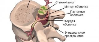
Muscles , tendons and their sheaths, ligaments, fascia, aponeuroses, and capsules play an important role in ensuring joint stability. Pathology of periarticular soft tissues can be considered as conditions associated with arthritis, and as an independent pathology. When describing soft tissue pathology, the following concepts are usually used:
- tendinitis – inflammation of tendon tissue;
- tenosynovitis/tenosynovitis – inflammation of the tendon tissue and tendon sheath;
- enthesitis/enthesopathy - inflammation of the tendon tissue at the site of its attachment to the bone;
- Bursitis is an inflammation of the bursae, thin-walled cavities lined with synovium that facilitate the movement of tendons and muscles over bony prominences.
Pathology of the ankle joint and foot, as well as damage to the periarticular soft tissues of this area are a common reason for visiting a doctor, accounting, according to domestic and foreign literature, from 6 to 21% of all pathologies of the musculoskeletal system.
The causes of soft tissue pathology in the ankle and foot area can be both external and internal factors. External include overload (change in the stereotype of physical activity), trauma (single or repeated microtrauma), local injection of glucocorticosteroids (GCS) into the thickness of the tendon, which can cause degeneration of tendon tissue; internal include congenital anomalies of joint structures leading to disruption of biomechanics, muscle imbalance surrounding the joint, physical inactivity (immobilization), impaired blood supply to certain areas of the tendons, age-related involution of the musculoskeletal system. Often there is a combination of several factors.
Pain syndrome with damage to the soft tissues of the ankle and foot usually has a clear localization.
The main causes of pain in the heel area are:
- Achilles tendinitis;
- enthesitis of the Achilles tendon;
- Achilles or retrocalcaneal bursitis;
- subcalcaneal bursitis;
- plantar fasciitis, heel spur.
And the hilly tendon is a continuation of the triceps surae muscle, which is formed from the gastrocnemius and soleus muscles. This rather powerful tendon attaches to the heel bone. Between the tendon itself and the bone, as well as between the tendon and the skin, there are synovial bursae.
The most common causes of chronic Achilles tendon pain are Achilles tendinitis, partial rupture, or bursitis. Typically, these diseases are manifested by the following symptoms:
- mild, aching pain in the tendon area after running or physical activity, which gradually intensifies;
- feeling of weakness in the leg;
- episodes of diffuse or localized pain in the tendon area immediately or several hours after running;
- swelling of the soft tissues in the tendon area;
- a feeling of stiffness (“cloggedness”) in the muscle that goes away as the tendon “warms up” during exercise.
Achilles tendonitis
Tendinitis of the Achilles tendon (Achilles tendonitis) often occurs in seronegative spondyloarthritis, in patients with joint hypermobility syndrome, and with severe flat feet. With Achilles' pain, swelling and pain occur when there is a load in the area of the tendon or where the tendon attaches to the heel bone.
The main clinical symptoms characteristic of Achilles tendonitis:
- pain in the heel, sometimes along the back of the shin;
- flexing the foot increases pain;
- the area of greatest pain is 2–3 cm above the junction of the tendon with the heel bone;
- the tendon may be swollen and thickened.
Retrocalcaneal bursitis
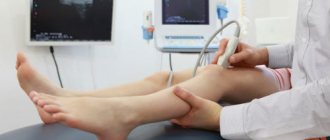
Posterior calcaneal bursitis is clinically similar to Achilles tendinitis, but the pain often becomes excruciating and intensifies significantly with walking and prolonged standing, and swelling or swelling often appears above the site of attachment of the tendon to the heel bone. Ultrasound examination of this area helps differentiate the conditions.
Achilles tendon enthesopathy
The most common cause of development of Achilles tendon enthesitis is seronegative spondyloarthritis.
Enthesitis is often caused by trauma to the entheses or overload of the tendons . Enthesitis is manifested by pain during movement in which the corresponding muscle is involved. Pain occurs more clearly when the affected muscle is tense. Swelling of the surrounding tissues and tenderness in the area of the involved enthesis are determined. The outcome of enthesopathy is, as a rule, ossification of the enthesis with the development of enthesophytes.
Plantar fasciitis
Plantar fasciitis, the most common cause of heel pain, is inflammation of the insertion of the flexor digitorum brevis muscle on the tuberosity of the heel bone. Overstrain of these structures due to flat feet, degenerative diseases of the musculoskeletal system, seronegative spondyloarthritis leads to reactive inflammatory production of bone tissue or the formation of heel spurs secondary to stretching of these structures.
The main symptom of plantar fasciitis is pain along the entire plantar surface of the foot when walking. Usually this pain appears during the first steps after the patient gets out of bed in the morning, or after sitting for a long time.
your feet hurt? Come, we will help you! appointment with a rheumatologist for an appointment and therapeutic blockade in Samara by calling the phone number on the contacts page.
Diseases of the spine and joints
Muscles react to disruption of the joints4 and vertebrae5 associated with them. Therefore, myalgia is one of the symptoms of diseases of the spine5,11 and joints of the limbs4. For example, with osteochondrosis or scoliosis (curvature of the spine), pain in the neck, chest or lower back is associated with overstrain of the paravertebral muscles5,11. And in advanced cases, when the vertebra compresses the nerve root emerging from the spinal cord, the pain can “radiate” to the arm or leg11.
Often myalgia with osteochondrosis is combined with a feeling of numbness or “crawling goosebumps”. At the moment of acute pain, a person freezes, taking a forced position11.
Diagnostics
The examination is usually carried out by orthopedic traumatologists. In case of purulent processes, patients are examined by a surgeon, in case of neurological pathology - by a neurologist. At the initial stage, the doctor finds out the history of life and illness, and conducts an external examination. The following additional studies are prescribed:
- X-ray of the hip.
A basic diagnostic method that allows you to identify all major bone pathologies: fractures, cancer, osteomyelitis, periostitis. In case of lesions of soft tissue structures, it is performed to exclude bone lesions and conduct differential diagnosis. - CT scan of bone.
It is used at the final stage of the examination to clarify the results of radiography, planning conservative therapy or surgical intervention. Makes it possible to accurately determine the size, configuration and structural features of the pathological focus in bone tissue. - Ultrasound.
Sonography of soft tissues in myositis and tendonitis reveals areas of inflammation or degeneration. Ultrasound scanning of the veins of the lower extremities is effective in assessing the condition of the veins and lymphatic vessels in patients with phlebitis and lymphangitis, and in identifying blood clots in thrombophlebitis. - MRI of soft tissues.
Recommended for ambiguous results of other diagnostic procedures. It is carried out to determine the localization of deep hematomas, confirm degenerative or inflammatory changes in tendinitis and myositis. - Electrophysiological studies.
For patients with pain of neurological origin, ENMG, ENG or EMG are indicated to clarify the level of nerve damage and study the condition of nerve and muscle tissue. - Lab tests
. They are performed to determine the severity of the inflammatory process in purulent diseases, assess the condition of organs and systems in malignant neoplasms.
Other reasons
Almost any disease of the internal organs can lead to myalgia11. When an organ is affected, it creates pain impulses that are partially transmitted to the muscles located nearby11.
Myalgia can also be caused by:
- endocrine diseases, such as thyroid hormone deficiency16,7;
- vascular pathologies that disrupt the nutrition of the muscles of the limbs15,16;
- chronic fatigue syndrome7;
- imbalance of microelements in the body16;
- taking medications that lower blood cholesterol levels12.
to come back to the beginning
Features of myalgia
Pain emanating from the muscles is usually deep6. Acute myalgia is protective in nature because it causes reactions aimed at eliminating the damaging factor11. Such reactions include, for example, muscle spasms4. But despite its protective nature, there is almost always a risk that the pain will become chronic11. There are 2 main causes of chronicity:
- Increased sensitivity. In response to irritation, the muscle releases substances that support inflammation. They further irritate pain receptors in the muscles. In response to frequent signals, the central nervous system lowers the pain threshold, so we can feel soreness in the muscle even when it is not strongly irritated11.
- Spasm. If pain and spasm persist, a “vicious circle” is formed: pain causes spasm, and spasm maintains pain1,5, 11.
Diagnosis of muscle pain
Determining why myalgia occurs is not always easy. Only a doctor can understand the causes and select treatment that helps get rid of disturbing symptoms or alleviate them. To find out the cause of myalgia, the doctor conducts a comprehensive examination, including a neurological one, prescribes laboratory tests, ultrasound, computed tomography and other research methods3.
Various specialists treat myalgia. Depending on its cause, a traumatologist, rheumatologist, neurologist or endocrinologist can help you.
How to treat leg pain?
It is impossible to effectively treat pain in the leg without attaching importance to or counteracting the processes of deformation of the bones of the spine. The processes of deformation of the bones of the spine, in turn, indicate a gradual change in the density of bone tissue and the entire musculoskeletal system with age in the patient, that is, these are reflections of the aging of bone tissue. It is extremely important to note here that no treatment for such pain gives a lasting result unless a special scheme is used to restore bone density, returning bone density to a younger, denser state. We have developed a special scheme that allows patients to get rid of these pains; it consists of special drugs that stimulate osteogenesis, which cause bone tissue not to weaken with age, but to strengthen. Such schemes are used for six months and they give excellent results, and if the patient begins to do such preventive treatment early, he does not develop a huge number of diseases that are associated with the aging of the musculoskeletal system. These diseases include, first of all, osteochondrosis of various parts of the spine, deforming arthrosis of the knee joints, hip joints, shoulder joints, arthrosis of the ankle joint, these also include heel spurs. You see a huge collection of diseases that affect an older person, can be ruled out and may never occur. If you pay attention to the first appearance of such pain, and take active measures to eliminate the causes of the development of this pain syndrome. Therefore, we recommend not to delay time if such symptoms appear, in order to figure out exactly what the cause of these pains is, contact our center and we will be able to correctly diagnose you, understand the nature of these pains, and most importantly, prescribe effective treatment for these problems, which will not allow the development of complications and the formation of independent diseases, in the form of arthrosis, osteochondrosis.
Pain in the legs due to problems with the musculoskeletal system
Particularly pronounced pain occurs in patients in cases where similar problems such as osteochondrosis lead to such deformation of the vertebrae that uneven pressure occurs on the intervertebral discs and these discs, squeezing the jelly-like contents of the intervertebral core in a certain direction, put pressure on the nerve roots, and already a herniated disc or Schmorl's hernia appears. Such pain can be unbearable and often lead to atrophy of the leg muscles, and all this can be avoided if, during menopause or even a little earlier, they think about the aging processes of their own musculoskeletal system, because doctors warned them about this possibility. In addition to such pains, there are, of course, pains that are caused by local processes, but as a rule, they are all associated with processes of deformation of the musculoskeletal system, for example, pain in the knee joint, pain in the knee joint is local pain and it occurs due to the fact that bone tissue in the area of the knee joint it is deformed and causes destruction and damage to the cartilage tissue on which the higher-lying bone stands. It is the pressure of the upper bone that leads to the wear and tear of the cartilaginous base on which the bone stands and which is the support of the joint, and this damage to the cartilage causes disturbances in the production of synovial fluid and forms dryness of the joint, which further leads to rapid depletion and destruction of the cartilaginous base of the joint meniscus, most often the internal meniscus, a degenerative rupture of the meniscus occurs; sections of destroyed cartilage begin to float directly in the form of fragments in the joint cavity and interfere with the normal flexion movement of the joint. Thus, such a situation leads to the development of such phenomena as inflammatory synavitis, and such fragments are called cartilaginous mice, and all this actually requires active medical intervention. If these fragments do not appear in large numbers, then naturally the problems of arthrosis can be eliminated by conservative measures, a liquid cartilage implant can be introduced into the joint, improving the structure of one’s own cartilage tissue, but still the basic treatment for all these problems, including arthrosis, is treatment with osteogenesis stimulants, without which it is impossible to expect reliable effective treatment for this problem.
In addition, pain in the legs can be naturally provoked by some local processes, for example, inflammation of the skin in the area of the ulcer, or, for example, neuropathic pain associated with damage to nerve fibers in diabetes mellitus, but this is a separate story and a separate nature of the pain.
To help understand this variety of pain, the variety of problems that arise from damage to the musculoskeletal system, from dilation or constriction of the vessels of the lower extremities, is the task of a specialist, and you need to start with a phlebologist who will help determine the nature of the pain and help outline a rational treatment plan . Often the veins themselves do not cause pain, but when they expand, they can irritate the inflamed nerve fibers. Thus, varicose veins themselves turn into a problem that increases the pain syndrome, which is based on deformation of the bones of the spine. Therefore, as you can see, there are a huge number of problems associated with the aging of the musculoskeletal system, advanced chronic venous insufficiency, venous stagnation in the subcutaneous venous system, the formation of diabetes mellitus and the effect of sugar surges on the nerve fibers in the foot area, all this can lead to various types of pain. syndrome in the legs. Therefore, such a concept as pain in the leg can only be clearly and clearly described by good specialists who have extensive experience in examining the lower extremities. We suggest that you quickly and without wasting time, when such symptoms appear, contact the center and find experienced specialists who can answer the main question, what is the cause of the pain in this leg, then the first step towards treatment will be taken, in the future you can outline a clear a treatment plan that is sure to give results. Depending on the speed of your treatment, the result can either be achieved after one course of treatment, or will require multiple, restorative courses that will last for several months, or even years. Therefore, do not waste time, because you have no other option other than proper treatment to maintain your quality of life normally, come to the center and carry out the necessary examinations. Sign up for a consultation