Joints are a complex osteochondral structure that bears the entire weight of the body. Small and large joints perform a motor function and regularly experience physical stress. Pain and swelling of the joints is a reaction to internal or external factors, which results in limited movement and change in shape. If swelling and pain appear in the joints, regardless of location, you should consult a doctor to avoid severe, irreversible consequences.
What is "edema"
Edema is a concentration of blood plasma from lymphatic and blood vessels in the intercellular space. If swelling is an exceptional phenomenon for the patient, then there is no cause for concern. Sometimes the body responds this way to overheating or the effect of certain medications. Otherwise, such a symptom is considered a signal of serious health problems.
What to do if the joints on your hand are swollen
Swelling of the joints of the hand quite often occurs against the background of degenerative-dystrophic changes. In the absence of pain, patients present characteristic complaints:
- swelling followed by stiffness of the finger joints;
- redness of the skin over the affected tissues;
- feeling of pulsation;
- discomfort in surrounding muscles;
- the appearance of fluid in local tissues.
After a thorough examination, all efforts are directed towards eliminating the root cause. Glucocorticosteroids and drugs with chondroprotective, anti-inflammatory, and muscle relaxant effects are prescribed. Thanks to them, degenerative processes slow down and the symptoms of inflammation are relieved. Be sure to use B vitamins.
A special place is occupied by the use of traditional medicine, which helps to alleviate the patient’s condition. For this purpose, it is effective to apply compresses, perform local baths, and apply ointments based on medicinal plants.
A pronounced anti-edematous and anti-inflammatory effect is achieved using various physiotherapeutic procedures:
- shock wave therapy;
- electrophoresis;
- paraffin therapy;
- ozokerite applications;
- mud wraps;
- warming up.
All active therapeutic manipulations are carried out after eliminating the signs of the inflammatory process.
Causes
Joint swelling can develop due to a variety of circumstances - from minor injuries to serious degenerative pathologies of the musculoskeletal system.
Mainly these are:
- changes in the biochemical composition of blood plasma and tissue fluid;
- hormonal disbalance;
- increased capillary permeability;
- difficulty in the outflow of venous blood and lymph;
- violation of gas exchange and excretory function of the kidneys;
- congestion in case of heart failure;
- critical ischemia;
- arthritis and arthrosis;
- viral infection and inflammation;
- sedentary lifestyle, etc.
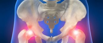
Swelling of the joints of the legs
Swelling of the ankle joints and small joints of the foot brings inconvenience and unpleasant symptoms. Patients suffering from swelling of the joints of the legs deny themselves nice shoes and cannot wear clothes that reveal their knees. Quite often the root cause of pathological changes are:
- all kinds of injuries;
- autoimmune diseases;
- deforming joint pathologies;
- degenerative processes;
- problems of the cardiovascular system;
- renal dysfunction;
- severe allergic reactions.
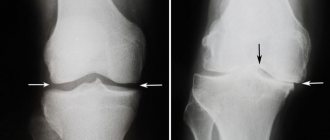
Swelling of the knee joint is often observed with injuries to the meniscus and cruciate ligaments.
To eliminate swelling of the leg joints, conservative, minimally invasive surgical methods and physical therapy are used. Of the medications it is advisable to prescribe:
- diuretics;
- antitumor agents;
- glucocorticosteroids;
- antibiotics;
- non-steroidal anti-inflammatory drugs;
- antihistamines;
- decongestant and cooling ointments, gels.
Minimally invasive procedures are performed with minimal disruption of the integrity of the skin and joint structure. A syringe is used to puncture the joint capsule and suck out the fluid from the joint cavity. The purpose of arthroscopy is to remove damaged osteochondral elements and restore the integrity of the cruciate ligaments.
Of the physiotherapeutic techniques, a pronounced therapeutic effect is provided by:
- UHF therapy;
- laser exposure;
- magnetic therapy;
- mud therapy;
- phonophoresis.
During the recovery stage, physical exercise is mandatory. A course of exercise therapy helps to normalize motor activity and restore metabolic processes in affected joints and surrounding tissues. Massage stimulates the movement of lymph and activates local blood flow. Massage sessions eliminate signs of swelling, relieve pain, and restore trophism of soft, cartilage and joint tissues.
Classification
First of all, it is necessary to establish the nature of the edema. It can be false and true.
False edema is observed in myxedema, systemic scleroderma and obesity and has a specific feature: when pressed with a finger, there is no trace or pit left on the skin.
In a situation with true edema, the hole does not disappear for a long time, which indicates the free movement of fluid in the tissues. With local edema, fluid accumulates in an unhealthy area of the body.
Pathologies
Unfortunately, patients do not always rush to seek medical help when joint swelling occurs.
However, a visit to the doctor is required if:
- unpleasant symptoms persist for about a week;
- at the same time, redness of the skin and an increase in body temperature are noted;
- swelling developed after joint puncture.
Pain and swelling in the joints occur due to congenital anomalies, various injuries, inflammatory processes, and degenerative changes (age-related). What diagnosis can a doctor make:
- Phlebeurysm;
Deep thrombosis and thrombophlebitis of the saphenous veins (occurs after childbirth, surgery on the abdominal organs, with a tumor, leukemia and some other diseases);- Postthrombophlebitic syndrome;
- Lymphedema;
- Arthrosis;
- Arthritis;
- Gout;
- Osteoarthritis;
- Psoriasis;
- Rheumatism.
Why do joints swell?
Rheumatoid arthritis
One of the most common types of arthritis is rheumatoid arthritis.
A distinctive feature is bilateral symmetrical damage to the joints. Pathology often debuts against the backdrop of changing seasons, hormonal changes, general infection, injury, and hypothermia. It manifests acutely or subacutely, and subsequently becomes chronic. Possibly a primary chronic course. Joint swelling is accompanied by pain, local hyperemia, hyperthermia, transient or permanent stiffness, and dysfunction. There are special forms of the disease - Felty's syndrome and juvenile arthritis. Felty syndrome develops on average 10 years after the first symptoms of the seropositive form of the disease appear. Damage to small joints in the arms and legs is observed. Hepatosplenomegaly, leukopenia, myocarditis, episcleritis, polyserositis are detected.
Juvenile rheumatoid arthritis develops in childhood or adolescence, resembles the pathology in adults, but differs from it in some ways. Predominant damage to the joints is possible, combined with pronounced systemic manifestations. The process often involves the large joints of the arms and legs. In addition to swelling, deformities and gait disturbances are detected. Sometimes hernial protrusions form.
Other aseptic arthritis
Diseases often occur chronically with alternating exacerbations and remissions. Swelling of the joints is combined with pain, stiffness of movement, and signs of local inflammation. The symptom is detected in the following types of arthritis:
- Psoriatic.
Possible asymmetric oligoarthritis, rheumatoid-like lesions, involvement of the distal joints of the hands. Swelling develops some time after the appearance of skin changes; less often, joint symptoms precede skin symptoms. - Gouty.
In the vast majority of cases, the first metatarsophalangeal joint is affected. The disease begins acutely with sharp pain, swelling, redness of the skin, and dysfunction. Subsequently, attacks gradually become more frequent. Migrating pain, mono- and oligoarthritis of large joints or hands are less common. - Reactive.
1-4 weeks after the onset of symptoms of a urogenital or intestinal infection, asymmetric swelling of the joints occurs. In the typical course (Reiter's syndrome), symptoms of urethritis and conjunctivitis are noted. Skin changes, lymphadenitis, and damage to internal organs are possible.
Swollen joints
Infectious arthritis
They are provoked by specific or nonspecific pathogenic microflora. Along with swelling, pain, redness, local hyperthermia, they are often accompanied by general symptoms: fever, chills, weakness, weakness, intoxication syndrome. The following types of infectious arthritis are distinguished:
- Nonspecific purulent.
Bacterial arthritis usually affects one large joint of the leg: knee, hip, ankle. The swelling increases and is combined with sharp jerking pains, general hyperthermia, and other signs of inflammation. Rapid destruction of the articular surfaces is possible. - Viral.
Swelling and pain are moderate or minor, migrating. Symptoms persist for several weeks or months, with a favorable outcome. - Fungal.
Develops against the background of immune disorders. There is a tendency to a long course, destruction of nearby bone, the formation of fistulas, and the formation of ankylosis. - Gonococcal.
The ankle, knee, elbow joints, and sometimes the hands swell. Multiple rashes appear on the skin and mucous membranes. Manifestations of urogenital infection (urethritis, cervicitis) may be smoothed or absent. - Syphilitic.
It is observed in both congenital and acquired syphilis. A symmetrical lesion of both knee joints and recurrent synovitis are revealed. Dactylitis may form with swelling and subsequent deformation of the fingers. - Tuberculous.
It occurs chronically and affects one of the large joints of the arm or leg. The swelling is initially mild, but subsequently progresses due to synovitis and the formation of cold abscesses. - Brucellosis.
The swelling is small, accompanied by short-term pain. There are pronounced symptoms of an infectious disease: fever, heavy sweats, chills, enlarged lymph nodes, liver, and spleen.
A complication of nonspecific or gonorrheal arthritis involving surrounding tissues is panarthritis. The pathology is accompanied by significant swelling of the joint, a symptom of fluctuation due to the accumulation of pus in the tissues, fever, a drop in blood pressure, and sometimes disturbances of consciousness.
Allergic arthropathy
Pain and swelling of the joints (usually minor) appear immediately after contact with the allergen or after a few days. Combined with skin rash, rhinitis, conjunctivitis, lymphadenitis, broncho-obstructive syndrome, and other manifestations of allergies. Swelling of the joints in some cases is detected with Quincke's edema in children. It is considered a permanent sign of allergic sepsis, which is also more often diagnosed in pediatric patients.
Arthropathy in infectious diseases
Arthropathy due to parasitic and infectious diseases is typically characterized by mild swelling and unstable symptoms that quickly subside with treatment of the underlying pathology. Swelling of the joints can be observed in diseases such as:
- Rubella.
The joints are involved symmetrically. - Parotitis.
The symptoms are similar to rheumatoid arthritis. - Chicken pox.
One or more joints swell for a short time. - Meningitis.
Swelling appears a week after the onset of the disease, one knee suffers, and polyarthritis is less common. - Viral hepatitis.
Swelling of the knees and hands precedes the development of jaundice. - HIV infection.
The knees and ankles are most often affected; swelling is associated with severe pain and limited movement.
Arthropathy in endocrine pathologies
Joint changes develop against the background of endocrine diseases or normal age-related changes. Occurs in the following cases:
- Climax.
Knees and feet are more likely to swell in overweight women. Stiffness, pain, and crunching are observed. - Diabetes.
The feet are affected, less often the knee and ankle joints, and even less often the hands are affected. The swelling occurs due to edema and progressive deformity. - Hyperparathyroidism.
Acute mono- or polyarthritis with short-term swelling of the joints is typical. - Hypothyroidism.
Large joints of the legs suffer. Swelling, joint and muscle pain are detected.
Other arthropathies
Swelling can occur due to intestinal diseases, vasculitis, taking certain medications, inflammation due to the formation of calcifications:
- Intestinal diseases
. Nonspecific ulcerative colitis and Crohn's disease are sometimes accompanied by swelling and other symptoms of arthritis of the legs. - Vasculitis
. Swelling of the joints and transient pain are observed with Wegener's granulomatosis, Henoch-Schönlein disease, Kawasaki disease, and Behçet's syndrome. - Hydroxyapatite arthropathy.
Calcium hydroxyapatite deposits form in the periarticular tissues. One or more joints are involved. Swelling and pain are minor. - Drug-induced arthropathy.
Develops with the use of NSAIDs, glucocorticoids, and anti-tuberculosis drugs. The location, severity of swelling, and the presence of other symptoms depend on the type of drug.
Arthrosis
For the initial stages of arthrosis, swelling is uncharacteristic; it occurs only during periods of exacerbation, and is more pronounced with the development of synovitis. As the process progresses, the swelling becomes permanent; changes in the contours of the joint are caused not only by swelling of the soft tissues, but also by bone deformations. The symptom is clearly visible with arthrosis of the knee joint, damage to the hands with the formation of Heberden and Bouchard nodes.
Injuries
The following traumatic injuries are accompanied by swelling of varying degrees:
- Injury.
The swelling is small, local, short-term. The contours of the joint are not changed, the functions of the leg or arm are preserved, some limitation of movements is possible. - Ligament damage.
The joint swells mainly in the projection of the damaged ligament. Bruises are also found there. The degree of edema increases in direct proportion to the severity of the injury. Functions are limited and instability may occur. - Dislocation.
The contours of the joint are grossly disrupted, movements are impossible, and function is lost. The patient experiences very intense pain. With a fresh dislocation, deformation visually predominates; in the absence of medical care, swelling quickly progresses. - Intra-articular fracture.
Diffuse swelling is accompanied by very sharp pain, deformation, and inability to support. Sometimes crepitus and pathological mobility are detected.
Hemarthrosis often occurs due to trauma. Due to the accumulation of blood, the joint becomes swollen and spherical. The swelling is diffuse, soft-elastic or elastic. Fluctuation is determined.
Other reasons
Swelling of the periarticular area is accompanied by the following pathologies:
- Soft tissue diseases
: bursitis, tendinitis, tendovaginitis, enthesopathies. - Parasitoses
: dracunculiasis, chinga. - Hematological diseases
: sickle cell anemia.
Local swelling of the wrist joint occurs with hygroma, and of the posterior surface of the knee - with Becker's cyst. Swelling and deformation are also detected in neoplasms originating from bone and cartilage tissue. May be caused directly by neoplasia, reactive inflammation, synovitis.
Diagnostics
One of the most important areas of the clinic’s work is the diagnosis of diseases manifested by edema. This makes it possible to detect any disturbances in the functioning of the musculoskeletal system and identify risk factors.
When conducting an initial general examination, the specialist pays attention to external deviations from the norm - swelling in the joint area, its nature and area of distribution.
To confirm the diagnosis, all necessary measures are prescribed, including:
- laboratory (blood and urine tests) diagnostics,
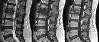
- Ultrasound of joints,
- MRI of joints.
The information content of hardware methods for studying all elements and tissues of articular joints helps to study in detail images of ligaments, tendons, cartilage, adipose tissue, bone marrow, blood vessels, etc.
Varicose veins - what is it and how to live with it?
As an advertisement
Varicose veins are a very common phenomenon. According to statistics, in Russia various forms of varicose veins occur in 30 million people. Women experience the disease more often than men1.
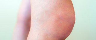
With varicose veins, the walls of blood vessels lose their elasticity and become thinner. The veins themselves become tortuous, and nodular protrusions appear under the skin. Due to the deformation of blood vessels, the venous outflow of blood is disrupted2.
Symptoms of varicose veins of the legs
With varicose veins, symptoms vary depending on the stage of the disease3.
I. At the initial stage
Varicose veins may not manifest themselves in any way, so diagnosing the disease is quite difficult. However, without treatment, over time, a feeling of heaviness and fullness in the legs, swelling, and calf cramps arise. Spider veins are added to the signs of varicose veins in women, but without accompanying symptoms it can simply be a cosmetic defect.
II. At the second stage
swelling and fatigue no longer go away after a night's rest. The skin on the legs may become less sensitive, dryness and flaking appear, sometimes reddish or dark spots, itching.
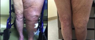
III. At the third stage
changes in veins and skin are visible to the naked eye. Venous vessels become swollen. There is constant pain in the legs, the skin in some places acquires a bluish tint. Patients are increasingly experiencing cramps in their calves. Systemic problems begin due to impaired blood flow, for example, headaches.
IV. At the last – fourth stage
manifestations of varicose veins progress. Trophic ulcers appear in places of inflammation on the skin. The risk of blood clots and thrombophlebitis – inflammation of the veins – increases.
Also, with varicose veins, the local temperature may increase. In the later stages, even minor bruises and wounds on the lower leg lead to minor hemorrhages4.
Why do varicose veins appear?
There are primary and secondary varicose veins5:
• primary varicose veins are caused by congenital, often hereditary, factors: insufficiency of the venous wall or valves that direct blood flow from the lower extremities to the heart;
• the development of the secondary form of varicose veins is influenced by a person’s lifestyle and occupation.
Representatives of professions with a static load are at greatest risk, when they have to sit or stand motionless for a long time, or lift heavy objects. These are, for example, surgeons, teachers, waiters. The disease is more likely to develop in obese people6.
Some causes of varicose veins apply only to women7:
• dyshormonal conditions, including those caused by taking hormonal contraception, replacement therapy;
• Pregnancy also increases the risk of the disease, but in most women, after childbirth, the veins return to a healthy state.
Is it possible to cure varicose veins?
Many changes in the veins are irreversible, so at the first signs of varicose veins you should consult a phlebologist. To make a diagnosis, a specialist will conduct an examination, perform hardware diagnostics and prescribe tests.
If the disease is severe, the main treatment will be surgical. Minimally invasive methods such as sclerotherapy or laser coagulation are possible8.
In the early stages of varicose veins, conservative treatment is sufficient, which may include:
• venotonics, improving blood flow and strengthening the walls of blood vessels. For example, Venolife duo® is a complex drug that helps strengthen capillaries and increase venous tone9;
• anticoagulants, which thin the blood and prevent blood clots from forming. Venolife® gel prevents blood clots, helps cope with swelling and pain in the legs, accelerates the healing of trophic ulcers10;
• anti-inflammatory drugs;
• compression underwear.
To prevent varicose veins, it is recommended to normalize weight, eat right and exercise. If you work sedentary or standing, you can wear compression socks or stockings.
THERE ARE CONTRAINDICATIONS. BEFORE USE, CONSULT A PROFESSIONAL.
Products by topic: [product](Venolife gel), [product](Venolife Duo).
1. Litynsky A.V., Polyakov P.I., Gorelik S.G. Modern problems of varicose veins of the lower extremities (epidemiology, diagnosis, treatment) and its features in people of older age groups // Scientific bulletins of Belgorod State University. Series: Medicine. Pharmacy. 2012. No. 5 (part 2). pp. 276-280.
2. Daudyaris J.P. Diseases of the veins and lymphatic system. // M.: “Medicine”, 1984. P. 59-97.
3. Ibid.
4. Petukhov V.I. The nature of functional disorders in patients with varicose veins and the possibilities of sclerotherapy // Bulletin of Vitebsk State Medical University. 2005. No. 2, volume 4. pp. 22-27.
5. Evdokimov A.G., Topolyansky V.D. Diseases of the arteries and veins: A reference guide for the practitioner. // 2nd ed. M.: Soviet sport, 2001. P. 182.
6. Clinical recommendations. Varicose veins of the lower extremities without chronic venous insufficiency // Ministry of Health of the Russian Federation.
7. Ibid.
8. Ibid.
9. Venolife Duo, instructions. // https://www.venolife.ru/instructionDuo.pdf
10. Venolife Gel, instructions. // https://www.venolife.ru/instructionGel.pdf
Which doctor treats
When starting treatment, the patient should seek advice from an orthopedic traumatologist and rheumatologist.
IMPORTANT! Swelling or swelling in the joint area is an alarming manifestation that requires immediate consultation with a specialist.
If a person notices swelling or swelling once, after an injury or painful impact on the joint, then it is necessary to contact an orthopedic traumatologist at our center for immediate help in this situation. However, if the patient complains of constant pain and swelling in the joint area, he should visit a rheumatologist. Make an appointment with a qualified orthopedic traumatologist or rheumatologist at the Kuntsevo Medical and Rehabilitation Center to understand the causes of joint swelling and eliminate swelling!
Sign up
Types of treatment
The prescribed set of therapeutic measures is individual for each patient, but is invariably aimed at directly relieving swelling and ensuring normal blood circulation in the tissues and lymphatic drainage from the problem area.
Depending on the established cause of edema, the doctor prescribes drug therapy (taking tablets and injections), the use of medicinal ointments and creams. Non-invasive procedures are also of decisive importance - physiotherapy, kinesitherapy, diet therapy.
The clinic’s specialists are professionally proficient in modern, effective therapeutic and surgical methods for treating pathologies. Among them is the unique ACP SVF therapy - the latest treatment method that involves the use of adipose tissue stem cells.
Treatment
Pre-hospital assistance
To reduce swelling, it is recommended to ensure rest and an elevated position of the limb. In case of injury, the leg is fixed with a splint, a bandage is applied to the arm, cold is applied, and a painkiller is given. For non-traumatic lesions with mildly expressed symptoms, short-term use of local anesthetics and anti-inflammatory drugs is acceptable. Progressive swelling, sharp pain, and deterioration in general condition are reasons to immediately consult a doctor.
Conservative therapy
For patients with traumatic injuries, dislocations are reduced, fractures are reduced, plaster casts are applied, and skeletal traction is applied. The rest of the patients are recommended a protective regimen, selected taking into account the characteristics of the disease. Drug therapy includes the following:
- Antibiotics
. Prescribed for infectious arthritis. First, broad-spectrum drugs are used, and subsequently antibiotic therapy is adjusted taking into account the sensitivity of the pathogen. In severe cases, combinations of two medications are recommended. - NSAIDs
. Reduce pain, reduce the severity of the inflammatory process. In the form of tablets and injections, they are usually used for a short time due to the negative effect on the gastric mucosa, liver, and kidney function. Application of gels and ointments is allowed in long courses. - Corticosteroids
. They have a pronounced anti-inflammatory effect and are the basis of drug therapy for collagenosis. If necessary, combined with immunosuppressants. - Chondroprotectors
. They improve the nutrition of cartilage tissue and promote its restoration by activating the synthesis of proteoglycans. They are prescribed intramuscularly, orally, and injected directly into the joint cavity. - Vitamin and mineral complexes
. Helps correct metabolic disorders, improve immunity, and increase the effectiveness of drug therapy for joint diseases.
Other medications include hormonal agents to correct endocrine disorders and chemotherapy drugs to combat malignant neoplasms. Drug therapy is supplemented with physiotherapeutic procedures: UHF, laser therapy, magnetotherapy, electrophoresis, and other techniques. Patients are prescribed massage and exercise therapy. Intra-articular and periarticular blockades are performed.
Lifestyle recommendations
To prevent swelling and pain in the joints, experts recommend:
- minimize the static load on the musculoskeletal system, and also alternate between “standing” and “sitting” positions;
- stop regularly wearing high heels and wear comfortable shoes (even better - with orthopedic inserts);
- try not to sit with your legs crossed;
- get rid of excess weight, if any;
- do not lift heavy objects;
- exercise and lead an active lifestyle;
- adhere to proper nutrition (do not eat fatty foods and alcohol, minimize the consumption of salt and sugar);
- maintain water balance;
- do not overcool the joints;
- rationally distribute time for sleep, work and rest.
You may find it useful:
- Rehabilitation program for rheumatological patients
- Rehabilitation program for joint diseases
- Rehabilitation program after injuries