Stiffness and pain in the groin area while walking are not pleasant sensations. Destruction of cartilage tissue in the hip joint progresses over a long time, so osteoarthritis is often diagnosed in the later stages, when conservative methods are no longer effective. How to prevent the development of the disease and what to do if the diagnosis is confirmed?
35-40% of cases of osteoartosis are lesions of the hip joint
Who is at risk?
Natural aging of cartilage tissue occurs after 45 years - it is at this age that the first signs of the disease appear. More women are affected: during menopause, calcium is “washed out” from the bones and accumulates in the cartilage, which contributes to its destruction.
Be especially attentive to the condition of the hip joint if:
- your age is approaching o;
- your relatives have already been diagnosed with this disease (the hereditary structure of cartilage tissue is a serious argument!);
- you regularly expose the joint to physical stress and risk of injury;
- are overweight.
In the “60+” category, every first person has osteoarthritis
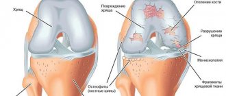
Features of the development of the disease
Treatment is selected by the doctor depending on the degree of neglect and symptoms of the lesion and has a degenerative course. The disease is characterized by the proliferation of bone tissue at the edges of the diseased joint (osteophytes). The joints themselves have significant deforming deviations.
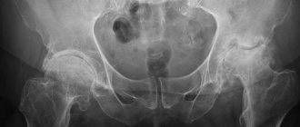
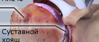
This is what the head of a joint looks like, affected by grade 3 osteoarthritis.
Comparison of the surfaces of two joints: on the left - healthy, on the right - affected by the disease.
People over 45 years of age are most susceptible to grade 1 and 2 osteoarthritis. At increased risk are athletes, as well as those men whose musculoskeletal system regularly experiences excessive physical stress.
Treatment of osteoarthritis cannot ensure a complete recovery of a person, especially if the disease is in the second or third form of neglect.
Timely initiation of therapy significantly reduces the risk of developing dangerous complications, relieves the patient of a number of unpleasant symptoms of the disease, and stops the destruction process. The sooner the examination is carried out and therapy is selected, the higher the chance of complete restoration of mobility.
What are the signs of osteoarthritis of the hip joint?
- Pain during movement is localized in the thigh, buttock or groin.
- It becomes difficult to rotate the hip, as well as to bring it to extreme positions of extension and flexion.
- Gradually, a person develops a comfortable hip position - a slight abduction of the bent leg to the side and its rotation outward. The leg shortens compensatoryly, which negatively affects gait.
- If osteoarthritis affects both hip joints, a characteristic “duck” gait is formed.
Even minor trauma in childhood lays the foundation for osteoarthritis in the future.
Postoperative rehabilitation period
Postoperative treatment consists of taking antibiotics and painkillers.
On the first day after surgery, the patient can move a little in bed. One day after hip cartilage replacement, you can sit. After two days you can move around with the help of crutches. After a week and a half, the sutures on the surgical incision are removed and the patient is discharged.
It is advisable to carry out the rehabilitation period after joint replacement surgery in a special center under the supervision of doctors. If this is not possible, then the patient should follow the doctor’s recommendations. Rehabilitation treatment of the articular area includes: massage, gymnastics.
The service life of prostheses ranges from 15 to 20 years. Depends on the patient’s weight and the loads in the area of the hip joint prosthesis.
Osteoarthritis of the hip joint is a dangerous disease, often leading to disability. You should not let the disease get worse; at the first symptoms that appear, consult a doctor.
Degrees of osteoarthritis of the hip joint
- The first degree is almost imperceptible and asymptomatic. The maximum manifestation is a weak aching pain after physical activity, which is often confused with fatigue. Changes are detected only by x-ray examination. Small bone growths are visible along the edges of the articular surfaces, and under the cartilaginous tissue the bone becomes denser. This is the first warning sign, but, unfortunately, rarely does anyone start treatment at this stage.
Have you been diagnosed with osteoarthritis at the first stage? This is great luck!
- The second degree is accompanied by an inflammatory process, swelling, and pain during physical activity. Progressive deformations of the joint begin, it ceases to function adequately - the pain intensifies. Osteophytes limit mobility, and characteristic clicking sounds appear during movements. The picture shows a noticeable narrowing of the joint space (severe osteosclerosis) - about a third of normal.
The second stage of osteoarthritis without treatment always progresses into the complicated third stage
- The third degree is extremely severe both in terms of the patient’s subjective sensations and in terms of objective indicators. The joint space narrows even more - by 2/3 of normal. The pain becomes very strong and constant. The image shows large marginal osteophytes.
In the third stage, painkillers practically do not help
- The fourth degree is the final stage of the development of osteoarthritis. The joint space practically disappears or is difficult to trace, the bones in the joints are severely deformed and compacted. The hip joint is completely immobilized.
Diagnostic measures
Confirmation of the diagnosis of coxarthrosis is carried out during a physical examination of the patient by a rheumatologist. Additional studies are prescribed to evaluate degenerative changes in joint tissue. Radiography allows you to determine the degree of coxarthrosis and the probable cause of the development of the pathology. Computed tomography and magnetic resonance imaging provide doctors with visual data about the pathological processes inside the joint. Images obtained during MRI record changes in bone structures. CT scan demonstrates the extent of soft tissue damage. This information allows doctors to determine the patient’s treatment strategy and exclude from the medical history other diseases that provoke limited mobility of the hip joint.
Principles of conservative treatment of osteoarthritis of the hip joint
Unfortunately, there is no “magic pill” for this disease. For therapy to bring relief, it must be comprehensive. Treatment has several goals:
- eliminate risk factors that can aggravate the course of the disease, such as physical activity, excess weight (if you lose weight and reduce the load on the joint, the pain will decrease);
- relieve pain to improve quality of life;
- improve joint function if possible;
- restore cartilage tissue;
- slow down the progression of pathological deformations.
Who is given disability
In severe hip arthrosis of the 3rd degree, a person gradually loses professional skills and cannot do housework. He is given the right to receive 1 or 2 disability groups. For this purpose, a medical and social examination (MSE) is appointed to assess the patient’s condition. X-ray images and the results of conservative treatment are studied.
The left joint is deformed.
What serves as the basis for receiving disability:
- rapid progression of arthrosis;
- ineffective use of medications, physiotherapeutic and massage procedures, exercise therapy for several months;
- severe or complete loss of mobility;
- the patient's inability to take care of himself at home.
Disability can be assigned in case of bilateral damage to the hip joint, as well as in case of endoprosthetics of two joints at once.
Drug therapy for this disease is identical to the treatment of arthrosis:
- painkillers;
- anti-inflammatory;
- chondroprotectors for nourishing cartilage tissue (available in capsules, tablets and ointments);
- means for improving microcirculation;
- additionally – glucocorticoid hormones.
Medicines are aimed at temporarily eliminating symptoms: it is impossible to stop the progression of the disease with their help alone. Therefore, physiotherapy is included in the complex scheme - thermal procedures, laser, reflexology, therapeutic massage, acupuncture. Be sure to limit the consumption of easily digestible carbohydrates and introduce dairy, greens, lean fish and meat, vegetables and fruits into the diet.
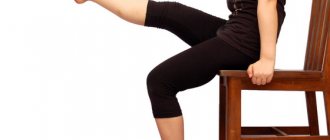
Gymnastics and physical therapy for osteoarthritis of the hip joint are selected individually by a physiotherapist. The choice of exercise depends on your general health and the degree of dysfunction in the joint. During the period of remission, you can perform, for example, the following simple exercises:
Traditional methods of therapy
The most effective alternative treatment will be in grade 1, when the disease has not yet severely damaged the articular joint, and the inflammatory process is not so advanced.
As for treatment of grade 2 or 3, traditional therapy only partially helps. In advanced forms, osteoarthritis provokes serious pathological changes in the joint, which are difficult to treat even with medication and surgery.
Traditional methods are dangerous: a patient without proper diagnosis is often treated with dummies and wastes time.
The following recipes will help improve a person’s well-being with deforming osteoarthritis of the hip joint:
- Squeeze juice from cabbage. Soak a gauze bandage with it. Use as a compress at night. The product will help relieve inflammation.
- Mix honey and calendula tincture. Soak a compress bandage in the resulting mixture.
- Pour 2 liters. elderberry 0.5 liters of vodka. Leave for 24 hours and rub the prepared mixture onto the sore joint.
- Take 2 liters. chopped nettle and pour 500 ml of boiling water. Infuse and apply for compresses.
- Take 1 kg of beef bones and pour 4 liters of water over them. Cook for four hours. Drink the resulting decoction during exacerbation of osteoarthritis.
How is osteoarthritis treated in complex cases?
In the final stages, traditional medicine knows only one way to restore lost function to the limbs. Joint replacement is a complex, expensive and traumatic operation that is hazardous to health, like any surgical intervention.
In many cases, there are contraindications to endoprosthetics
How effective are intra-articular injections?
Osteoarthritis of the hip joint always develops against the background of a deficiency of synovial fluid or a change in its natural viscosity. To compensate for its deficiency, special agents are injected into the joint:
- preparations based on hyaluronic acid;
- hydrogel-based preparations, for example synthetic viscoprosthesis Noltrex, with a much longer duration of action and a powerful anti-inflammatory effect.
Unlike drugs with hyaluronic acid, which are subject to rapid resorption, Noltrex fragments inside the joint very slowly, so its effect continues for 12-18 months after the course of treatment.
Today, in the later stages of the disease, they mainly resort to surgical prosthetics. However, modern medicine is ready to offer an effective way to stop the development of osteoarthritis. Synovial fluid endoprostheses restore the natural environment of the joint and protect it from further deformation. A course of intra-articular injections "Noltrex" slows down the progression of the disease as much as possible and returns a person to the ability to live a full life!
Causes of coxarthrosis
Doctors distinguish two types of arthrosis - primary (occurs for unknown reasons) and secondary (formed against the background of other pathologies). Secondary coxarthrosis results from the following diseases:
- hip dysplasia;
- congenital hip dislocation;
- Perthes disease;
- necrosis of the femoral head;
- infectious lesions.
Often, degenerative processes in joint tissues are formed under the influence of injuries - dislocations, hip fractures or gonarthrosis.
The likelihood of developing coxarthrosis increases under the influence of a number of factors. These include:
- increased sports loads;
- the patient is overweight;
- metabolic and hormonal imbalance;
- kyphosis and scoliosis;
- flat feet;
- old age.
A patient's sedentary lifestyle can also cause degenerative changes in joint tissues.