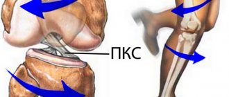
Surgical treatment of patients with chronic rupture of the Achilles tendon is a pressing problem at present [1, 5, 7, 9]. Despite the variety of clinical and instrumental diagnostic methods, every fifth patient with early subcutaneous rupture of the Achilles tendon is not referred on time or does not seek specialized help [2, 6, 15]. This work is devoted to restoring the function of the ankle joint with diastasis of the ends of the Achilles tendon of more than 10 cm. As a rule, it is formed when the rupture has been for a long time (more than a year) [3, 8, 10, 11]. The main objectives of surgical treatment in such cases are to restore the continuity of the calcaneal tendon, create normal physiological tension of the calf muscle, and restore the weight-bearing ability of the foot [12-14].
The existing variety of plastic options does not allow for a qualitative restoration of the strength of the triceps surae muscle due to pronounced retraction, hypotrophy and, as a consequence, a decrease in its contractile ability.
The purpose of the study is to improve the results of treatment of patients with chronic ruptures of the Achilles tendon and severe diastasis of its ends through the use of a new method of surgical treatment.
Material and methods
In the period from 2005 to 2021, 37 patients with chronic rupture of the Achilles tendon were treated in the traumatology and orthopedic department No. 1 of the Samara State Medical University Clinic. The diastasis between the ends of the Achilles tendon in the neutral position of the ankle joint exceeded 10 cm. Due to the retraction of the triceps muscle, it is not possible to sew the tendon end to end; it is necessary to resort to plastic surgery of the diastasis formed between its distal and proximal ends.
In group 1 (control), we included 21 patients who underwent autoplasty with reversal flaps (according to Chernavsky, Krasnov), V-Y-plasty. The 2nd group consisted of 16 patients who underwent a new method of surgical treatment proposed by us [4]. In all subjects of the main and control groups, there was a significant difference (more than 4 cm) in the circumference of the symmetrical areas of the upper third of the legs, which indicated severe hypotrophy of the triceps muscle. Both groups were comparable in age, gender, mechanism of injury, time of visit to the traumatology and orthopedic department No. 1 of the Samara State Medical University Clinic, where their surgical treatment was carried out.
Before the operation, the patients were examined in the biomechanics laboratory of the Samara State Medical University Clinic (electromyography, podometry, measurements of the strength of the “flexors” of the feet), and an ultrasound diagnosis of the injury area was performed.
A team of authors from the Department of Traumatology, Orthopedics and Extreme Surgery named after. acad. RAS A.F. Krasnov received RF patent No. 2537888 dated November 13, 2014 for the invention of a new method of surgical treatment of patients with chronic rupture of the Achilles tendon with pronounced diastasis of its ends (more than 10 cm), which consists of transposition of the tendons of the peroneus brevis and tibialis posterior muscles to the distal end of the Achilles tendon in the optimal tension [4].
The proposed method is illustrated with graphic material. In Fig. 1 shown
Rice. 1. Isolation of the peroneus brevis and tibialis posterior muscles. 1 - proximal end of the Achilles tendon; 2 - distal end of the Achilles tendon; 3 - tendons of the peroneus brevis muscle; 4 - peroneus muscle; 5 - tendons of the tibialis posterior muscle; 6 - tibialis posterior muscle. proximal end (1) and distal end (2) of the Achilles tendon. The tendon (3) of the peroneus brevis muscle (4) was isolated and held. The tendon (5) of the tibialis posterior muscle (6) was isolated and held.
In Fig. 2:
Rice. 2. Relocation and plastic surgery of the Achilles tendon defect. 1 - proximal end of the Achilles tendon; 2 - distal end of the Achilles tendon; 3 - tendons of the peroneus brevis muscle; 4 - peroneus muscle; 5 - tendons of the tibialis posterior muscle; 6 - tibialis posterior muscle; 7 - sutures fixing the ends of the Achilles tendon. the peroneus brevis tendon (3) and the tibialis posterior tendon (5) are passed through the triceps muscle, covering the defect between the proximal end (1) and the distal end (2) of the Achilles tendon, stretched to optimal tension and fixed with sutures (7) parallel to each other to the distal end (2) of the Achilles tendon.
Clinical example
Patient P
., 53 years old, was admitted for surgical treatment to the traumatology and orthopedic department No. 1 of the SamSMU Clinic.
Life history: originally from the village of Sernovodsk. Works as a truck driver. Medical history: injured 20 years ago as a result of playing football. After visiting the clinic at the place of residence, a diagnosis was made: “bruise of the left ankle joint.” After the injury, he walked with crutches for 3 weeks due to pain. There is a decrease in the strength of plantar flexion of the foot over time. I applied for a consultation at SamSMU Clinics due to my inability to drive freight transport.
During local examination, wasting of the muscles of the posterior surface of the left leg was visually noted. Positive left calf compression test (Thompson sign). Circumference in the upper third of the legs: on the right 35 cm, on the left 30 cm. Strength of the plantar flexor muscles of the feet (according to the Lovvet scale) [2]: on the right 5 points, on the left 2 points.
Additional examination methods
According to the results of electromyography, there is a sharp decrease in the amplitude and frequency of contractions of the triceps muscle of the left leg in comparison with the healthy side (contraction frequency on the left 15 per 1 s, on the right 150 per 1 s; contraction amplitude on the left 25 mV, on the right 135 mV).
Podometry results revealed a pronounced asymmetry of gait.
Ultrasound conclusion: old injury of the Achilles tendon on the left with the presence of connective tissue in the area of the rupture, diastasis between the ends is 11 cm.
Surgical treatment is recommended.
Due to the severe hypotrophy of the triceps muscle of the left leg and the long history of the injury, it was decided to transpose the tendons of the peroneus brevis and tibialis posterior muscles to the distal end of the Achilles tendon in order to restore one of the most important functions of the ankle joint - plantar flexion [5, 6] .
Operation stages
Access to the Achilles tendon defect and its distal end was made. The distal end is isolated from scars. The Achilles tendon defect is visualized (Fig. 3).
Rice. 3. Isolation of the distal end of the Achilles tendon.
A linear skin incision 3 cm long was made along the outer surface in the middle third of the leg to isolate the tendon of the peroneus brevis muscle (Fig. 4).
Rice. 4. Isolation of the tendons of the peroneus brevis muscle.
Next, a 3 cm long incision was made along the inner surface in the middle third of the leg to isolate the tendon of the tibialis posterior muscle (Fig. 5).
Rice. 5. Isolation of the posterior tibial tendon.
After isolation, the peroneus brevis tendon and the tibialis posterior tendon are passed subcutaneously and brought into the defect area. Next, with optimal tension, the tendons are sutured to the distal end of the Achilles tendon (Fig. 6).
Rice. 6. Transposition of the peroneus brevis and tibialis posterior tendons to the distal end of the Achilles tendon.
After the operation, the patient was placed in a plaster cast along the anterior surface of the left shin from the upper third to the fingertips with a plantar flexion angle at the ankle joint of 20°. Recommended: walking without support on the left leg for up to 5 weeks. Then he underwent a course of rehabilitation treatment.
As a result, after 3 months the patient was able to lift his body weight on two feet, and after 6 months - to lift his body weight on one foot.
According to the results of podometry 6 months after surgery, no gait asymmetry was detected. Strength of the plantar flexor muscles of the feet (according to the Lovvet scale): on the right - 5 points, on the left - 5 points. The result of surgical treatment in patient P
. rated good.
Treatment of old Achilles tendon rupture
Treatment of chronic Achilles tendon ruptures is a difficult problem in the practice of an orthopedic traumatologist. Late diagnosis and inadequate treatment of ruptures of the Achilles tendon leads to the fact that it either fuses with significant elongation, or a synovial cyst forms at the site of the rupture. In both cases, there is a pronounced weakening of the force of the plantar impulse and, as a result, gait disturbance and lameness. This makes playing sports and sometimes even regular walking impossible. Surgical treatment of chronic ruptures of the Achilles tendon is a major traumatic operation, with a high risk of postoperative complications, such as infection, aseptic necrosis of the wound edges, and repeated ruptures of the Achilles tendon. The difficulties of surgical treatment are associated with the fact that the Achilles tendon undergoes degeneration, and the gastrocnemius muscle contracts significantly, and matching the edges of the tendon after excision of scar tissue becomes extremely problematic. To solve this problem, several types of tendon grafting are traditionally used, which allows you to “lengthen” the tendon. Below are examples of treatment for such complex pathologies.
Patient X. 65 years old. She suffered a ruptured Achilles tendon after a long history of Achilles tendonitis. Surgical treatment was not offered to the patient due to her advanced age and concomitant pathology. In this photograph, the Achilles tendon defect is visible to the naked eye.
The patient came to our clinic 6 months after the injury. At such times during surgery, the problem of adapting the ends of the tendon always arises. There is a need to perform tendon plasty and augmentation of the suture with an autograft or transposition of the flexor pollicis longus tendon. It is also possible to use inverted flaps, Chernavsky plastic, or an autograft from the semitendinosus tendon.
With such large defects, difficulties arise both with the adaptation of the ends of the tendon and with the lack of local tissue to replace the defect. In this case, we decided to perform VY plasty, transposition of the flexor pollicis longus tendon with the addition of plasty with local tissues.
The use of VY plastics and tissue of the flexor pollicis longus tendon made it possible to form a powerful conglomerate of tissue at the site of the tendon defect, which should subsequently be rebuilt into a new Achilles tendon.
The postoperative management of such patients differs little from the standard management of patients with Achilles tendon ruptures. However, the risk of infectious complications and marginal necrosis of the skin flap is, of course, much higher than with a standard Achilles tendon suture.
Another option for repair of healed Achilles tendon ruptures is the use of an inverted or reverse flap.
A clinical example of the use of this method in a case of chronic rupture of the Achilles tendon in a 52-year-old patient.
2 months have passed since the injury; the injury occurred during sambo training; when I suddenly jerked from a place, I felt the sensation of being hit on the shin with a stick, after which I was unable to continue training.
The patient did not receive treatment, walked with full weight-bearing, as a result of which a significant lengthening of the Achilles tendon occurred. The active scar process in the area of damage did not give the sensation of a “palpable defect”; in addition, due to the developed muscles of the synergists, the patient retained active plantarflexion of the foot, which led the doctors down the wrong path and led to such a late request for specialized help.
Weakness, pain, lameness, and the inability to stand on the toe of the injured leg forced the patient to seek help 2 months later from the injury.
Upon examination, noticeable atrophy of the gastrocnemius muscle and pastosity in the area of the Achilles tendon were noted. The passive plantarflexion test revealed significant differences in foot position in a relaxed position.
An MRI examination reveals a large area of tendon degeneration, significantly larger than the size of the defect.
Considering the size of the defect that will form after removal of scar tissue, it was decided to use a reverse flap to lengthen it.
When planning the operation, it is important to take into account the location of the sural nerve, n.suralis, as well as to bypass the area of the poorest blood supply to the skin, in order to avoid its necrosis.
To perform Achilles tendon plasty with an inverted or reverse flap, extensive access is required, the length of which can exceed 20 cm. When performing access, it is necessary not to separate the skin, subcutaneous fatty tissue and paratenon from each other in order to avoid skin necrosis.
In case of such a large access, it is better to use separate interrupted sutures; in the area of maximum skin tension, longitudinally oriented releasing incisions can be used. In the early postoperative period, we recommend the use of vascular and metabolic drugs, antibiotic prophylaxis and the use of anticoagulants for at least 1 week after the intervention. All this makes it possible to avoid marginal skin necrosis even with such an extensive and traumatic approach.
Appearance of the posterior surface of the leg immediately after suturing the wound.
Appearance of the posterior surface of the leg 2 weeks after surgery. The sutures have been removed, there is no dehiscence of the wound edges, no necrosis is detected.
4 weeks after the operation, the satisfactory condition of the postoperative suture is determined, the foot is in a position of moderate hypercorrection of plantarflexion. From this moment, immobilization is carried out in a Walking boot orthosis, the patient is allowed a dosed axial load.
The rehabilitation protocol after this type of plastic surgery should be more gentle. Moving the foot to a neutral position is possible after 6 weeks; wearing a Walking Boot with plastic clasps is recommended until 12 weeks after surgery. Resumption of forced physical activity is not advisable before 12 months.
results
When analyzing the results of treatment of patients in the control group ( n
=21), which performed plastic surgery of the Achilles tendon defect zone (according to Chernavsky, Krasnov, V-Y-plasty), we noted that the strength of the plantar flexor muscles of the feet (according to the Lovvet scale) on the damaged side averaged 3.43 points, in main group - 4.63. Muscle strength of 5 points in the main group was in 62.5% of patients, and in the control group in 14.3%.
When analyzing podometric indicators, we studied such time characteristics of the step as the duration of the double step, the time of transfer of the foot, the duration of single and double support, the sequence of contact of the parts of the foot with the support. The comparison was carried out on different limbs according to the asymmetry coefficient of the subject’s gait, which was defined as the ratio of the larger period of support to the smaller one minus one and multiplied by 100%. Based on this indicator, gait asymmetry of up to 5% is normal; from 5 to 10% - hidden lameness; more than 10% - obvious lameness
Gait asymmetry up to 5° in the main group was 81.25%, and in the control group - 14.3%. Gait asymmetry of 5-10% in the main group was 18.75%, in the control group - 71.43%. Asymmetry of more than 10% was not detected in the main group, but in the control group it was 14.27%.
The data obtained indicate that the best gait indicators were in the main group.
As can be seen from the obtained functional indicators, patients in the control group showed a significant decrease in the strength of the plantar flexor muscles of the feet on the injured side and persistent gait asymmetry in the late postoperative period. This is evidence that even high-quality replacement (plasty) of the Achilles tendon defect with severe hypotrophy of the triceps muscle cannot fully compensate for the function of proper active flexion in the ankle joint, and was the basis for the development of a new method.