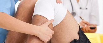
One of the most complex structures of parts of the human body are joints, both large and small. The structural features of the knee joint make it possible to consider it the most susceptible to various injuries, such as fractures, bruises, hematomas, arthrosis, and rupture of the posterior horn of the medial meniscus.
This is justified by the fact that the bones of the joint (femur, tibia), ligaments, menisci and patella, working together, ensure normal flexion when walking, sitting and running. However, large loads placed on the knee during various manipulations can lead to a rupture of the posterior horn of the meniscus.
A rupture of the posterior horn of the internal meniscus is an injury to the knee joint caused by damage to the cartilage layer located between the femur and tibia.
Knee
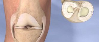
Knee The knee has a complex structure.
The joint includes the surfaces of the femoral condyles, the tibia, and the patella. For better stabilization, shock absorption and load reduction, paired cartilaginous formations called medial (internal) and lateral (external) menisci are localized in the joint space. They have the shape of a crescent, the narrowed edges of which are directed forward and backward - the front and rear horns. The external meniscus is a more mobile formation, therefore, with excessive mechanical stress, it moves slightly, which prevents its traumatic damage.
The medial meniscus is secured by ligaments more rigidly; when exposed to mechanical force, it does not shift, as a result of which damage more often occurs in various parts, in particular in the area of the posterior horn.
Causes
Damage to the posterior horn of the medial meniscus is a polyetiological pathological condition that develops under the influence of various factors:
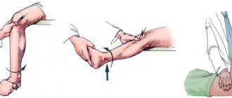
- The impact of kinetic force on the knee area in the form of a blow or fall on it.
- Excessive flexion of the knee, leading to tension in the ligaments that secure the menisci.
- Rotation of the femur with the tibia fixed.
- Frequent and long walking.
- Congenital changes that cause a decrease in the strength of the knee ligaments, as well as its cartilage.
- Degenerative-dystrophic processes in the cartilaginous structures of the knee, leading to their thinning and damage. This cause most often occurs in older people.
Finding out the causes allows the doctor not only to select the optimal treatment, but also to give recommendations regarding the prevention of recurrence.
Rehabilitation
After the basic therapeutic course, restorative procedures and measures are prescribed. They involve special physical training in a physical therapy complex (PT). In this case, the load on the knee increases in stages, and this allows the cartilage structures to adapt to it in order to avoid repeated damage.
Due to the fact that the joint is in a state of functional rest for quite a long time, there is a risk of developing contractures (connective tissue adhesions), so rehabilitation with exercises is necessary to prevent the development of this complication. The duration of the course of such measures depends on the type of treatment performed, as well as on the severity of the damage. Typically it varies over a period of time from several months to six months.
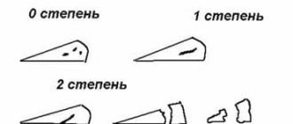
degree of meniscus damage
degree of meniscus damage
Depending on the main causative factor that led to the development of the pathological condition of the cartilaginous structures of the knee, traumatic and pathological degenerative damage to the posterior horn of the medial meniscus is distinguished.
According to the criterion of the duration of the injury or pathological violation of the integrity of this cartilaginous structure, fresh and old damage to the posterior horn of the medial meniscus is distinguished. Combined damage to the body and posterior horn of the medial meniscus was also identified separately.
What are the conditions of stay at the Gelenk Clinic?
Private room at the Gelenk Klinik - Gundelfingen, Germany
During your stay at the Gelenk Klinik, you are usually in a private room, which has a shower and toilet. In addition, we provide towels, bathrobes and slippers. You can also use a safe and minibar. All rooms are equipped with TV. You only need to take medications, comfortable clothes and nightwear with you. Patient care is provided around the clock. The medical staff and physiotherapists at Gelenk Clinic will always answer all your questions. Generally, the length of hospital stay is 3 days. Your relatives can stay at a nearby hotel. Our staff will be happy to take care of your room reservation.
Manifestations
Clinical signs of damage to the posterior horn of the medial meniscus are relatively characteristic and include:
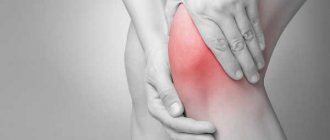
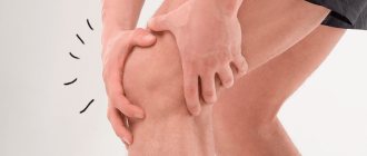
- Pain that is localized on the inner surface of the knee joint. The severity of pain depends on the cause of the violation of the integrity of this structure. They are more intense with traumatic injury and sharply intensify while walking or descending stairs.
- Violation of the condition and functions of the knee, accompanied by a limitation in the full range of motion (active and passive movements). When the posterior horn of the medial meniscus is completely torn off, a complete block in the knee may occur against the background of severe pain.
- Signs of inflammation, including hyperemia (redness) of the skin of the knee area, swelling of soft tissues, as well as a local increase in temperature, which is felt after touching the knee.
With the development of the degenerative process, the gradual destruction of cartilaginous structures is accompanied by the appearance of characteristic clicks and crunches in the knee during movements.
How to prepare for the procedure
Special preparation is not required before MRI with further classification of the condition of the Stoller meniscus. The study is subject to the same contraindications as MRI, and primarily the presence in the body of metal elements and various electrical stimulators and insulin pumps, the operation of which can be affected by the magnetic field of the device.
There will be no pain or discomfort during the examination. The only thing that the patient needs to take into account is that the procedure is lengthy. If the patient is very nervous before the examination, it is recommended that he take valerian or another herbal sedative.
Diagnostics
Diagnostics
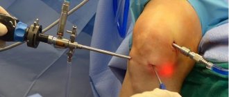
Arthroscopy also makes it possible to carry out therapeutic manipulations under visual control after additional introduction of special microinstruments into the joint cavity.
Damage to the posterior horn of the medial meniscus - treatment
After an objective diagnosis has been carried out, determining the location and severity of the violation of the integrity of the cartilaginous structures of the joint, the doctor prescribes a comprehensive treatment. It includes several areas of action, which include conservative therapy, surgical intervention, and subsequent rehabilitation. Mostly all activities complement each other and are assigned sequentially.
Treatment without surgery
If partial damage to the posterior horn of the medial meniscus (grade 1 or 2) has been diagnosed, conservative treatment is possible. It includes the use of drugs of various pharmacological groups (non-steroidal anti-inflammatory drugs, vitamin preparations, chondroprotectors), the performance of physiotherapeutic procedures (electrophoresis, mud baths, ozokerite). During therapeutic measures, functional rest for the knee joint must be ensured.
Cost of arthroscopic knee surgery in Germany
In addition to the cost of surgical treatment of a torn meniscus, it is worth considering the additional costs of diagnostics, doctor's appointments and aids (eg elbow crutches), amounting to approximately 1,500 to 2,000 euros. If you are planning to undergo outpatient physical therapy treatment after meniscal tear surgery, we will be happy to provide you with an estimate of the costs.
Information regarding the cost of hotel accommodation and possible additional treatment at a rehabilitation center can be found on the corresponding website.
Surgical intervention
Surgical intervention
Surgical intervention
The main purpose of the operation is to restore the anatomical integrity of the medial meniscus, which allows for the normal functional state of the knee joint in the future.
Surgery can be performed using an open approach or arthroscopy. Modern arthroscopic intervention is considered the technique of choice, since it is less traumatic and can significantly reduce the duration of the postoperative and rehabilitation period.
Forecast
The effectiveness of conservative therapy depends on many factors that are taken into account by the doctor before prescribing it, these include:
- Patient's age - in older people, regeneration processes occur more slowly.
- The severity of damage, determined using additional imaging techniques and classified according to Stoller.
- Localization of changes - horn rupture gives a favorable prognosis in relation to damage to the meniscal body.
- The general condition of the human body, as well as the presence or absence of concomitant somatic pathology.
- The length of time that elapsed from the moment of injury until the start of treatment.
To determine the effectiveness of conservative measures, a repeat control objective study is prescribed. If the treatment for a torn internal meniscus is ineffective, the doctor decides whether surgery is necessary.
Modern methods of timely treatment of a partial tear of the internal meniscus without surgery make it possible to obtain a favorable long-term result in terms of restoring the functional state of the damaged structures.
Source