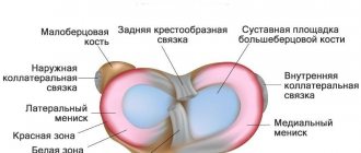
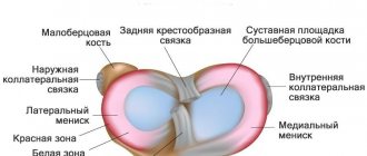
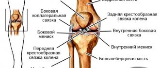
Two cartilaginous formations located in the cavity of the knee joint, serving to cushion, stabilize, distribute load and reduce friction of the articular surfaces, are called menisci. One of them, located on the inside, less mobile and more susceptible to injury, is called medial. The second, located on the outside, is lateral. Athletes, dancers, ballet dancers, and people leading an active lifestyle often consult a doctor about pain, swelling, limited mobility in the knee, and even blockage—the inability to straighten the leg. All these are symptoms of a meniscus tear. Let's take a closer look at them.
In case of injury to the medial cartilage, the following is observed:
- At the time of injury;
- immediate sharp pain;
- rapid swelling of the joint, increase in its temperature;
- blockade - “jamming” of the knee in a slightly bent position;
- in severe forms of damage, muscle wasting and pain on palpation of the joint space are observed.
- In the absence of medical assistance, after 14 days or later:
- enlargement of the joint cavity;
- the sound of a click when the limb is extended;
- dull pain that gets worse when walking down;
- repeated blockades;
- inflammation of the periarticular bursa.
If the lateral cartilage is damaged, the following symptoms of a meniscus tear of the knee joint are determined:
- In the first hours after damage:
- pain when straightening the leg in the knee;
- inflammation of the joint.
- Within 2-4 weeks after injury:
- constant pain;
- inflammation of the joint capsule;
- blockade;
- increased pain during extension, displacement, compression.
To determine the nature of the damage, diagnostic motor tests (Eple, McMurry tests) are used. Often after an injury, upon visual inspection, it is difficult to find out the type and location of the damage, so it is necessary to use hardware diagnostics. X-rays, even performed in several projections, and ultrasound are not always informative. There is a risk of misdiagnosis. To accurately determine the nature of the damage, MRI, CT or endoscopic arthroscopy are used.
Types of meniscus tears:
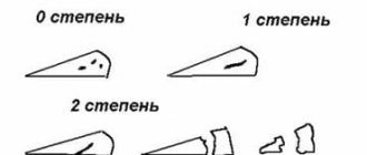
- longitudinal: patchwork and in the form of a “watering can handle”,
- paracapsular,
- horizontal;
- degenerative;
- damage to the cartilage body;
- separation of the anterior or posterior horn.
The main causes of injuries are impacts, sharp turns, falls, rapid straightening of the lower leg, and intense strength training. In older people, the cause of rupture can be degenerative changes in tissues, leading to serious consequences even with light loads. 80% of visits to orthopedic traumatologists with meniscus injuries are related to sports (running, jumping, football, skateboarding, skiing). 75% of patients are men under 40 years of age. In 80% of cases, the internal meniscus suffers, since it is less mobile than the external one.
If you consult a doctor in a timely manner, in mild cases it is possible to avoid surgery and treat the patient with therapeutic methods. If a cartilage injury is accompanied by a bone fracture, tearing off fragments of the meniscus, its crushing, rupture and displacement, if conservative methods do not produce results, surgery will help.
How to identify a meniscal injury
The reasons why a meniscus injury occurs can be not only falls and other mechanical impacts. Common precursors to damage are:
- gout;
- intoxication of the body of any etiology;
- rheumatism;
- age-related changes.
In addition, minor injuries to the meniscus that do not lead to a tear, but are associated only with its stretching or thinning, will eventually cause a tear. If you do not pay attention to the thinning or gradual breakdown of cartilage tissue in time, this can lead to deformation and arthrosis, which ultimately leads to disability.
Meniscus injury is a common problem that traumatologists encounter in their practice. Men are three times more likely to suffer a torn meniscus than women. The average age at which the peak of knee ligament injury occurs is 23-25 years.
At the time of injury, the victim experiences pain not only in the knee area, but along the entire length of the limb. Only after two weeks the pain will be localized in the sore knee. The main signs of a meniscus tear are:
- acute pain;
- hyperthermia at the site of injury, meaning the knee may become hot compared to the rest of the body;
- enlargement of the knee due to severe swelling;
- a loud crunching sound during flexion and extension of the joint, including without load;
- severe weakness of the femoral muscle;
- lumbago when trying to put pressure on the leg.
Despite the severity of pain in the knee joint, these same signs can indicate not only a meniscus tear. In the same way, ruptures of ligaments and muscles manifest themselves.
Symptoms of a pinched meniscus
Pain and stiffness in the knee are the main symptoms of pinching.
The main symptom is a blocking of the knee joint that periodically appears during certain movements (the inability to straighten the knee), accompanied by acute localized pain on the outside or inside.- With meniscitis, effusion (fluid from blood vessels) may accumulate in the joint cavity, sometimes with a small amount of blood. Therefore, swelling or redness of the skin near the knee may occur. The process is often accompanied by an increase in temperature.
- After an injury in which pinching occurs, a person develops a feeling of fear and uncertainty when walking, a sensation of a foreign body in the knee area.
- Unpleasant painful sensations appear upon palpation and pressure.
First aid to a patient
The victim may experience severe pain from a couple of days to a week. If the severity of pain is moderate, you can do without contacting a traumatologist. To relieve pain and reduce suffering, you need:
- Apply ice to the sore knee. This will relieve swelling. Cold during prolonged contact causes blood vessels to constrict, thereby reducing local hyperthermia.
- You can reduce the intensity of pain with the help of non-steroidal anti-inflammatory drugs, including ibuprofen, diclofenac, analgin.
- To reduce the risk of worsening the situation, it is important to ensure immobility of the injured limb using a splint. It is better if the knee is elevated in relation to the healthy leg.
When the acute period is over and the patient can see a traumatologist, the specialist will assess the extent of the damage and prescribe a course of treatment and care.
Exercise therapy for rehabilitation after a meniscus tear
- The patient lies on his stomach. Keeps legs straight. The limb with a damaged meniscus should be lifted up carefully and held in the air for about 20-30 seconds. It is necessary to perform this exercise 4-5 times.
- We stretch our arms forward. First, bend your healthy leg 90 degrees at the knee. Raise your bent leg off the floor and hold it there for about 10 seconds. Then repeat the same with the injured limb. Select the angle so that there is no pain. It is necessary to repeat the exercise 2-3 times with each limb.
- The ankle joints must be alternately bent and extended in different directions, as well as made in circular movements.
- You should press your feet against the support, bending your knees. You need to press hard, alternately with increasing pressure. If pain occurs, the exercise should be stopped.
- Lying on your stomach, you need to straighten the injured leg, straining and stretching it for a few seconds, and then relax. If severe pain occurs, exercise should also be stopped.
- In a lying position, the patient tries to grab various objects with his feet and move them from one place to another.
- Lying on his back, the patient takes turns bringing his knee to his chest, bending it and pressing it to the body with his hands.
- Lying on your back, arms are extended along the body at the seams. At the same time, two legs are raised and pressed to the stomach.
- The patient sits down, resting his hands back. In this case, the knees must be brought together and spread apart, bending the legs at an angle of 45 degrees.
After your condition improves, you can add work with exercise equipment. But in this case, it is necessary to maintain constant monitoring by the attending physician, which will reduce the risks of complications.
It is recommended to walk more often, and gradually add light jogging. The most important thing is not to overload the knee in the early rehabilitation period. The load should be given gradually.
The Traumatology and Orthopedics Clinic offers professional services from qualified medical specialists. The resulting injury is diagnosed using modern equipment, so treatment is selected on an individual basis.
The recovery period after treatment plays an important role in an active future. Physical therapy methods have a positive effect on the progress of rehabilitation. It is possible to evaluate the positive results together with specialists from Elena Malysheva’s clinic.
Home care
Any home therapy should be discussed with your doctor, because it is only an auxiliary component of drug treatment. Traditional medicine can only be indicated for relieving pain and swelling:
- Compresses of a hypertonic solution draw out excess fluid, thereby relieving swelling. A solution is prepared from a tablespoon of salt dissolved in 250 ml of water. The cloth is soaked in salt water and applied to the injured knee.
- Peppermint rubs. For this, essential oils of clove, mint, camphor and eucalyptus are taken. After mixing the ingredients, take a few drops of the product and rub it into the injury site with slow and careful movements. Menthol will create a feeling of freshness and coldness, which will alleviate the patient's condition.
- Pine baths. They can improve blood flow to the injured knee and relieve pain. In addition, pine needles have a restorative effect on the body. To prepare baths, you can use pharmaceutical pine mixtures, which just need to be brewed immediately before preparing the bath. If it is not possible to find pine needles, you can use pine salt.
Treatment
Depending on the degree of damage, conservative and surgical treatment is possible.
To relieve pain, a “joint blockade” injection in the form of an anesthetic is used. This helps relieve pain, but not for long. Some patients manage to unblock the joint on their own, through some movements. As a rule, a migrating meniscal fragment re-blocks the joint.
For mild cases of the disease, when the meniscus is not completely torn off, drug treatment may be used. In some situations, blood accumulates in the joint (hemarthrosis), but most often fluid accumulates there. If there are a large number of accumulations, a puncture is required to remove them.
When swelling swells, cooling bandages are applied. For general drug treatment, NSAIDs are prescribed in the form of ointment or cream for topical use or tablets for oral administration, as well as hormones and chondroprotectors.
During treatment, the patient requires rest and the possibility of fixing the joint. For this purpose, a plaster cast, tapes or bandage are used.
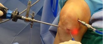
If the meniscus or part of it is completely torn off and moves freely in the joint, surgical intervention is required. Arthroscopy is often sufficient. But sometimes they resort to a complete menisectomy followed by installation of an implant.
Gymnastics to restore knee function
Any gymnastics is indicated only if the acute period has already ended and you can work with the knee joint without pain. As a rule, gymnastics is prescribed after wearing a plaster cast or splint for a long time. To return the knee to working capacity, you need to begin to gradually warm it up according to a certain method.
Clinics offer physical therapy sessions where, under the supervision of a specialist, patients recover from serious injuries. If a doctor has diagnosed a meniscal tear, any exercise will be postponed until the tissue has completely healed.
Exercises begin with minimal loads. First, these will be exercises for flexion and extension of the sore knee. You need to do it while sitting on a high chair. 10 lifts twice a day is enough. Gradually, the load can be increased, in accordance with the doctor’s recommendations. If you experience any pain, you should immediately report it to a specialist to adjust your rehabilitation plan.
Postoperative recovery
There are 4 phases of recovery after arthroscopy. Let's consider them from the point of view of events that happen to the human body and its subjective sensations.
Phase 1. Lasts one day after arthroscopy. Knee pain gets worse. The tone of the muscles of the front surface of the thigh, primarily the quadriceps (quadriceps muscle), sharply decreases. During this period, loads on the operated limb are recommended, primarily affecting the hip.
Phase 2. Lasts 3 days after surgery for meniscus damage. The pain decreases. The patient's motor mode is expanded. The range of motion in the knee increases. It is recommended to walk for a few minutes, 3 or 4 times a day.
Phase 3. Continues in the first 3 weeks after surgery. The person does not have pain at this time. Atony of the thigh muscles disappears. Therapeutic gymnastics is used. Isometric exercises are done, an exercise bike is used for 15 minutes, three times a day.
Phase 4. Begins 21 days after surgery for a meniscal injury and lasts an average of 6 weeks. A person can lead a full life. However, full weight-bearing on the operated limb is still not recommended. It will become possible only after the completion of the last phase of rehabilitation.
In addition to this division, concepts such as inpatient and outpatient rehabilitation stages are also used. The first lasts about 2 weeks. The second is from the moment of discharge from the hospital until the ability to carry out maximum load on the knee. Its duration is about 2 months.
Treatment of the meniscus with the involvement of medicine
When fragments of a meniscus tear are displaced, as well as in other difficult situations, surgical intervention may be required. To intensively relieve inflammation, injections of corticosteroids and hormones are used directly into the knee. This allows you to instantly relieve pain and alleviate the patient’s general condition. If the tissue of the knee joint is damaged, medications will be prescribed aimed at restoring cartilage tissue. These include Alflutop. It is injected directly into the joint or intravenously. Separately, there are drugs such as Teraflex and Honda. They promote the natural restoration of cartilage tissue and increase the elasticity of the meniscus.
To support the body and help the meniscus recover after heavy loads or injuries, you need to have meat dishes in your daily diet, as well as cod, eggs, legumes and dairy products.
Diagnostics
The clinical picture of meniscopathy is similar to most other pathologies of the knee joint, so there is a need for a long-term differential diagnosis by a traumatologist. Diagnostic measures include:
- General inspection. The preliminary onset of the disease, its causes are assessed, and an anamnesis is collected.
- Physical examination and tests. The condition of the injury is determined by palpation, and tests are performed to assess possible limitation of movements.
- X-ray. The condition of the bone structures is assessed and serious injuries are excluded.
- CT and ultrasound. Needed to determine the main diagnosis.
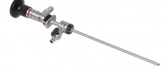
- Arthroscopy. A diagnostic and treatment method that allows minimally invasive surgery to cure most pathologies of the knee joint and its structures.
In addition, you will need: a general blood and urine test, biochemical studies, C-reactive protein, rheumatic tests. Only a specialist will be able to make an accurate and correct diagnosis, as well as prescribe treatment.
Rejection of bad habits
Rejection of bad habits
- Functional rest - during the entire stage of the main course of therapy, as well as partially during rehabilitation, the knee should not be subjected to stress. To ensure rest, a special motor regimen is used for the patient, and a tight bandage is applied to the joint using an elastic bandage or plaster splint.
- Diet – Dietary recommendations include eating high-calorie, highly digestible foods with sufficient fat, protein, carbohydrates and vitamins. Also, organic compounds that are directly involved in the synthesis of the intercellular substance of cartilage tissue must enter the body with food.
- Quitting bad habits - during the main course of therapy, it is advisable to give up smoking and drinking alcohol-containing drinks, since nicotine and alcohol are vascular toxins, they significantly slow down the processes of tissue regeneration (healing).
The implementation of such general recommendations (with the exception of functional rest) is desirable at the rehabilitation stage.
Drug therapy
In almost all cases of conservative treatment without surgery, medications of various groups are used. The most widespread are:
- Hormonal anti-inflammatory drugs based on adrenal hormones and glucocorticosteroids. They are used in the development of a pronounced inflammatory reaction in the form of intra-articular, intravenous or intramuscular injections.
- Non-steroidal anti-inflammatory drugs - reduce the intensity of the process, the synthesis of mediators of the inflammatory reaction by cells of the immune system, thereby reducing the severity of manifestations. These drugs are mainly used in the form of injections or tablets for oral administration. Additionally, local therapy may be prescribed in the form of a cream or ointment.
- Chondroprotectors are a large group of medications, the active substance of which is the main structural component of cartilage tissue. These drugs have the ability to slow down degeneration processes, as well as restore damaged cartilage tissue.
- Vitamins are an essential component of drug therapy, helping to improve the course of metabolic (metabolic) processes in the body and accelerating healing.
Medicines are used over a long period of time, which is determined individually by the attending physician.
Physiotherapeutic procedures
Physiotherapeutic treatment involves the use of physical factors that have a therapeutic effect. Most medical clinics use procedures such as electrophoresis with topical medications, treatment with a magnet, ozokerite, and mud therapy.
They help reduce the intensity of inflammation and also accelerate the healing of damaged tissue. As a rule, physiotherapeutic procedures are combined with the prescription of drug therapy.
Causes
There is a stereotype according to which meniscus damage is possible only in those who are fond of sports. In reality this is not the case. Over 50% of all meniscal injuries occur literally “out of the blue.”
Injury can occur due to an unsuccessful step or movement, while walking, hitting your knee on a hard surface, or under other similar circumstances. Meniscus injuries occur in those who like to ski, run, jump, and also in those who like to squat.
As a result, a meniscal injury is an injury that can occur to anyone, regardless of gender or age.
The risk group includes people with weakened ligaments and people who are prone to obesity.
Therapy without surgery
Any intervention involving tissue dissection is invasive, so modern traumatologists and orthopedists try to carry out treatment without surgery whenever possible. Conservative treatment is possible in certain cases:
- Diagnosed incomplete rupture with the absence of cartilaginous fragments.
- Localization of damage - due to certain anatomical features, an incomplete tear of the lateral meniscus of the knee joint is diagnosed; treatment in this case is possible without surgical intervention.
- The patient has absolute contraindications to surgery - usually conservative therapy is prescribed to elderly patients with pathological changes in the cartilaginous components.
Therapy without surgical intervention can be carried out in a hospital or on an outpatient basis, but only if the patient follows medical prescriptions in a disciplined manner.
