Author of the article: Victoria Stoyanova, category 2 doctor, head of the laboratory at the diagnostic and treatment center (2015–2016).
Article publication date: 03/16/2013
Article updated date: 01/27/2020
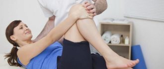
The knee joint is one of the largest and most complex joints in our body. Unfortunately, he is prone to injury. A sprained or torn ligament can occur as a result of a fall, blow, overuse, or unnatural movements. How dangerous is this and what can it mean? Injuries, of course, are best avoided, but if a knee sprain occurs, treatment started in a timely manner will avoid unpleasant consequences.
The photo shows the structure of the knee joint
General information
One of the most common types of injuries is sprained ligaments , most often of the ankle or knee joint.
The main cause of sprains is sudden movements in the joints that exceed their normal amplitude. It is important to distinguish between sprains of ligaments and tendons, because the latter are connections between muscles and bones, while ligaments are the elastic connecting link of bone formations. Moreover, they are capable of regeneration, therefore, when restored, they are given time to grow on their own, even if they are completely torn.
The structure of the ligamentous-capsular apparatus
Most often, simple sprains, tears of the lateral (medal), cruciate ligaments, and damage to the meniscus , and complex injuries are also possible, combining damage to the internal ligaments and the meniscus, including the cruciate ligaments. For example, a sprained tibia is a severe injury to all or almost all the ligaments of the knee joint.
Diagnostics
First of all, of course, the doctor will ask the victim about the nature of the injury and perform a visual examination to determine the severity and which joint structures are damaged. Of the additional methods, the following studies help obtain the most complete information about the joint:
- MRI (magnetic resonance imaging) - takes a series of layer-by-layer images in different projections, in which all structures are clearly visible, including cartilage, ligaments and tendons.
- Computed tomography (CT), a method similar in information content, is often considered as an alternative to MRI.
- An ultrasound of the knee (ultrasound) will allow the doctor to examine the joint area in detail. This is one of the most accessible methods, but requires professional training of a doctor in the field of ultrasound diagnostics, otherwise unreliable results are possible.
- Arthroscopy is an endoscopic method for examining the joint cavity from the inside. It is classified as minor surgical operations. They are used for the purpose of both diagnosis and treatment - for example, when other methods cannot determine the nature of the injury and the degree of damage. With the help of arthroscopy, ligaments are sutured, bleeding is stopped and other therapeutic procedures are performed.
- X-ray in several projections in case of damage to the ligamentous apparatus of the knee is used as an auxiliary method, since it does not allow us to see the ligaments and soft periarticular tissues. Looking at X-rays, the doctor can judge the nature of the damage only by indirect signs.
Pathogenesis
Usually the lateral ligaments of the knee are susceptible to injury - the rupture occurs in the area of the joint space or at the place of their attachment, causing pain on the part of the damaged ligament. This is caused by a violation of their physiological elasticity, when the tension in the ligament is too strong, this leads to tearing, rupture or separation of the ligament from its place of attachment. Pathologically and anatomically, the sprain is close to a bruise , but is supplemented by disintegration and rupture of individual fibers.
Damage to the ligaments, menisci and other structures of the knee joint causes rapid atrophy of the thigh muscles, a decrease in their tone, strength and ability to voluntarily contract the quadriceps muscle, therefore it is extremely important to carry out the correct treatment to restore full extension of the joint, as well as to ensure that it stays in this position sustainability.
Important! Despite the good regenerative properties of ligament tissue, no replacement tissue formed during restoration can fully provide the same strength and function of the natural ligament, and it will differ both in structure and size. This can lead to stiffness, stiffness, instability and a high likelihood of re-injury, therefore, to obtain the best result, it is necessary to carry out adequate treatment and restoration of the anatomical integrity of the ligaments under the supervision of a qualified physician.
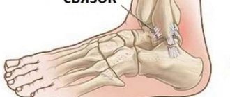
Ligament structure
The knee joint is a vulnerable organ. The knee contains various types of tissue, menisci and cartilage, ligaments and muscles.
The weak side is the connecting apparatus, which connects four bones:
- fibula;
- femoral;
- tibial;
- kneecap.
The knee joint contains the following ligaments:
- Cruciform or cruciform. Thanks to them, the shin does not move forward or backward. Located in the center of the knee joint, the building material is collagen fibers. The bunches are intertwined with each other, resembling a cross, which is why they have this name.
- Collateral. The femur and tibia are connected to each other. There are two types: medial (located on the inside of the knee joint) and lateral (located on the opposite side). Maintain stability of the joint during leg movement and protect against unnecessary stress.
- The ligament itself unites the patella with the tibia into one whole. It is especially vulnerable in athletes who run or jump.
Causes
In addition to sudden movements and actions with a large amplitude, which are unusual for the knee joint, a sprained ligament can cause:
- forced rotation of the lower leg or foot outward, and the hips inward;
- lifting heavy weights, such as during powerlifting;
- sports such as running, jumping, basketball, hockey, football and others;
- falling on the knee or being hit, for example, as a result of an accident;
- hereditary disease - Ehlers-Danlos syndrome , which causes defects in collagen and increases the risk of injuries such as dislocations , sprains and strains.
Prevention
To reduce the risk of re-injury or initially reduce the chance of a knee sprain when playing sports, adhere to the following rules:
- Use knee protection: wear special equipment designed for your specific sport.
- Always wear comfortable sports shoes with spring soles when training.
- Strengthen your leg muscles by performing special exercises.
- When doing strength training, increase the load gradually. Use recommendations only from experienced instructors.
Now, knowing about the causes and consequences of knee sprains, you will learn to avoid traumatic situations, and if something happens, you will be able to provide first aid to yourself and others.
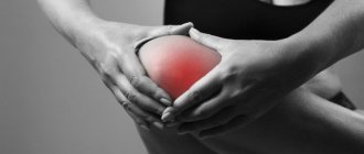
Symptoms of a knee sprain
Depending on the degree of injury, the mobility of the joint changes. A moderate injury leads to significant limitation of mobility, while a more serious injury makes the joint excessively mobile and unstable, causing pathological instability. Such moderate-to-severe injuries occur with a characteristic sound - a pop, which indicates rupture of the fibers in the ligament.
Symptoms of a knee sprain are usually local and include:
- local (in the knee area) redness or hematoma , a bruise may appear on the 2-3rd day, and slightly below the location of the injury;
- increasing tissue temperature in the stretched area;
- the occurrence of gradually increasing swelling of the knee joint, possibly a significant increase up to a size resembling an “elephant look”;
- pain – significantly increasing when the knee is turned in the direction of sprain, as well as when palpating and pressing on the area of the stretched ligaments and their attachment to the bones;
- constant crunching and clicking sounds;
- lameness.
In addition, pain may occur when trying to step on a leg with an injured knee. The pain can be so severe that a person can neither lean on his leg nor take a step.
Treatment
Restoring full leg function is very important, especially for an athlete. But, unfortunately, this is not always possible. Modern methods of low-traumatic operations make it possible to restore the joint in case of many injuries, but sometimes this takes quite a long time - it may require not one, but several consecutive operations, followed by stage-by-stage rehabilitation. It all depends on the severity and nature of the injury.
Treatment for mild sprains is often limited to the following:
- Immobilization - tightly bandaging the knee with an elastic bandage or using a special bandage (brace). This provides immobility and rest to the joint.
- Cooling - applying an ice pack or a special cooling pack to the affected area. This reduces pain and tissue swelling.
- Providing the injured limb with rest, that is, excluding any movements in the joint and support on the sore leg for the first time after the injury. Crutches should be used to walk.
- By placing the knee in an elevated position in bed. The victim should spend most of the time lying down, raising the sore leg above the level of the heart - this helps reduce swelling.
For mild sprains, other than these measures, nothing more is usually required. Although sometimes the doctor prescribes anti-inflammatory and painkillers, as well as thermal physiotherapeutic procedures.
Moderate severity of injury requires more serious measures. Immobilization is carried out using a plaster cast or bandage for a period of 3 to 4 weeks. Drug therapy is prescribed using anti-inflammatory and restorative agents, thermal physiotherapy, and after removing the bandage, massage and therapeutic exercises.
Treatment for a severe sprain usually begins with surgery to restore the structure of the joint. After the operation, a plaster cast is applied for a period of 2 months or more, depending on the severity of the injury. A patient in the acute stage of injury is in a traumatology or orthopedic hospital, after which he is transferred to a dispensary register with an orthopedic doctor at his place of residence.
After discharge from the hospital, a set of rehabilitation measures is required to develop the knee and restore the full range of movements in it. Rehabilitation includes a course of massage, physical exercise, swimming in the pool, exercise equipment, and thermal treatments.
Tests and diagnostics
To recognize a rupture of the internal ligaments, it is necessary to conduct an examination and, using the method of palpation/light pressure on the area of the outer surface of the knee, damaged ligaments, retract the lower leg to identify an increase in the valgus position of the knee joint. This symptom of shin deviation can be visualized using radiography. In case of a stretch - an incomplete rupture, a gap of approximately 2-3 mm is visible on the film between the femoral condyle and the tibia, while the rupture will allow the tibia to be retracted much more easily, and the void recorded on the radiograph will be much wider.
In case of incomplete rupture of the cruciate ligaments - anterior or posterior, which are less common - the internal collateral ligaments and/or the internal meniscus may be damaged. Rupture of the anterior cruciate ligaments leads to subluxation in the front, and posterior cruciate ligaments, respectively, in the back, and is characterized by an anterior or posterior “drawer” symptom.
To determine the extent of change in knee instability, you may also need to:
- conducting electromyography, rheovasography, podography and other functional and biomechanical examinations;
- biopsy and arthroscopy to detect damage to specific anatomical structures.
Knee ligament disease
The knee ligaments are very vulnerable and are susceptible to various diseases.
Tendinitis (inflammation of the ligaments). Mostly athletes suffer from this disease as a result of prolonged physical exertion. It is characterized by inflammation of the ligaments and disruption of the musculoskeletal system, accompanied by severe pain, which later becomes constant and lasts even at rest.
Ligamentosis (ligamentitis):
- Inflammation and damage to ligaments caused by infectious diseases and injuries.
- I am concerned about the aching pain, the intensity of which is constantly increasing.
- The knee ligaments are under constant tension. It becomes difficult to bend and straighten the leg. The patient begins to limp on the affected limb.