What Causes a Knee Sprain?
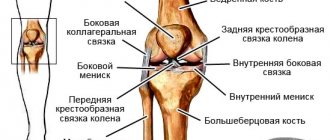
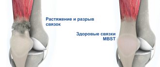
The knee joint has 4 ligaments that allow it to move. The ligaments themselves are made up of collagen and elastin, components that allow them to stretch and cushion the joint under stress. The knee twists when the ligaments do not have time to adapt to the load and stretch. As a result, microtraumas and ruptures occur, causing severe pain.
A sprain under the knee can be caused by the following reasons:
- jumping from heights;
- Fall with a heavy load on the knee joint;
- strong physical activity;
- sprain, fracture and other joint injuries;
- sudden careless movement of the knee.
Athletes are at risk because they struggle with increased physical stress on a daily basis. Lifters and runners are more likely to suffer from sprains than others.
Symptoms and signs
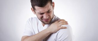
The symptoms of a knee sprain are very similar to other knee injuries, with the main symptom being severe, limiting, and exclusionary pain. The pain is localized in the area of the cervical tendon, radiating into the joint itself and into the calf muscles.
In case of injury, it is extremely difficult to distinguish a sprained knee from a broken ligament, but there are a number of signs that help in this:
- Moving stiffness of the joint, accompanied by increased pain at the slightest touch to the site of injury.
- Feeling of burning pain spreading throughout the entire limb.
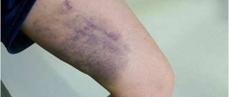
- Change in skin color around the joint and below the knee, caused by the presence of a hematoma and associated swelling.
- Crunching, clicking, cracking in the joint when moving.
- Coldness in the lower extremities, located in the toes.
How is diagnosis performed?
When visiting a doctor, he will most likely ask how the injury was sustained and visually diagnose the knee joint.
Signs of ligament damage:
- constant pain independent of palpation;
- atypical clicking and crunching in the joint, causing pain even with minor movements;
- difficulty changing posture, reduced knee mobility;
- tissue swelling;
- bruises that appear several hours after the injury;
- atypical deviation of the tibia to the side due to rupture of the collateral ligaments;
- pathological mobility of the lower leg, bent at the knee due to rupture of the cruciate ligaments;
- instability of the joint, its stiffness, while the patient complains of “looseness” of the knee, or it “falls out” during movements.
To clarify the location of the damage, the doctor prescribes an X-ray or ultrasound examination (ultrasound), magnetic resonance imaging (MRI).
Additional diagnostics provide information about possible fractures or injuries to the tissues of the knee.
Knee sprain: degrees
Although a hamstring sprain can heal spontaneously, the injured person will require diagnosis and treatment to speed up the process. Trauma should not be taken lightly as its manifestations can have more serious connotations. Based on personal feelings, it is impossible to determine exactly which ligament is damaged and how to stop it.
There are 3 degrees of twisting, the symptoms of which have their own characteristics:
- 1st degree - characterized by the presence of microcracks in the ligament without violating its integrity. The pain is moderate, the person can stand on his leg, but there is no stable support.
- Second degree - accompanied by partial damage to the bag and an increase in the area of fiber rupture. The pain is intense, limiting, intensifies with movement, mobility is limited.
- At the third stage, a complete rupture of the ligament occurs at the attachment site, accompanied by acute stabbing, pressing pain, tendon swelling and hematoma. It requires complex treatment, as it has many negative consequences.
It is impossible to diagnose the degree of damage to the ligamentous fibers solely by pain symptoms.
Knee sprain: diagnosis
Without diagnostics it is impossible to make an accurate diagnosis. If there is severe pain indicating a sprain, it is recommended to undergo procedures such as:
- X-ray - the image shows the presence or absence of bone fractures, knee dislocations and other effects that accompany the injury.
- Arthroscopy is an examination of the joint from the inside, allowing you to assess the extent of damage not only to the ligaments, but also to other elements of the knee.
- Ultrasound - performed if there is a suspicion of fluid accumulation and increased inflammation inside the joint.
During palpation examination, the surgeon assesses the severity of pain, its exact location and joint mobility. Based on these data, conclusions are drawn about the nature of the injury and the degree of sprain, as well as treatment methods. Without this data, it is almost impossible to restore full mobility of the knee, since each degree of damage has its own therapeutic characteristics.
Read also[edit | edit code]
- Knee-joint
- Child's knee joint
- Anatomy of the knee joint
- Knee injury
- Sports knee injury - treatment
- Crunching in the knees
- Taping the knee joint
- Knee orthoses
- Massage for joint sprains
- Massage for damaged menisci of the knee joint
- Hyperextension of the knee joint
- Patellar ligament rupture
- Avulsion of the tibial tuberosity
- Meniscus tear: symptoms and treatment
- Meniscus tear in children
- Osteochondrosis dissecans
- Injuries and damage to the quadriceps femoris muscle
- Lateroposition of the patella
- Incarceration of the peripatellar synovial fold
- Patellar bifurcation
- Knee ligaments Anterior cruciate ligament rupture
- Anterior cruciate ligament rupture in children
- Posterior cruciate ligament rupture
- Chondromalacia patella: symptoms and treatment
Treatment for knee sprains
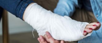
It is important to know what to do in case of a sprain, as proper first aid will help to avoid many problems. negative health consequences. The knee must be immobilized and bandaged with an elastic bandage.
When transporting the patient, do not lean on the injured leg and shift the main weight to the healthy limb. Place an elastic bandage on your bent knee.
You can apply dry cold to the area of pain, which will reduce bruising and pain. If the joint is clearly deformed, quickly increases in volume and acquires an unnatural color, the patient should be immediately taken to a medical facility.
Further therapy is carried out taking into account the degree of ligament torsion and the individual characteristics of the patient.
How to provide first aid for a sprain?
When sprained, it is important to urgently diagnose the injury in order to quickly reduce tissue swelling and the risk of hematoma formation.
First aid methods include:
- creating complete peace;
- cooling the injured joint by applying a cold compress or ice wrapped in a cloth;
- fixation of the lower leg and knee by tightly bandaging or using a splint.
As soon as possible, consultation with a traumatologist or surgeon is necessary. When transporting a patient, it is necessary to ensure maximum immobilization of the limb.
Knee sprain: treatment
Treatment for a first-degree knee dislocation involves mobilizing the joint and limiting the patient's movement, as well as wearing a flexible kneecap to reduce pain. The doctor prescribes a number of medications that can eliminate pain, swelling and hematoma. The most effective of them are:
- Heparin ointment;
- Diclofenac and preparations containing this substance;
- Heparil gel.
To relieve pain, NSAIDs are prescribed to reduce discomfort, inflammation and fever: Nurofen, Ibuprofen, Ketoprofen, Meloxicam. Peppermint and chamomile tea can help reduce anxiety due to stress.
The second degree of damage will require complex treatment, including:
- Pharmacological treatment: ointments, creams, gels with analgesic and regenerating effects, analgesics for oral use.
- Using a limited mobility splint or patch when the affected leg is above the level of the head.
- Physiotherapeutic procedures that accelerate regeneration processes: magnetic therapy, medicinal electrophoresis.
Ointments for knee sprains are selected individually, as some ingredients can cause allergic reactions.
Stages of assistance
Before considering what the sequence of providing pre-medical first aid for a sprain is, we note what absolutely cannot be done. After a blow, the first reflex movement is to rub the bruised area. It is better to refrain from this, since rubbing activates blood circulation, and if the vessels are damaged, then hemorrhage into the surrounding tissue will increase, which will lead to the growth of a hematoma.
The same applies to heating the damaged area. In the first two days, it is forbidden to apply warm heating pads to the injury site, take a hot bath or shower, or make warming compresses. This can be done only after 48 hours, when the threat of hemorrhage from damaged vessels has passed.
First aid steps for a sprain include resting the injured joint, cooling, and compression. Painkillers are also used if necessary. We briefly covered the stages of first aid for a sprained ligament victim, and now we will look at each of them in detail.
Peace
The first step in helping with muscle and tendon sprains is to keep the injured joint immobile. This allows you to achieve two goals - to stop the development of pathological processes caused by injury and to reduce pain.
The victim must be laid down, placing the joint in a position in which pain is minimal. In this case, it is desirable that the injured area is above the level of the heart. This reduces blood supply to the injured area. If there is a suspicion of a fracture or ligament rupture, it is recommended to immobilize the limb using splints. But this must be done after the stages of bandaging with an elastic bandage and cooling. Splints are applied on both sides of the injured area, extending beyond it, and attached to the limb using bandages. It is necessary to place a soft cloth under the splints to avoid damaging the skin.
Compression
The next stage of first aid for an injured person with sprained muscles and ligaments is the application of a tight bandage, which, on the one hand, provides fixation of the joint, and on the other hand, slightly compresses the damaged vessels and reduces internal hemorrhage. It is best to use a compression bandage for this purpose, but if this is not available, you can make a tight bandage with a regular bandage or strips of clean fabric. If there are wounds on the skin, they must be covered with several layers of sterile dressing material before applying a tight bandage. The bandage should be tight, but in moderation, it should not interfere with blood circulation in the limb, because when a ligament is sprained, providing first aid, everything should be done to improve, and not worsen, the condition of the injured person. If, some time after applying a tight bandage, the skin of the limb turns blue or, conversely, becomes too pale, the bandage must be loosened.
Cooling
First aid for a sprain also includes applying cold to the injured area. This helps stop internal bleeding. Immediately after the injury, cold exposure should be 25–30 minutes, and then for two days, cold should be applied for ten minutes every two hours, excluding sleep time. When using cooling agents, wrap them in a cloth and make sure the dressing does not get wet, especially if there are wounds on the skin.
Recovery
Recovery after following all the doctor’s recommendations will take longer than in the initial stages of the injury, but the prognosis in this case is favorable. If there are no associated injuries to the posterior part of the knee sprain, the person will be able to walk fully in 3-4 weeks.
A third-degree knee sprain is characterized by tearing of the fibers and requires surgery. During surgery, the damaged fibers are cut out and the ligament is firmly attached to the bone, restoring mobility to the knee.
Then a cast is applied for 3-4 weeks and pharmacological treatment is carried out. Complex painkillers and drugs that normalize the synthesis of collagen and elastin in the body are prescribed.
In the absence of appropriate treatment, a thickening forms at the site of the torn ligament, which is provoked by natural recovery processes. Subsequently, this can cause limited mobility, constant pain during exercise and the development of inflammatory processes in the ligamentous apparatus.
Lameness, inability to walk long distances, and impaired activity may occur.
Recovery period
There are several areas of treatment for damaged tissues of the knee joint.
Sprain and rupture of knee ligaments - treatment:
- the use of analgesics or NSAIDs to relieve pain and inflammation: Analgin, Ketanov, Indomethacin, Dikloberl, Movalis;
- local application of warming ointments: Diklak, Voltaren;
- the use of warm compresses with medications;
- use of a bandage;
- plaster fixation for incomplete ligament rupture.
The recovery period can take from 1.5 to 3-4 months. This period depends on the age of the patient and the severity of the injury.
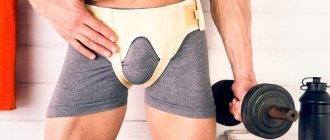
To reduce the load on the injured knee when walking, crutches are used, and for complete fixation, specially selected braces are used.
Rehabilitation
Once a sprained knee muscle has progressed to the acute phase and the person can put weight on the affected leg without experiencing severe pain, a number of rehabilitation techniques can be used. They will help prepare the muscles for exercise, gradually developing them. This, in turn, will reduce the likelihood of a second injury and also allow you to recover faster.
Exercise therapy can speed up the rehabilitation process
Exercise therapy should begin with the simplest ones, gradually increasing the load. When normal leg movements, bending and bending do not cause pain or discomfort, you can begin exercising on exercise machines. Bicycles and elliptical trainers are best suited for this.
Swimming in a pool can be a good option as it reduces the stress on your knee joints and improves their efficiency. The duration of exercises and load are increased gradually, dividing the rehabilitation program into 1-2 months.
Standard stages of rehabilitation
In order to restore the functions of the injured limb as quickly as possible after treatment for a ruptured and sprained ligament, the attending physician may recommend:
- massage;
- performing physical therapy exercises;
- physiotherapy.
The goal of using these methods is to restore the previous range of motion.
Muscle strengthening is carried out through the use of exercise bikes, treadmills, and special equipment.
Stages of rehabilitation:
- It takes about a week. Includes flexion and extension exercises, leg swings, and careful walking with the help of crutches.
- Takes 2 weeks. Supplemented with squats and toe raises.
- It takes a month. Previously recommended exercises are supplemented by abducting the legs to the side and up, using an elliptical trainer.
- It takes 1.5 months. Includes swimming, exercise on an exercise bike, and exercises with weights.
- It takes six months. Includes exercises to strengthen leg muscles and maintain ligament tone.
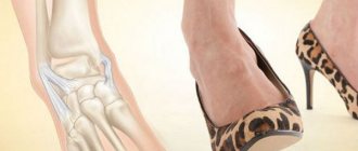
To minimize the likelihood of sprains and ruptures of ligaments, you need to wear comfortable shoes, strengthen your leg muscles, and warm up before sports training.
Losing excess weight and using protective equipment while playing sports or working will reduce the risk of injury.
Knee sprain: tendon
Tendons can become stretched due to overuse of the knee, causing pain, swelling and stiffness. Treatment at home is only permissible if a first-degree injury is diagnosed.
Grade 2 and 3 knee sprains require careful supervision by a specialist. Treatment of sprains is a complex of measures that includes not only taking medications, physical therapy and physiotherapy, but also wearing a special knee brace. Lack of treatment leads to many irreversible processes, reducing joint mobility.
Treatment of knee ligament rupture
Knee ligament rupture
- the most common injury among patients of all ages, resulting from twisting and sudden movements in a bent joint or a strong blow to the knee, as well as to the thigh, shin. Most often, such damage occurs in athletes when playing basketball, volleyball, football, martial arts and skiing, but it can also occur in everyday life, for example, due to a fall. Main Causes of Injury
The functions of the knee joint stabilizer are performed by four ligaments: two lateral ligaments, which prevent external deviations of the tibia and inward displacement, as well as cruciate elements, which are located inside the joint and ensure its rotational stability.
The mechanism of injury is usually indirect:
- a sharp change in direction of movement and twisting of a limb;
- unsuccessful jump and landing on one leg;
- falling while playing one of the sports.
However, ruptures of the cruciate ligament of the knee joint can also occur due to direct impact. About a third of such pathological conditions are caused by road traffic accidents, when sharp contact with the dashboard of a car with the front part of the knee causes displacement of the tibia and rupture of the posterior ligament of the knee joint at its thinnest part.
Injury to the PCL is also possible when a person is struck from the front of the knee at the moment of its full extension, when the foot has assumed a fixed position on a hard surface. The most rare occurrence is damage to the internal lateral process, more typical of breaststroke swimmers experiencing excessive valgus loads. Types and degrees of damage
There are three main degrees of violation of the integrity of the ligaments:
- 1st degree. The patient complains of moderate localized pain, virtually no swelling or swelling, and no hemorrhage. The mechanical integrity of the dense formation is preserved, only part of the fibers is torn;
- 2nd degree. The main complaints relate to limited walking and pain in the injured area. A large number of fibers are damaged. There is a slight instability in the knee;
- 3rd degree. The patient complains of severe pain. There is marked swelling of the affected area and bruising, and instability in the knee. All fibers are torn.
Due to the excessive pressure and high range of motion created by uncontrolled landing on one leg, a complete or partial tear of the knee ligaments, popularly called a “sprain,” can occur.
Symptoms of damage
Violation of the integrity of the stabilizer of the knee joint is accompanied by sharp pain; patients can hear a characteristic crunching sound during its destruction. You can often hear the phrase that the knee “came out and snapped back into place.”
The main symptoms of knee ligament rupture are:
- acute pain at the time of injury, which can persist for a long time;
- skin redness, swelling, extensive subcutaneous hematoma;
- limitation of functions, feeling of instability.
Due to damage to the nerve endings, fluid accumulation and severe pain in the knee, the patient cannot lean on the limb immediately after the accident; he spares his leg and limps. Often there is a specific fear associated with the “displacement” of the knee. Diagnostics
Severe pain and lack of stability in the joint are always a reason for concern and consultation with a specialist. During the examination, the orthopedic traumatologist will assess the presence of edema and fluid in the joint and conduct the necessary tests (ligamentous and meniscal). The patient may also be recommended an MRI of the damaged joint, ultrasound and X-ray examinations to prevent bone tissue defects. In the case of a partial rupture of the cruciate ligament of the knee joint, the mobility of the joint may be somewhat limited, and in the event of a rupture, the amplitude is too large due to a change in the position of one of the bones, so simple movements such as flexion and extension of the knee in the joint are impossible for the patient. Since the symptoms of a closed fracture and sprains may be the same, additional diagnostics may be required to make an accurate diagnosis.
Rupture of the cruciate ligament of the knee joint: treatment
To completely restore the damaged ligament, it is necessary to use accurate diagnostic measures and the correct approach to treatment. This will avoid aggravation of symptoms, the development of undesirable consequences in the form of early arthrosis and joint destruction, and also facilitate subsequent treatment.
The first time after an unsuccessful jump or fall, the following measures must be taken:
- provide rest to the injured area, protecting the limb from unnecessary stress;
- apply cold to relieve pain, inflammation and bruising;
- apply a tight bandage that will immobilize the damaged joint and relieve swelling;
- place a pillow under the limb to improve venous outflow, reduce swelling and pain threshold;
- take analgesics and anti-inflammatory drugs.
If these measures are carried out in a timely manner at home, the swelling will be removed as quickly as possible, and the injured leg will receive the necessary rest. Conservative treatment of partial rupture of the knee ligaments is based on the use of an elastic bandage and analgesics, but for an accurate diagnosis a referral to a specialist is required. The full course of treatment is about 4 weeks, and full recovery will require at least 10–12 weeks.
When the cruciate ligaments of the knee joint are completely torn, plastic surgery is required. In 20% of situations, you can get by with reconstructive surgery, but in the remaining 80% of cases, dissection occurs and such ligaments can no longer be sewn on. Then they resort to plastic replacement of the ligament, using the patient’s existing tendon and special implants to fix the manufactured ligament at its attachment points. Rehabilitation after a complete rupture of knee ligaments can take more than six months.
Treatment for a torn anterior cruciate ligament
Rupture of the anterior cruciate ligament Upon admission to the traumatology department, the patient is sent for radiography to exclude bone damage. The knee joint is punctured to relieve symptoms, eliminate pain and accumulated blood.
Among the main purposes: local cold, elastic bandaging, ensuring an elevated position of the leg, fixation with an orthosis, lasting up to 7 days. As pain decreases and swelling subsides, the patient can try to lean on the limb after injury and perform flexion and extension in the knee joint. An MRI is performed to make an accurate diagnosis.
If the patient feels instability in the joint or displacement forward, his lower leg twists when walking and during professional sports, such a decrease in functionality becomes the main indication for reconstructive surgery. Otherwise, swelling and pain may increase, which not only affects a person’s quality of life, but can also lead to abrasion of the articular cartilage, secondary meniscal rupture and rapid development of osteoarthritis.
Conservative treatment in this situation has no prospects, since due to insufficient blood supply, the torn fibers do not heal independently.
The patient may be recommended arthroscopic reconstruction with the production of a new tendon ligament and the use of various grafts. For this purpose, the tendons of the tender and popliteal muscles can be used - the ideal option, which is most suitable for all the characteristics of the strength of the ligament. The fixing structures used are RIGID-FIX, ENDO-BUTTON, BIO-RCI and BIO-INTRAFIX.
The arthroscopic technique has the following benefits for the patient:
- low-traumatic treatment;
- shorter rehabilitation period;
- return to previous sports activities within 7–8 months after surgery.
Let us recall that previously a rupture of the anterior cruciate ligament of the knee joint for an athlete could result in the end of his professional career. Nowadays, this damage no longer poses a threat to his sporting future and can be easily repaired thanks to the mastered technique.
The first few days after the operation, the athlete is in a hospital setting, where his condition is monitored by the attending physician. Subsequently, treatment is carried out on an outpatient basis; the need to visit the center arises only when dressing is performed.
During the rehabilitation period, the patient is recommended to spare the leg with the installed autograft and limit the load on it. To achieve this, immobilization of the operated leg is ensured. Muscle strengthening and changes in range of motion are carried out gradually under the supervision of a rehabilitation therapist.
Treatment for a torn posterior cruciate ligament
Damage to the knee ligaments, symptoms and treatment When the PCL is damaged, a smaller amount of blood accumulates in the joint cavity than with a rupture of the anterior ligament and menisci. But this is still a reason to perform x-rays in several projections so as not to miss bone damage.
Next, conservative therapy is carried out:
- ensuring an elevated position of the leg;
- applying cold and bandaging;
- Limiting exercise and taking painkillers.
If, after the pain subsides and a return to the previous rhythm of life, the patient continues to complain of instability in the joint, this may indicate a ligament rupture. An MRI is required to clarify the diagnosis. Using radiography, it is possible to detect a separation of the posterior ligament from the place of its fixation with a bone fragment. If this occurs early after injury, the patient is prescribed refixation using cannulated screws.
In case of partial rupture of the anterior ligament of the knee joint, conservative rehabilitation therapy is carried out, which allows creating the necessary conditions for strengthening the muscles and fusion of the fibers of the ligament of the damaged joint.
With prolonged sensations of lack of joint fixation, when there is a complete rupture of the PCL, conservative treatment may be ineffective. This is an indication for surgical intervention, including strengthening with synthetic threads and arthroscopic plastic surgery.
Treatment for rupture of the lateral ligament of the knee joint
The treatment tactics used in this case are conservative, even if we are talking about the third degree of destruction of the collateral ligament. Appointed:
- elevated position of the limb;
- exposure to cold;
- elastic bandaging;
- avoiding overload of the injured limb;
- analgesics;
- using a brace equipped with strong side inserts.
Radiography is mandatory, and in a controversial case, under load. An MRI is required to establish the definitive nature of the rupture and identify other joint injuries.
Tears and sprains of the external collateral ligament are somewhat less common than injuries to the internal ligament. The most likely cause of such an injury is a sharp blow to the shin or knee area. Usually the opposite leg protects the knee joint from such trouble, but if, for example, there is an attempt to hit the ball with a volley, the knee becomes unprotected from direct impact directed from the inside to the outside. It is also possible that an indirect injury occurs when the leg twists during a sudden change in the direction of the athlete’s movement.
It may take about 1 month until the damage is repaired, provided the listed conditions are met. In rare cases, the ligament may fail to heal and weak stability in the joint may remain. Then plastic surgery is performed using an autograft.
Complete rupture of the collateral ligament with injury to other joint structures (menisci and ligaments) always requires surgical intervention. If the damage is fresh, the ligament can still be sutured, but to ensure complete stability of the joint, plastic surgery with the installation of an autograft is usually used.
Recovery period
As part of the rehabilitation period, anti-inflammatory, antibacterial and symptomatic therapy is carried out.
To reduce swelling, cold applications are used, indicated within two weeks after plastic surgery for 15–20 minutes. Patients are advised to spare the leg and use crutches for 7 days or more. The sutures are removed on the 14th day.
The joint is fixed using an orthosis, which is worn for two weeks, after which an elastic bandage or compression hosiery is used (up to 1 month).
In the following weeks, the patient needs to limit the load on the leg. You are allowed to give up crutches only three weeks after surgery. It is during this period that when the ligaments of the knee joint are torn, exercise therapy, active muscle development and restoration of range of motion can be used.
The patient can begin sports training after 3 months, and sports without any restrictions - after 9–12 months.
The New Step Sports Medicine Rehabilitation Center offers its services for the recovery of patients after sprains and ruptures of knee ligaments. We can undergo all types of diagnostics, including MRI, special X-ray and ultrasound examinations. Patients are examined in a short time, after which an individual treatment and rehabilitation program is selected for the patient. The center provides massages, physiotherapy, classes on special simulators and therapeutic exercises.