The knee is the largest and perhaps one of the most complex joints in the human body. On the one hand, it must ensure flexion and extension of the leg, its mobility, in all directions, maintain coordination and correct position of the body in space. On the other hand, the knee joint, as one of the connecting parts of the lower extremities, must be as stable and strong as possible in order to withstand the weight of the human body, and not be deformed or injured under intense loads. Nature took care of this balance, having thought through the anatomy of the knee joint to the smallest detail: there is not a single superfluous detail in the structure of this joint, therefore every, even the most minor failure or injury leads to a serious limitation of the normal functions of the entire limb. How does the knee work, what does its functionality depend on, and how to maintain the health of this most complex and extremely important joint, avoiding injuries and age-related changes? A little medical education will help you find answers to such pressing questions of modern orthopedics!
Knee joint: anatomy and physiology of the muscular system
Alternate contraction and relaxation of the muscles causes the knee to move in three planes, thereby ensuring mobility and stability of the lower limb. That is why the main classification of the muscular system is based not on the anatomy or localization of each group, but on the functions assigned to it:
- Knee flexion. This movement is ensured by the balanced and complete work of the largest muscle group of the knee joint. It includes the biceps, semitendinosus, semimembranosus, popliteal, gastrocnemius, plantaris, sartorius and gracilis muscles.
- Extension of the joint. This function is assigned to only one, but the largest muscle of the leg - the quadriceps. It consists of the direct, lateral, medial and intermediate wide muscle fibers.
- Pronation is the inward movement of the leg. Limited “rolling” of the lower leg towards the internal axis is ensured by the popliteal, semitendinosus, gracilis, sartorius, semimembranosus, and medial head of the gastrocnemius muscles.
- Supination is an outward movement. Turning the tibia outward is possible due to the contraction of the biceps and lateral head of the gastrocnemius muscle.
Innervation of tissues adjacent to the knee joint
The nerve fibers of the knee joint are a complex interconnected network, which ensures the full functioning of the lower extremities. Despite the fact that the innervation network of the knee is not very developed, each of its elements plays a key role, which means that at the slightest failure the entire system of joint mobility is “switched off”.
The nervous system, localized in the knee area, is represented by the following fibers:
- The meniscal nerve bundles penetrate the tissue along the periphery of the body of the cartilage itself, along the blood vessels of the knee. These nerves contribute to the formation of soft and soft fibers, maintaining normal innervation of joint tissues.
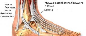
- The tibial nerve, through its articular branches, provides sensation to the posterior surface of the knee.
- The peroneal nerve innervates the anterior part of the knee, including the cap.
Anatomy of the blood vessels of the knee
The two key blood vessels located in the area of the knee joint are localized on the posterior surface, that is, under the knee (which is why both the vein and the artery are called popliteal in anatomical reference books). The artery transiently carries blood from the heart to the lower parts of the leg - the leg and foot, and the vein of the same name, in turn, returns depleted blood to the heart. However, these vessels do not represent the entire circulatory system of the knee: many vessels of smaller diameter depart from them, connected to each other by a network of anastomoses. Thanks to them, nutrition is provided to the muscles and tissues adjacent to the knee joint.
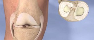
Anatomical features
Bones that form the knee:
- femur;
- tibia;
- knee cap.
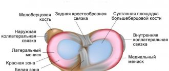
Inside the knee joint there are cartilage plates called menisci. They are located on the border of the femur and tibia. The menisci essentially divide the knee joint into two equal parts. The joint is of the condylar type, which means that the joints of the bones between which the meniscus is located are represented by condyles.
Joint movement is carried out in the following directions:
- vertical (sagittal) plane – movements are performed in flexion and extension directions;
- frontal plane;
- horizontal plane - movements are performed with a bent joint.
In addition, the knee joints perform sliding and rolling.
The following elements are involved in the formation of the human knee:
- epiphyses of bones, which are located on both sides of the meniscus;
- joint capsule and cavity;
- bursa;
- cartilage plate;
- knee ligaments.
Let's talk about these elements in more detail.
Epiphyses of the femur and tibia
The femoral epiphysis forms the knee on the superior side, and the tibial epiphysis forms the inferior side. The epiphysis is presented in the form of an expanded end section, which participates in the formation of the knee along with the bone on the adjacent side.
The thickenings of the epiphyses of the femur - the condyles - are convex in shape, and the condyles of the tibia are concave in shape. Menisci are located between the surfaces of the joints, since they are not symmetrical. Cartilaginous plates are able to equalize this discrepancy.
The thickenings of the epiphyses of both bones are covered with cartilage. Cartilage tissue is represented by hyaline substance, which consists of collagen substance.
The structure of the human knee joint includes the following elements:
- chondrocytes are the main cells of cartilage;
- tissue fluid;
- organic matter;
- germ layer, which promotes the restoration of cartilage tissue.
During mechanical action when walking, the load is evenly divided between the germ layer, chondrocytes and collagen fluid.
Read also: Shoulder joints
Hyaline cartilage does not exceed five millimeters in thickness. Despite the constant friction of the joint, the cartilage always remains smooth. Cartilage has elastic properties that have a shock-absorbing effect.
The patella, the sesamoid bone, also plays an important role in the formation of the knee. The kneecap is located inside the tendon, or rather in its thickness, the femoral muscle. The patella is directly involved in the process of straightening the leg.
If you look at the kneecap from the inside, it is covered with massive cartilage, which facilitates easy movement of the joints of both the femur and tibia.
The patella improves muscle function and performs a blocking function.
Joint cavity
The articular cavity is represented by a closed space, which looks like a gap. The cavity is limited by the inner layer of the articular capsule, as well as the surfaces of the joints of the femur and tibia.
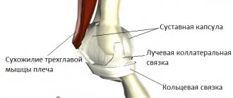
Joint capsule
The main function of the joint capsule is protection. It protects the human knee joint from increased mechanical influence from the outside.
If you look at the capsule from the inside, it is covered with a synovial membrane. The tension of the joint capsule of the knee joint is weak. Thanks to this, motor activity can be performed in different planes with different strengths.
If you look at the posterior section of the articular capsule, in this place it is slightly thicker and is distinguished by the presence of numerous holes, which are a kind of canals. Blood vessels pass through them.
The capsule contains the following shells:
- synovial It lines the inner surface of the joint capsule. The membrane does not cover only the articular surfaces of the epiphyses of the femur and tibia. The synovium produces synovial fluid. The liquid contains small vessels, therefore it is a good nutrition for cartilage tissue. Also, thanks to the shell, joint mobility increases. The shell provides good protection against mechanical stress. If an inflammatory process of bone tissue occurs, the synovial membrane prevents the pathological process from spreading further and damaging the joint cavity. Villi are outgrowths that form the synovial membrane. They help increase the surface area of the synovial membrane, and also take part in the production of intra-articular fluid;
- fibrous. The membrane covers the outside of the joint capsule. The main component that makes up the fibrous membrane is collagen substance. The membrane reaches the periosteum.
Synovial bursae
They are located under the muscles and near the tendons. To prevent muscles and tendons from rubbing when moving, the bags are filled with synovial fluid.
Synovial bursae come in different types:
- suprapatellar type of the bursa. The bursa is located between the femur and the tendon. Next to it is the cavity of the knee joint;
- subpatellar type of the bursa. Located between the patella and kneecap;
- prepatellar type of the bursa. It is located between the skin and the patella. During physical activity, the bag slides along the patella;
- semi-membranous appearance of the bag. Located between the semimembranosus and gastrocnemius muscles;
- popliteal view of the bag. The bulging capsule of the knee is the bursa of the popliteus muscle.
Pathological processes of the musculoskeletal system can be the result of congenital pathology or disorders during intrauterine development, as well as injuries. In addition, orthopedics provides information on diagnostic methods and therapeutic measures for diseases of the musculoskeletal system.
Let's talk about the sections of orthopedic medicine:
- outpatient section. Outpatient medicine is a fairly important section, since in most cases patients undergo outpatient treatment;
- children's and teenage industry. The main objective of this section is the prevention and timely elimination of congenital pathologies;
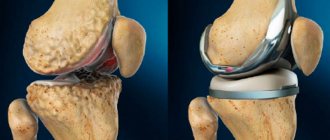
- operative orthopedics. In this case, the main method of dealing with the problem is surgery;
- implantation orthopedics (replacement or prosthetics);
- sports section;
- traumatology.
Physiology and pathology of the knee: chain reaction to injury
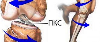
Knee injuries are considered one of the most difficult in orthopedics, and for good reason: every muscle or ligament fiber, every cartilage or bone affects the functionality and mobility of the joint. Even a minor deviation, for example, mild inflammation of a ligament or a bruise, can trigger destructive processes, the treatment of which will require long-term and serious therapy.
As you know, the surfaces of bones cannot be connected like a puzzle, providing full mobility. Therefore, if the functioning of the ligamentous apparatus, muscles or meniscus, which hold the joint in a physiological position, is disrupted, the cartilage tissue begins to gradually wear out. As a rule, such destruction becomes clearly expressed only in the final stages: at first, sensations during the pathological process can be attributed to the consequences of dislocation or overwork. That is why any pain, atypical sound during flexion/extension, or discomfort during exercise requires a detailed diagnosis of the knee joint and timely qualified assistance.
Joint capsule
The connective tissue that hermetically seals the cavity of the knee joint is called the joint capsule. It is attached to: firstly, the femur, secondly, the tibia, and thirdly, the patella. Since the fit of the bone structures in the knee joint is not tight, a so-called gap, or in other words, an articular cavity, is formed in the articular capsule. Normally, there should be a small amount of serous fluid in the joint cavity. This fluid is essential for the normal functioning of the knee, working simultaneously as a lubricant, moisturizer and nutrient medium.
The biological mechanics of the knee joint are comparable to the operation of door hinges, with the addition of forward movement. In addition to this, the knee joint bears the load of the entire body weight.
The main aspect for the full functioning of the knee apparatus is body weight control and maintaining an active lifestyle. The optimal load for people of any age is walking for 20–30 minutes at a speed of 5–6 km per hour. If a person does not have the opportunity to walk every day, then the best option to replace this kind of load would be a trainer without impact loads on the knees. Such training tools include exercise bikes and electromagnetic ellipsoids.