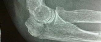
Elbow joint
Type of joint Synovial hinge joint (trochlear hinge joint).
Articulation The superior surface of the head of the radius articulates with the head of the humerus. The trochanteric notch of the ulna articulates with the trochanter of the humerus (which forms the “ball-and-socket device” and a major stabilizing factor).
Articular Capsule The relatively loose articular capsule extends from the coronoid and olecranon fossa of the humerus to the coronoid and olecranon processes of the ulna and to the annular ligament surrounding the head of the radius. The capsule is thin anteriorly and posteriorly, allowing flexion and extension, but is strengthened on the sides by collateral ligaments.
Ligaments Ulnar (medial) collateral ligament. Three strong strips that strengthen the medial side of the capsule. Radial (lateral) collateral ligament. A strong triangular ligament that strengthens the lateral side of the capsule.
Stabilizing tendons The tendons of the biceps brachii, triceps brachii, brachialis and other muscles located in the forearm. These tendons cross the elbow joint and provide additional security.
Movements: Flexion and extension only.
Elbow joint: side view
Elbow joint: medial view
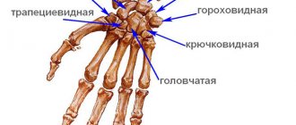
Hand joints and intercarpal ligaments
The wrist joint, articulatio radiocarpea, is formed by the carpal articular surface of the radius
and
the distal surface of the articular disc, representing
Articular capsule, capsula articularis,
thin;
is attached along the edge of the articular surfaces of the bones that form this joint. The joint is strengthened by the following ligaments:
• radial collateral ligament of the wrist, lig. collaterale carpi radiate.
Some of the bundles of this ligament reach the polygonal bone. The ligament inhibits adduction of the hand;
• ulnar collateral ligament of the wrist, lig. collaterale carpi ulnare.
The ligament inhibits abduction of the hand;
• dorsal radiocarpal ligament, lig. radiocarpeum dorsale.
The ligament inhibits flexion of the hand;
• palmar wrist ligament, lig. radiocarpeum palmare,
starts from the base of the lateral styloid process of the radius and the edge of the carpal articular surface of the same bone;
goes down and medially, attaching to the bones of the first and second rows of the wrist - scaphoid, lunate, triquetrum and capitate. The ligament inhibits the extension of the hand. The pisiform joint, articulatio ossispisifosmis,
connects the pisiform bone to the triquetrum.
Articular capsule, capsula articularis,
is fixed along the edge of the articular surfaces of the bones.
The joint cavity can communicate with the cavity of the wrist joint. The joint has the following ligaments:
• pisiform-hook, lig. pisohamatum;
• Pisiform metacarpal.
These ligaments are a continuation of the tendon of the flexor carpi ulnaris, m. flexor carpi ulnaris, in the thickness of which there is a large sesamoid bone - the pisiform bone, os pisiforme.
Sesamoid bones
They are small bone or fibrocartilaginous rounded formations located in the thickness of the tendons. These bones cause the corresponding muscle tendon to be lifted and create a more favorable angle of action on the bone.
Carpal bones
They form intercarpal joints among themselves, articulationes intercarpeae, and between the first and second rows of carpal bones there is a midcarpal
The joint is strengthened by the following ligaments:
• dorsal intercarpal ligaments, ligg. intercarpea dorsalicr,
• palmar intercarpal ligaments, ligg. intercarpea palmaria.
Some of the bundles of these ligaments start from the capitate bone and diverge in the form of rays to the bones of the first and second rows of the wrist, forming the radiate
carpi radiatum.
In addition to these ligaments, there are also interosseous intercarpal ligaments, ligg. intercarpea interossea.
Movements in the joint are sharply limited, so the midcarpal joint is classified as a low-moving joint. The carpometacarpal joints, articulationes carpometacarpeae, form the distal surfaces of the bones of the second row of the wrist and the base of the metacarpal bones.
There are two carpometacarpal joints.
• one is formed by the polygonal bone and the first metacarpal bone (thumb);
• the other is located between the polygonal bone, trapezoid, capitate and hamate bones, on the one hand, and the II-V metacarpal bones, on the other. Carpometacarpal joint of the hand, articulatio carpometacarpea pollicis,
formed by the distal saddle-shaped articular surface of the polygonal bone and the saddle-shaped articular surface of the base of the first metacarpal bone.
Carpometacarpal joint of the thumb
is a type of biaxial joint -
the saddle joint, articulatio sellaris.
Carpometacarpal joints of the II—V metacarpal bones
formed by the flat articular surfaces of the distal side of the polygonal bone, as well as the trapezoid, capitate and hamate bones and the proximal articular surfaces of the bases of the II-V metacarpal bones facing them.
The carpometacarpal joint of the fifth metacarpal bone is close in
the saddle joint, articulatio sellaris.
The articular capsule, capsula arlicularis, is attached to the edge of the articular surfaces of the bones and is tightly stretched.
The cavity of the carpometacarpal joint communicates with the cavity of the intercarpal, midcarpal and intercarpal joints. The ligamentous apparatus of the carpometacarpal joints includes
the palmar and dorsal
carpometacarpal
carpometacarpea patmaria et
dorsalia,
which on the corresponding side are stretched between the bones of the wrist and metacarpus.
The carpometacarpal joints are mechanically one whole - the solid base of the hand. They are inactive and belong to flat joints, articulationes planae. Intermetacarpal joints, articulationes intermetacarpeae, are formed by the lateral flat surfaces of the bases of the II-V metacarpal bones.
Articular capsule, capsula articularis,
attached along the edge of the articular surfaces.
The joint cavities in the proximal part communicate with the carpometacarpal joints.
The intermetacarpal joints include two groups of ligaments:
• four dorsal metacarpal ligaments, ligg. metacarpea dorsalia,
and three
palmar metacarpal ligaments, ligg.
metacarpea palmaria. These ligaments are located on the dorsal and palmar surfaces of the joints; they are stretched from the dorsal and palmar sides between the bases of the metacarpal bones;
• interosseous metacarpal ligaments, ligg. metacarpea interossea,
- located between the bases of the metacarpal bones.
The intermetacarpal joints are flat, low-moving joints.
Metacarpophalangeal joints, articulationes metacarpophalangeae, formed by the articular surfaces of the heads of the metacarpal bones
the articular surfaces of the bases of the first phalanges
facing them Articular capsules, capsulae articulares,
spacious.
They are strengthened in the lateral sections by collateral collateralia,
which start from the depressions on the ulnar and radial surfaces of the heads of the metacarpal bones and are attached to the lateral and partly palmar surfaces of the bases of the proximal phalanges.
Some of the fibers of these ligaments, starting from the lateral surface of the heads of the metacarpal bones, are directed to the palmar surface of the bases of the proximal phalanges, where they intersect with bundles of the same fibers of the opposite side. These ligaments are called palmar ligaments, ligg. palmaria
On the palmar surface of the joints, between the heads of the II-V metacarpal bones, deep
metacarpea transversa profunda.
The first metacarpophalangeal joint (thumb) belongs to the trochlear joints, gingfymus,
and the metacarpophalangeal joints of the II-V fingers are spherical
The interphalangeal joints of the hand, articulationes interphalangeae manus, are located between the adjacent phalanges of each finger. The articular surface of the head of each phalanx has
Ligamentous apparatus of the interphalangeal joints of the hand
represented by palmar
palmaria,
which come from the lateral surfaces of the blocks and are attached: some to the lateral surface of the bases of the phalanges (collateral ligaments), and others to their palmar surface.
The first (thumb) finger has one interphalangeal joint; interphalangeal joints of the II-V fingers are located between the proximal and middle phalanges and are called proximal interphalangeal joints;
the joints between the middle and
distal the distal
The interphalangeal joints are typical representatives of the trochlear joints, ginglymus.
Proximal radioulnar joint
Type of joint : Synovial hinge joint.
Articulation The disc-covered head of the radius rotates within a ring formed by the radial notch on the ulna and the annular ligament of the radius. Note : the synovial cavity of this joint continues (connected with) the cavity of the elbow joint.
Movements Pronation and supination of the forearm.
Proximal (upper) radioulnar joint: front view
Connections in the free part of the upper limb
The joints in the skeleton of the free part of the upper limb are represented by the shoulder joint (articulatio humeri), elbow (articulatio cubiti), proximal and distal radioulnar joints (articulatio radioulnaris proximalis and articulatio radioulnaris distalis), wrist joint (articulatio radiocarpea) and joints of the hand skeleton - midcarpal, carpal - metacarpal, intermetacarpal, metacarpophalangeal and interphalangeal joints.
The shoulder joint (Fig. 31, 32) is formed by the connection of the glenoid cavity of the scapula with the head of the humerus. The articular cavity of the scapula is surrounded by the articular lip (labrum glenoidale), which has a fibrocartilaginous structure. The labrum increases the relatively small (compared to the head of the humerus) size of the glenoid cavity of the scapula, and also serves to absorb possible sudden movements in the joint.
The head of the humerus, shaped like a third of a ball, provides greater mobility of the joint around all three axes, and also allows for circular movements. The thickness of the cartilage covering the articular surfaces decreases from the center to the edges. The articular capsule, or bag (capsula articularis) (Fig. 31, 32), is attached to the scapula along the outer edge of the articular labrum, and to the humerus along its anatomical neck, leaving the greater and lesser tubercles of the humerus outside the joint cavity.
The joint capsule is strengthened by ligaments, which are thickened areas of its fibrous layer; The coracohumeral ligament (lig. coracohumerale) (Fig. 32), which runs from the base of the coracoid process, is of greatest importance. Most of its fibers are woven into the capsule, a smaller part reaches the greater tubercle.
| Rice. 31. Shoulder joint, frontal section 1 - joint capsule; 2 - glenoid cavity of the scapula; 3 - head of the humerus; 4 - articular cavity; 5 - tendon of the long head of the biceps brachii; 6 - articular lip; 7 - inferior inversion of the synovial membrane of the joint |
On the outer side, front and back, the muscles and tendons of the muscles of the shoulder and shoulder girdle are adjacent to the joint capsule. On the inferomedial side, the joint capsule does not have muscles that strengthen it, as a result of which there is a high probability of inferomedial dislocations occurring in the joint. The synovial membrane of the joint (consisting of subsynovial and synovial layers) forms three inversions that expand the joint cavity. The largest of them - recessus axillaris - is located in the lower part of the joint and is clearly visible when the shoulder is adducted (Fig. 31). The elbow joint is a complex joint formed by the connection of the humerus with the ulna and radius in the common capsule.
| Rice. 32. Capsule and ligaments of the shoulder joint 1 - acromion of the scapula; 2 - coracoid process of the scapula; 3 - coracohumeral ligament; 4 - greater tubercle of the humerus; 5 - tendon of the long head of the biceps brachii; 6 - joint capsule |
There are three joints in the elbow joint: the humeroulnar, humeroradial and proximal radioulnar.
The trochlear humeroulnar joint is formed by the block of the humerus (Fig. 33, 34) and the trochlear notch of the ulna (Fig. 33). The ball-and-socket joint consists of the head of the condyle of the humerus and the head of the radius (Fig. 34). The proximal radioulnar joint connects the articular circumference of the head of the radius with the radial notch of the ulna (see section “Bones of the forearm”).
The humeral-ulnar joint allows flexion and extension of the arm at the elbow. The cylindrical upper radioulnar joint allows only rotational movements, that is, movements around the vertical axis - pronation and supination (in this case, the radius rotates along with the palm).
| Rice. 33. Elbow joint vertical section 1 - block of the humerus; 2 - joint cavity; 3 - olecranon; 4 - trochlear notch of the ulna; 5 - coronoid process of the ulna |
The fibrous fibers of the elbow joint capsule are attached to the periosteum of the humerus anteriorly above the radial and coronoid fossae, posteriorly above the ulnar fossa, and in the lateral sections to the base of both epicondyles. On the bones of the forearm, the articular capsule is fixed along the edges of the articular cartilage on the ulna, and on the radius it is attached to its neck. At the back, the capsule of the elbow joint is less strong.
| Rice. 34. Front view of the elbow joint 1 - articular capsule; 2 - ulnar collateral ligament; 3 - head of the condyle of the humerus; 4 - block of the humerus; 5 - coronoid process of the ulna; 6 - head of the radius; 7 - interosseous membrane of the forearm |
The joint is strengthened by the radial (lig. collaterale radiale) and ulnar (lig. collaterale ulnare) collateral ligaments (Fig. 34, 35), which pass from the epicondyles of the humerus to the ulna.
The proximal radioulnar joint is formed by the radial notch of the ulna, located on the lateral side of its upper epiphysis, and the head of the radius. The annular ligament of the radius (lig. annulare radii), attached to the ulna, covers the neck of the radius, thus fixing this connection.
| Rice. 35. Ligaments of the elbow joint 1 - joint capsule; 2 - ulnar collateral ligament; 3 - radial collateral ligament; 4 - annular ligament of the radius |
The distal radioulnar joint (Fig. 36) is rotational and cylindrical in shape. The ulnar notch of the radius and the articular circumference of the head of the ulna, which form it, are separated by a triangular-shaped cartilaginous articular disc. The apex of the disc is attached to the styloid process of the head of the ulna, and the base is attached to the ulnar notch of the radius. The joint provides adduction and abduction of the hand (its movement in the sagittal plane).
| Rice. 36. Joints and ligaments of the hand, dorsal surface 1 - ulna; 2 - radius; 3 - distal radioulnar joint; 4 - articular disc; 5 - wrist joint; 6 - midcarpal joint; 7 - intercarpal joints; 8 - carpometacarpal joints; 9 - intermetacarpal joints; 10 - metacarpal bones |
The wrist joint (Fig. 36) is ellipsoidal and connects the lower epiphysis of the radius and the articular disc (discus articularis) (Fig. 36) of the ulna with the bones of the proximal row of the wrist. Since the head of the ulna is located at some distance from the wrist, the free space is filled with cartilage (fibrocartilago triangularis), which serves as an articular surface for the triquetral bone. The carpal articular surface of the radius and the distal surface of the articular disc form the articular fossa of the radiocarpal joint, and its head is the scaphoid, lunate and triquetrum bones of the wrist. In approximately 40% of cases, the cartilage has a gap through which the radiocarpal joint can communicate with the lower radioulnar joint.
Movements in the joint occur around two axes: the hand can move in the sagittal plane (towards the radius or ulna), as well as bend and bend, rotating around the frontal axis of the wrist joint.
The joint capsule is strengthened by the palmar radiocarpale ligament (lig. radiocarpale m. palmare), radiocarpal ligament of the dorsum of the hand (lig. radiocarpale m. dorsale), ulnar and radial collateral ligaments (lig. collaterale carpi ulnare and lig. collaterale carpi radiale).
There are six types of joints in the hand: midcarpal, intercarpal, carpometacarpal, intermetacarpal, metacarpophalangeal and interphalangeal joints (Fig. 37, 38).
The midcarpal joint (articulatio mediocarpalis), having an S-shaped joint space, separates the bones of the distal and proximal (except for the pisiform bone) rows of the wrist. The joint is functionally combined with the wrist and allows the latter to somewhat expand the degree of freedom. Movements in the midcarpal joint occur around the same axes as in the radiocarpal joint. Both joints are strengthened by the same ligaments.
Intercarpal joints (articulationes intercarpales) connect the lateral surfaces of the carpal bones of the distal row, and the connection is strengthened by the radiate ligament of the wrist (lig. carpi radiatum) (Fig. 38).
Carpometacarpal joints (articulationes carpometacarpales) connect the bases of the metacarpal bones with the bones of the distal row of the wrist. With the exception of the articulation of the trapezius bone with the metacarpal bone of the thumb (I), all carpometacarpal joints are flat, their degree of mobility is small. The connection of the trapezoid and first metacarpal bones is trapezoidal and provides significant mobility of the thumb. The capsule of the carpometacarpal joint is strengthened by the palmar and dorsal carpometacarpal ligaments (ligg. carpometacarpea palmaria et dorsalia) (Fig. 37, 38).
| Rice. 37. Ligaments of the wrist joint and joints of the hand, dorsal surface 1 - ulnar collateral ligament of the wrist; 2 - radial collateral ligament of the wrist; 3 - wrist ligament of the back of the hand; 4 - dorsal carpometacarpal ligaments; 5 - dorsal metacarpal ligaments; 6 - metacarpal bones; 7 - collateral ligaments; 8 - metacarpophalangeal joints; 9 - lateral ligaments of the interphalangeal joint |
| Rice. 38. Ligaments of the wrist joint and joints of the hand palmar surface 1 - ulnar collateral ligament of the wrist; 2 - palmar radiocarpal ligament; 3 - radial collateral ligament of the wrist; 4 - radiate carpal ligament; 5 - palmar carpometacarpal ligaments; 6 - palmar metacarpal ligaments; 7 - metacarpal bones; 8 - collateral ligaments; 9 - metacarpophalangeal joint of the fifth finger; 10 - deep transverse metacarpal ligament; 11 - lateral ligaments of the interphalangeal joint |
Intermetacarpal joints (articulationes intermetacarpales) are flat, with low mobility. They are composed of the lateral articular surfaces of the bases of the metacarpal bones (II-V), strengthened by the palmar and dorsal metacarpal ligaments (ligg. metacarpea palmaria et dorsalia) (Fig. 37, 38).
Metacarpophalangeal joints (articulationes metacarpophalangeales) (Fig. 37) are ellipsoidal, connect the bases of the proximal phalanges and the heads of the corresponding metacarpal bones, strengthened by collateral (lateral) ligaments (ligg. collateralia) (Fig. 37, 38). These joints allow movements around two axes - in the sagittal plane (abduction and adduction of the finger) and around the frontal axis (flexion-extension).
Interphalangeal joints (articulations interphalangeales) are block-shaped, connecting the heads of the superior phalanges with the bases of the inferior ones. The interphalangeal joints provide flexion and extension of the fingers and are strengthened by collateral ligaments.
The heads of the metacarpal bones do not have an articular connection with each other; they are connected (from the palmar side) by the deep transverse metacarpal ligament (lig. metacarpeum transversum profundum) (Fig. 38).
Intermediate radioulnar joint
joint type .
Articulation Connects the interosseous border of the radius with the interosseous border of the ulna through the interosseous membrane. In addition, a thin fibrous strip called the oblique chorda interosseus of the forearm connects the ulnar tuberosity to the proximal end of the radial shaft.
Function Increases the surface of origin of the deep muscles of the forearm; Helps connect the radius and ulna and transmits forces to the ulna up the arm along the radius.
Distal and intermediate radioulnar joint: a) front view; b) coronal view
Wrist joint
Joint type Synovial elliptical.
Articulation The distal surface of the radius and articular disc (the same disc is described in the distal radioulnar joint, see previous page) connects to the proximal row of carpal joints: scaphoid, lunate and triquetral.
Movements Movements are carried out in combination with the intercarpal joints: flexion, extension, adduction (ulnar displacement), abduction (radial displacement) and rotational movement.
Joints of the hand
The wrist joint (articulatio radiocarpea) is complex (Fig. 1). The shape of the articular surfaces is elliptical. It is formed by the articular surface of the radius, the articular disc and the proximal row of carpal bones (scaphoid, lunate, triquetrum). An articular disc separates the distal radioulnar joint from the radiocarpal joint. Movements around the frontal axis - flexion and extension, and around the sagittal axis - abduction and adduction are possible.
The wrist joints, intercarpal joints (articulationes intercarpales), connect the bones of the wrist. These joints are strengthened by interosseous and intercarpal ligaments (ligg. interossea et intercarpea), palmar and dorsal intercarpal ligaments (ligg. intercarpea palmaria et dorsalia).
The pisiform joint (articulatio ossis pisiformis) is the joint between the pisiform bone, located in the extensor carpi ulnaris tendon, and the triquetrum bone.
Carpometacarpal joints (articulationes carpometacarpals) are complex. They articulate the second row of carpal bones with the bases of the metacarpal bones. II-IV carpometacarpal joints belong to flat joints. They are strengthened by palmar and dorsal ligaments.
The carpometacarpal joint of the thumb (articulatio carpometacarpea pollicis) is formed by the trapezium bone and the base of the first metacarpal bone; This is the saddle joint. Movements in the joint are carried out around two axes: frontal - opposition (opposition) and reverse movement (reposition) and sagittal - abduction and adduction.
Intermetacarpal joints (articulationes intermetacarpals) are located between the bases of the II-V metacarpal bones.
Metacarpophalangeal joints (articulationes metacarpophalangeae) are formed by the heads of the metacarpal bones and the fossae of the bases of the proximal
phalanges of fingers. The metacarpophalangeal joints of the II-V fingers have a spherical shape. The joints are strengthened by ligaments. Movements in them are possible around the frontal axis - flexion and extension, the sagittal axis - abduction and adduction; Rotational movements are also possible, and in the first metacarpophalangeal joint - only flexion and extension.
The interphalangeal joints of the hand (articulationes interphalangeae manus) are formed by the heads and bases of the middle phalanges, the heads of the middle and the bases of the distal phalanges. These are block-shaped joints in shape. Ligaments run along the lateral surfaces of the joint. Movements in the joint are possible around the frontal axis - flexion and extension.
Rice. 1. Joints and ligaments of the hand: a - front view: 1 - distal radioulnar joint; 2 - ulnar collateral ligament of the wrist; 3 - pisiform-uncinate ligament; 4 - pisiform-metacarpal ligament; 5 - hook of the hamate; 6 - palmar carpometacarpal ligaments; 7 - palmar metacarpal ligaments; 8 - deep transverse metacarpal ligaments; 9 — metacarpophalangeal joint (opened); 10 — fibrous sheath of the third finger of the hand (opened); 11 - interphalangeal joints (opened); 12 - tendon of the muscle - deep flexor of the fingers; 13 - tendon of the muscle - superficial flexor of the fingers; 14 - collateral ligaments; 15 - carpometacarpal joint of the thumb (opened); 16 - capitate bone; 17 - radiate carpal ligament; 18 - radial collateral ligament of the wrist; 19 - palmar radiocarpal ligament; 20 - lunate bone; 21 - radius; 22 — interosseous membrane of the forearm; 23 - ulna
b - frontal cut of the left wrist joint and joints of the wrist bones), front view: 1 - radius bone; 2 - wrist joint; 3 - radial collateral ligament of the wrist; 4 - midcarpal joint; 5 - intercarpal joint; 6 - carpometacarpal joint; 7 - intermetacarpal joint; 8 - intercarpal ligament; 9 - collateral ulnar ligament of the wrist; 10 - articular disc; 11 - distal radioulnar joint; 12 - ulna
Human anatomy. S. S. Mikhailov, A. V. Chukbar, A. G. Tsybulkin; edited by L. L. Kolesnikova.
Metacarpal joint of the thumb
Joint type Synovial saddle joint.
Articulation Between the trapezium bone and the base of the first metacarpal bone (thumb).
Movements: Flexion, extension, abduction and adduction. With extreme flexion, the first metacarpal rotates medially so that the palmar surface of the thumb is opposed to the pads of the fingers. Conversely, slight lateral rotation occurs when the thumb is fully extended. Combining these movements creates an approximate rotational motion of the thumb.
Intermetacarpal joints
Joint type Synovial flat.
Articulation Between adjacent sides of the bases of the 2-5 metacarpal bones.
Movement Restricted movement between adjacent metacarpals.
Radiocarpal (wrist), intercarpal, metacarpal and intercarpal joints
Metacarpophalangeal joints
Joint type A series of flat synovial joints.
Articulation Between the head of the metacarpal bone and the base of the proximal phalanx. Note : The joint capsule is incomplete on the dorsal side, where it is replaced by an extension of the extensor longus tendon.
Movements Flexion and extension. Abduction and adduction (only possible with extension, but with slight mobility in the thumb). Combined movements can cause rotational motion.
Anatomy of the human midcarpal joint - information:
Midcarpal articulation, art. mediocarpea, located between the first and second rows of carpal bones, minus the pisiform bone, which is a sesamoid. The glenoid cavity of this joint is the distal surface of the first row of carpal bones. The proximal surface of the second row of the wrist forms the articular head. Both wrist joints (radiocarpal and midcarpal) have independent articular capsules attached to the edges of their articular surfaces.
The capsule of the wrist joint is strengthened by auxiliary ligaments on the radial and ulnar sides: lig. collaterale carpi radiate, going from the styloid process of the radius to the scaphoid bone, and lig. collaterale carpi ulnare, extending from the styloid process of the ulna to the os triquetrum and os pisiforme. On the palmar side of the wrist joint there is a lig. radiocarpeum palmare, which, starting widely from the styloid process and from the edge of the articular surface of the radius, is attached to the os scaphoideum, lunatum, triquetrum et capitatum by several bundles. On the dorsal side, the capsule of the wrist joint is reinforced by the lig. radiocarpeum dorsale, which goes from the radius to the bones of the first row of carpal bones.
At the point of attachment of the ligaments of the wrist joint to the bones, the latter include blood vessels and nerves, damage to which during operations entails pathological changes in the bones. Capsule art. mediocarpea also includes the last four carpometacarpal joints, which communicate with each other.
Besides art. mediocarpea, individual carpal bones connected to each other by interosseous ligaments, ligg. intercarpea interossea, in places articulate with each other with articular surfaces facing each other. Such joints are called intercarpal joints, articulationes intercarpeae. The intercarpal joints are reinforced by a number of short ligaments, running mostly transversely from one bone to another on the dorsum, ligg. intercarpea dorsalia, and palmar, ligg. intercarpea palmaria, sides. On the palmar side, in addition, there are bundles diverging from the capitate bone to the neighboring bones, lig. carpi radiatum.
Movements in the wrist joints occur around two mutually perpendicular axes passing through the head of the capitate bone, around the frontal (flexion and extension) and around the sagittal (abduction and adduction). These movements are inhibited by ligaments that are located perpendicular to the axes of rotation and at their ends, namely: collateral - at the ends of the frontal axis, dorsal and palmar - at the ends of the sagittal axis. Therefore, the former inhibit abduction and adduction around the sagittal axis, and the latter inhibit flexion and extension around the frontal axis. As in all biaxial joints, circumductio is also possible here, in which the ends of the fingers describe a circle.