The atlanto-occipital joint is formed by the condyles of the occipital bone and the concave articular fossae of the atlas. Both pairs of articulating surfaces are enclosed in separate articular capsules, but they move simultaneously, forming a single combined joint.
Stability of the atlanto-occipital joint is ensured by:
- two joint capsules;
- posterior atlanto-occipital membrane;
- anterior atlanto-occipital membrane.
Atlanto-occipital joint: what is it, where is it located?
AZ node
The Atlanto-occipital node (AZ) is a cartilaginous node that connects two important bones of the body: the skull and the spine. It is located at the top of the neck. It can be easily felt with your fingers near the head.
The movements of the articular microcapsules occur at the same time. This indicates the combined operation of the node. The connection between the surface of the cartilaginous unit and the protrusions of the occipital fossae forms the condyles. Therefore, such a joint is also called a condylar joint.
Anatomical features
An important role in the functionality of the cervical spine is played by its anatomical structure. The main element that is crucial for the entire spine is the atlanto-occipital joint. This articulation is formed by the cartilaginous surfaces of the condyles of the occipital bone and the upper articular cavities of the first cervical vertebra (atlas). A synovial capsule is attached to the edge of the joints, and additional stabilization is provided by two ligamentous structures:
- Anterior atlanto-occipital membrane - stretched from the anterior edge of the occipital foramen and the anterior arch of the atlas.
- Posterior atlanto-occipital membrane - connecting, respectively, the posterior edge of the foramen with the posterior arch of the vertebra.
The first cervical vertebra does not have its own body, so its opening is as wide as possible, which is necessary for the passage of the spinal cord. The vertebral artery and nerve fibers going into the cranial cavity pass through the posterior membrane.
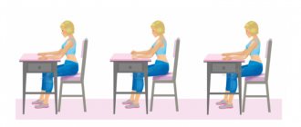
The atlanto-occipital joint is also called the upper joint of the head, which in its structure is combined (paired). This means that movements are carried out in them simultaneously - flexion and extension of the head, slight bends to the sides. The joint contains most of the receptors responsible for the function of upright walking and ensuring correct posture.
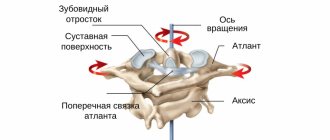
The peculiarity of the cervico-occipital region is the presence of another important joint - the median atlantoaxial joint. It is formed by a special process of the second vertebra (tooth) and a depression on the inner surface of the anterior arch of the atlas. In addition, an additional articular connection is formed between the surface of the tooth and the transverse ligament. Due to its structure, the atlantoaxial joint allows the head to rotate to the sides.
The mobility of the craniocerebral segment is ensured by two joints - the atlanto-occipital and atlanto-axial.
Atlanto-occipital joint: structure, anatomy
Structure of the AZ node
Between the atlas junction and the axial vertebral circle there are 3 cartilaginous nodes:
- Two lateral nodules, which are formed by the cartilage pits in the atlas region and the spinal pits.
- In the middle there is a tooth that connects to the arched atlas and transverse tissue. Around this tooth there is a ring, which consists of fibrous and bone tissue. All this together forms a cylindrical assembly that rotates.
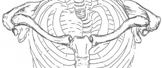
See what the atlas node looks like from the back and to the right - in the picture above. In the picture below, you will see what the knot looks like when you look at it from above and below.
Top and bottom view of the AZ unit
2 bundles consisting of fibrous cells extend from the edge of the transverse tissue. One is directed upward, and the other is directed downward. Together they create two lines crossed in the middle. This ligament is important for the functional features of the atlanto-occipital ganglion: the first directs the movement of the odontoid bone, and the second keeps it from injury. Thanks to these elements, the spinal and medulla oblongata fluid is protected.
AZ node
The auxiliary function is performed by the ligamentous node, which extends from the top of the tooth. On the side of the vertebral circles, the node is covered with a special membrane membrane, which extends from the slope of the bone at the back of the head.
How is the examination carried out?
MRI of the craniovertebral junction is performed without special preparation. The patient is only asked to remove metal objects (glasses, knives, pens, keys) from his pockets and remove jewelry. If clothing has metal parts, it must be removed and changed into a disposable sterile suit/gown. The patient is placed on the tomograph table on his back and moved inside the machine. The procedure lasts 30 minutes.
The office has climate control. The patient can choose the desired lighting and visual effects, music. Constant audio communication with medical staff is maintained.
After the examination and software processing, the doctor describes the images obtained and gives the necessary recommendations. If desired, the photographs are copied onto the patient’s electronic media or printed on film.
Contraindications Preparation
You can see prices for services
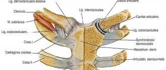
Atlanto-occipital joint: ligaments, muscles
Ligaments and muscles of the AZ node.
A stable and constant position of the head, as well as its mobility, is ensured by the following ligaments:
- The membrane, which is located in the front, stretches from the front of the occipital bone to the 1st link of the spinal column.
- The membrane that is located at the back is structured in the same way as the front one, but it is an articulation of bones behind the node.
Muscles and ligaments are responsible for the following movements:
- Mobility of the head relative to the neck.
- Fixation of the back of the head and cranium in the desired position.
- The special structure allows the circulatory system and nerve roots to be located unhindered.
- Providing space for the functioning of the central nervous system.
- Nerve endings in this area allow us to stand upright and walk.
The functional features of this node may be impaired after injury. Such injuries can even be fatal. If the bones are displaced by even a few tenths of one degree, this can lead to deformation of the spinal cord. Even if a person remains alive after injury, there is a high probability that he will be paralyzed.
Blood supply and innervation
At the top of the spinal column is the third (atlas) of the four segments of the vertebral artery. Through the transverse opening in the posterior membrane of the atlanto-occipital joint and the foramen magnum, the blood vessel passes into the cranial cavity. Inside the skull, the vertebral artery branches into a series of large blood vessels that supply various areas and tissues of the brain.
Innervation (connection of organs and tissues with the central nervous system) allows the body to function harmoniously. It has a two-way direction: centrifugal and centripetal.
Centripetal communication is carried out through signal transmission by receptors of peripheral nerve fibers from the organ to the spinal cord and then to the brain. The responses of the central nervous system organs are carried by centrifugal nerve fibers along the same chain. The atlanto-occipital joint and adjacent tissues are supplied with an extensive network of nerves.
Movements, functions of the atlanto-occipital joint
Functional feature of the AZ node
In the AZ node, movement is performed around two axes. With the help of one axis we can nod our head forward and bend it back. The second axis allows you to tilt to the right and left. The front of one of these axles is located slightly higher than the rear. This oblique position allows for simultaneous tilting of the skull to the side with a slight rotation in the other direction.
Motor capacity of the AZ unit
The top of the tooth-like bone is held in a constant position during head rotations by ligamentous nodes that regulate movement and protect the spinal cord from concussions. Motor ability in the area of the connections of the skull and vertebrae in the neck is performed with a small amplitude. Deeper movements occur when the entire neck is involved. The cranial-vertebral ligaments are well developed, as we walk straight and also hold our heads straight.
Osteokinematics
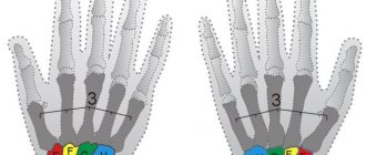
Flexion/extension
- Swing-type movement.
- Total amplitude: 15-30° (10° flexion, 20° extension).
- Axis: transverse axis through the external auditory canal.
Lateroflexion (side bending)
- “Arc-shaped” swing + rotation (for example, lateroflexion to the right occurs simultaneously with rotation to the left).
- Total amplitude: 15° (8-10° in each direction).
- Axis: oblique sagittal axis through the nose (approximately).
- The movement is associated with rotation at the atlantoaxial joint.
Rotation
- It occurs during lateroflexion due to inclined articular surfaces (their anteromedial orientation) and alar ligaments.
- Overall amplitude: 5-7° in each direction.
- Axis: vertical axis in front of the foramen magnum.
Blood circulation of the atlanto-occipital joint
Arteries of the AZ node
The first vertebra in the neck area has a special structure. It is thin and wide at the same time. This allows the upper part of the spinal cord to fit freely inside. Behind the atlanto-occipital ganglion is the vertebral artery, as well as many nerve roots that transmit information from the brain.
Blood circulation in this area should be free. If it is violated, then the following negative effects can occur:
- Headaches, various types of migraines, attacks of hypertension.
- Lack of nutrition in the brain area.
- Constant nausea, vomiting, dizziness.
- Temporary loss of consciousness, fainting.
- Confused consciousness, tinnitus, spots before the eyes.
If blood circulation in this area is impaired for some reason, then the brain suffers. It does not receive the required amount of nutrients. In this case, medications are prescribed in the form of vitamins with the addition of microelements.
What bones are involved in the formation of the atlanto-occipital joint?
The bones of the AZ node
of the Atlanto-occipital node surround the condyles of the occipital bones and the articular apparatus. The following bones of the skull are involved in the formation of this node:
- Frontal
- Parietal
- Occipital
- Temporal
- Wedge-shaped
The spine originates in this area. The two upper cervical vertebral circles, the atlas and the axis, have a specific anatomical structure that differs from the structure of all other vertebrae. The first cervical vertebral bone consists of arches that are connected not by bones, but by bone thickenings. The second cervical vertebra, as mentioned above, has a tooth-like process, which is fixed with the help of ligamentous tissues.
The atlanto-occipital joint has a special structure that is both strong and vulnerable. In addition to excellent mechanical strength, this section must have excellent flexibility and motor ability. All this is achieved by special joints, articular tissues and a special arrangement of ligaments.
Pathological changes in the joint and their symptoms
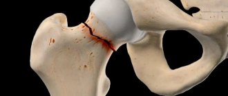
Damage to the atlanto-occipital joint occurs due to:
- spinal injuries, a strong blow to the head, an unsuccessful jump into the water, an accident (dislocation, fracture of the atlas, torn ligaments);
- dystrophic changes in the structure of joint tissues against the background of progressive arthrosis;
- disorders of metabolic processes in the body (calcification of cartilage and ligaments).
Dislocations
Dislocation (disturbance of the conformity of adjacent articular surfaces) does not always have pronounced painful manifestations. The reason to contact a specialist is:
- discomfort when moving caused by prolonged tension in the neck muscles;
- swelling in the area of injury.
Fractures
A fracture of the anterior arch of the atlas is accompanied by acute pain and significant limitation of motor function of the head and neck. It is dangerous due to the possibility of damage to the spinal cord and blood vessels. Reduced sensitivity of the occipital part of the skull and neck, discomfort when swallowing and trying to open the mouth are grounds for urgent medical attention.
Joint rupture
The integrity of the ligaments is disrupted when the head is thrown back sharply as a result of strong mechanical stress . Partial rupture of the ligamentous apparatus between the atlas and occipital bone causes severe pain in the area of damage. Complete disruption of the integrity of the ligaments of the atlanto-occipital joint can lead to death.
When compiling an anamnesis, the doctor clarifies the diagnosis during the examination of a patient with suspected spinal injury. MRI results play a decisive role.
Important! Timely provision of medical care helps to avoid the development of serious neurological pathologies.
Arthrosis
This is inflammation. Pain and deformation of the joint signal the development of a common pathological process - arthrosis (damage to the structure of the cartilage tissue of the articular surfaces). The result of a progressive disease is complete or partial loss of functionality of the atlanto-occipital joint.
Arthrosis occurs as a result of:
- mechanical damage (trauma) to the cervical spine;
- metabolic disorders caused by endocrine diseases (for example, diabetes);
- inflammatory process (with rheumatoid arthritis or tuberculosis);
- congenital pathology of the joint structure (dysplasia).
The diagnosis is made based on:
- medical history;
- characteristic features of the pain syndrome (pain persists at rest, intensifies at night);
- visual signs of deformation of the joint and adjacent tissues;
- X-ray examination data;
- MRI.
Ligamentous calcification
Calcification of the atlanto-occipital ligament, like the transverse ligament of the atlas, is characterized by the deposition of insoluble calcium salts in the articular tissues due to disruption of the body's metabolic processes.
The disease impairs the elasticity of the ligaments and can lead to their rupture even with minor physical activity.
Important! The basis for the treatment of pathologies of the atlanto-occipital joint is its fixation in a stationary state using an orthopedic collar.