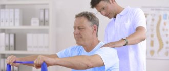
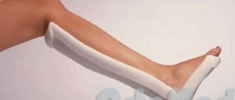
Some patients need rehabilitation after a fracture of the radius - this is a set of measures that is carried out to quickly heal the injury site. Recovery includes several stages; in the early period, complete immobilization of the limb under the supervision of an orthopedic surgeon is indicated. When a callus forms, the cast is removed, and the program includes measures to restore function and self-care to the patient. A visit to a sanatorium or rehabilitation in Greece will help you consolidate all basic skills.
Anatomy of the forearm and causes of fractures
The forearm consists of two bones - the ulna and the radius, which perform a supporting function. Most of the flexors originate from the first, and the extensor muscles (rectifiers) originate from the second. After an injury, displacement of fragments often occurs due to muscle spasm, which pulls the ends to the side under the influence of pain. If you do not provide assistance to the patient in time, the fusion will occur incorrectly, and the limb will lose its usual functions. High-quality rehabilitation after a displaced fracture of the radius can only be carried out in a specialized center, where the patient’s condition will be monitored over time.
If you turn your hand palm up, the radius bone will be on the side of the index finger. Its fractures most often occur in the lower sections for three main reasons:
- an unsuccessful fall on the hand - when landing reflexively, we lean on the hand, which is why a fracture occurs;
- osteoporosis – due to destruction, bone tissue loses its strength and is easily damaged;
- degenerative-dystrophic changes - with age, all tissues of the body wear out, including bones.
Most often, such injuries occur in older people or at a young age in accidents. These criteria determine how long rehabilitation takes after a fracture of the radius. At a young age, bones heal faster, so 2-3 months may be enough. Patients over 50-60 years of age may require about a year to recover.
Some features of the development of fractures of the phalanges of the fingers
A fracture of the phalanx of a finger is quite painful. A plaster splint is applied to the damaged phalanx for up to a month. In case of a displaced fracture, you first need to fold and fix all the fragments - with knitting needles or bone pins.
Many people do not take the consequences of such a seemingly minor injury very seriously. But long-term immobilization and neglect of the subsequent course of rehabilitation are the main reasons why the finger does not bend after a fracture. This can be observed quite often in people who have suffered a similar fracture.
The main method of restoring the natural motility of the phalanx is physical therapy. In this case, you should use not only the injured finger, but also the entire hand.
First of all, you need to rub your hands and do a massage: clasp your fingers and rotate them with closed hands. The next exercise is to clench and unclench your fingers into a fist and dangle your fingers outstretched in the air.
After this, you can perform several exercises on the table surface: put your palms on the table and bend them, moving up the table, lightly tap your fingers on the table.
Among the useful exercises on how to develop a finger after a fracture of the phalanx, the most ordinary things from everyday life are listed: sort buttons by size or small beads by color, type text on the keyboard, write text by hand, embroider a pattern or make an applique.
For complete rehabilitation, it is not necessary to perform only specially designed gymnastics; simple exercises for the development of fine motor skills will do.
However, many of these everyday activities do not help in solving the problem of how to develop a little finger after a fracture. In this case, using an expander or a set of special exercises recommended by a doctor or instructor can help.
Restoration activities require regularity and moderation. Injured phalanges should not be overexerted. Treatment should be combined with massage and physiotherapy. People who are faced with the problem of how to develop a finger after a displaced fracture should listen especially carefully to the recommendations.
Rehabilitation
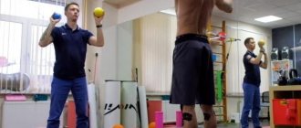
The program is designed to strengthen bones, restore lost functions, restore self-care and work activity. It is also important to work with a psychologist, then the patient will be able to adapt socially. In order to recover quickly, treatment of musculoskeletal diseases should be carried out on a clinical basis.
Diet
Proper nutrition is the basis for rapid healing of bone tissue. Such rehabilitation after a fracture of the radius includes eating plenty of food that contains magnesium, calcium and vitamin D.
The diet should include:
- fish and seafood;
- milk, cottage cheese, sour cream and cheese;
- eggs, broccoli, bananas;
- figs, peas, legumes.
The diet should be varied, you should eat 5-6 times a day in small portions. You should not abuse chocolate, alcohol and coffee; such products interfere with the digestion and absorption of certain substances. To create a daily schedule, you can contact a nutritionist.
Physiotherapy
Exercises should be selected by a doctor; loads should be treated with caution, because they can cause harm. Any approaches should be done at rest, with the forearm lying on the table. At the initial stages of rehabilitation after a fracture of the radius, the following movements can be performed:
- alternately lifting each finger and then the entire palm;
- spreading your fingers to the sides and returning to the starting position;
- carefully clench your fist with moderate force and relax your hand;
- circular movements with each finger;
- “Take a pinch of salt and add salt.”
The purpose of such a warm-up is to restore function and prevent muscle atrophy due to immobilization in a cast. When performing exercise therapy, blood circulation improves, blood vessels dilate, and nutrition in the affected area is normalized.
At a later date, you need to visit a doctor. If the doctor sees that the bones have become stronger, he will prescribe additional exercises:
- circular movements with the brush;
- rotation of the forearm in and out;
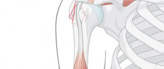
- flexion at the elbow joint and straightening;
- circular shoulder warm-up.
Push-ups, pull-ups, working with dumbbells, barbells or exercise machines are strictly contraindicated. The orthopedist will recommend such loads only when you are sure that a strong bone callus has formed at the fracture site.
How to relieve swelling
The accumulation of fluid after the removal of the cast is normal, if you do not take into account the discomfort it causes to the person. At a consultation with a surgeon, you can get recommendations on what creams and ointments will help get rid of it. To provide the limbs with rest and thermostable conditions favorable for relieving swelling, as well as to strengthen the joints and muscles, a regular bandage is prescribed to be applied to the site where the plaster is removed.
There are a number of popular tips for solving this problem. However, whether it is worth listening to them after such a serious intervention as the application of a plaster splint, or whether it is better to trust professionals in the field of medicine - everyone decides for himself.
Massage
In addition to exercise therapy, massage is prescribed; you only need to visit the office of a professional specialist with a medical education. Timely warm-up will provide a rush of blood and improve tissue trophism, preventing muscle wasting. Sessions are scheduled 2-3 times a week, the average course is 10-14 days. The doctor recommends the timing.
If the patient is in a cast, the collar area is warmed up to improve blood flow to the immobilized limb. After removal, the forearm is warmed up, the duration of the sessions and the intensity of the warm-up increases as the wound heals.
Physiotherapy
To speed up rehabilitation after a fracture, physical treatment is prescribed; even if the radius is damaged with displacement, such methods show high effectiveness. The patient may be prescribed:
- electrophoresis;
- UHF;
- warming up;
- ultrasound treatment;
- thermal procedures with paraffin, etc.
Such restoration will ensure rapid fusion of bones. To form a strong callus, the patient should not forget to eat properly and do therapeutic exercises.
Tekar therapy
For complete rehabilitation of the arm after a fracture of the radius, the patient will benefit from a new method of radio wave therapy. With this effect, energy is transferred without radiation, which ensures deeper penetration. After several sessions, pain and swelling are reduced, and tissue regeneration is accelerated.
The waves also affect the muscles, preventing their atrophy. This method of recovery will perfectly complement the recovery program and shorten the rehabilitation period.
What can you do during the immobilization stage?
Even while wearing a cast or bandage, you can begin to perform simple activities aimed at restoring blood circulation and muscle tone, relieving inflammation, swelling and pain.
Massage
Massage can be performed even when there is still a plaster or bandage for the hand in case of a fracture. For a non-displaced fracture, rehabilitation can begin on the second or third day after applying the plaster.
If the bandage allows, you can perform a set of simple exercises every day: carefully move your fingers free from the cast, squeeze and unclench them yourself or with the help of your healthy hand. Massage can also be performed on the collar area.
All these measures taken together will allow you to restore normal blood circulation and innervation, muscle mobility and elasticity of ligaments during a long period of forced immobility.
Physiotherapy
Not only exercises after a broken hand can speed up the rehabilitation process. Good results can be achieved with physiotherapy.
At the stage of wearing a plaster cast, it is possible to carry out hardware ultraviolet irradiation of the limb above the site of bone tissue damage. After half a month, the injury site itself can be irradiated.
It is also recommended to use interference current treatment with bromine and novocaine preparations to relieve severe pain and electrophoresis.
Symptoms of metacarpal fractures
Metacarpal fractures usually occur after a fight, car accident, or fall. Less commonly, these are open injuries (circular saw, axe, production machine). Symptoms include:
- pain (most intense in the fracture area);
- edema;
- shortening of the finger;
- deformities (for example, lack of a “knuckle”);
- subcutaneous hemorrhage.
Application of ointments
Ointments with comfrey, regardless of whether they are industrially produced or made independently, can also have a beneficial effect on the hand after a fracture. In order to prepare this remedy, you need to grind the dry grass and roots of this plant into powder, then mix everything with oil in the proportion of one spoon to half a glass of oil. Then the product is placed in a water bath for one hour and cooled. You need to apply the prepared ointment to the damaged area twice a day and rub in with light massaging movements. Next, we will consider the main rehabilitation methods.
Diagnosis of metacarpal fractures
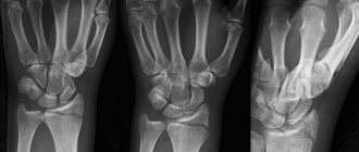
Any significant hand injury requires examination by a traumatologist and radiography. The patient may think that this is a bruise, but there may be a fracture, for example, of the scaphoid bone, which can create a lot of problems. A simple x-ray will not take much time, but the patient will already know for sure whether it is a bruise or a fracture.
Questioning the Patient: Most fractures are a direct result of trauma. Complains of pain, swelling, limitation of movements, subcutaneous hemorrhage in the injured arm. Typically, the patient will clearly acknowledge the injury and explain how he received it. Based on this, the doctor can already guess the diagnosis and location of the fracture. The patient may also complain of numbness if the blood supply is disrupted due to compression of the vessels by edema after serious extensive trauma (compartment syndrome or compartment syndrome).
Examination by a traumatologist: examination may reveal visible deformities if the fracture is clearly displaced. If the displacement is small or not at all, the anatomy of the hand may be completely normal. Local swelling and pain on palpation (touch) will be in the projection of the fracture. Possible decrease in grip strength.
Also, during the examination, the doctor pays attention to possible rotation of the fingers (rotation around an axis). You can evaluate this moment by bending your fingers into a fist. The fingers should be lined up without turning around and the nails should be parallel.
Instrumental examination methods:
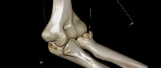
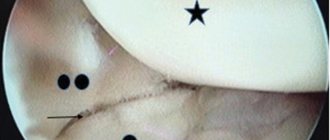
Radiography. To diagnose a fracture of the metacarpal bone(s), radiography is performed in three projections: direct (antero-posterior), lateral (sagittal) and oblique (3/4).
CT (computed tomography) is used in complex cases when the fracture is comminuted or intra-articular, as well as to confirm the diagnosis of fracture nonunion.