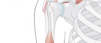
The head of the radius is located near the elbow joint. Its main function is to provide so-called rotational movements in the forearm (palm up - palm down).
A fracture of the radial head occurs mainly from a fall on the elbow. Pain appears in the area of the elbow joint along the outer surface; pain intensifies when trying to perform rotational movements in the forearm.
Fractures of the radial head are intra-articular fractures, and therefore require more attention compared to fractures of the diaphysis (middle third of the bone). And a lot depends on the person himself - the more disciplined the patient is in developing his arm, the better it will recover. If the fracture is displaced, then the sooner surgical treatment is performed after the injury, the greater the likelihood of complete restoration of limb function.
Anatomy of a radial head fracture
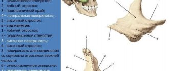
How does the elbow joint work? The radius articulates with the humerus and proximal ulna. This joint allows flexion and extension of the forearm, as well as pronation (turning the palm down) and supination (turning the palm up) of the forearm.
The head of the radius is covered with articular cartilage. This ensures sliding of the articular surface in two planes, which is extremely important for the elbow joint. Thus, articular fractures with post-traumatic arthrosis can lead to a mechanical obstacle to movement.
The head is also an important stabilizer of the elbow joint.
A fracture of the radial head, in addition to severe pain, is characterized by:
- significant decrease in elbow joint mobility, including passive and rotational movements,
- hemarthrosis,
- deformations of the outer surface of the elbow joint.
If a fracture of the head of the radial bone of the elbow joint is suspected, it is necessary to exclude the possibility of fracture-dislocation of the head of the radial bone, accompanied by a violation of the interosseous membrane. Therefore, if a fracture is suspected, adjacent joints should also be examined.
Most radial head fractures are isolated, but are sometimes accompanied by the following injuries:
- fracture of the coronoid process of the ulna
- rupture of the collateral ligament of the elbow joint
- interosseous membrane rupture
- Fracture-dislocation Goliazzi
Such an injury, which is also accompanied by a fracture of the head of the humerus, can occur with damage to the medial collateral ligaments and a fracture of the ulna bones with their shortening.
Symptoms
When the head is injured, the patient experiences the following symptoms:
- Pain syndrome. Sharp pain occurs in the upper part of the arm, which can radiate to the fingers and hand.
- It becomes difficult to move your hand. In some cases, this becomes impossible altogether.
- Pathological mobility of the damaged area. Thus, the elbow may move to the side, which is abnormal for the joint.
- A hematoma forms in the damaged area. Blood accumulates in the joint cavity, which leads to swelling. Hemarthrosis is very dangerous.
- Neurological symptoms result from nerve damage. In this case, the fingers become numb or there is a tingling sensation in them.
- Bleeding. Occurs with an open fracture.
Important! Symptoms become more pronounced with open fractures, when blood vessels, ligaments, muscles and nerve endings rupture.
The nature of the manifestations directly depends on the degree of injury. So, if the olecranon process also comes under attack, then pain occurs behind the elbow and can spread to the shoulder. With parallel injury to the neck of the radius, pain, on the contrary, occurs in front of the joint and radiates down the limb.
Diagnosis is carried out using various methods, including CT
Diagnosis of radius fractures
Diagnosis of an elbow injury begins with interviewing the patient. The doctor must find out what the mechanism of injury was. Assess whether there are visible deformities, swelling, crepitus of bone fragments (crunching), subcutaneous hemorrhage (if the fracture is 2 or more days old), i.e. symptoms characteristic of a fracture.
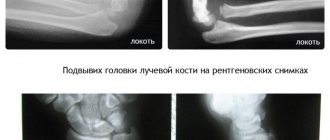
In almost all cases of injury to the elbow joint, radiography is performed. As a rule, in the frontal and lateral projections, although in the oblique projection the head of the radial bone is very well visualized. If the injury was minor and on radiographs we see the normal location of the head of the radial bone, then the diagnosis of a fracture is questioned.
Radial head fractures can often be nondisplaced and therefore easily missed on plain radiographs. Even if a fracture is not visible on an x-ray, this does not mean that it does not exist. After 7 days, control radiographs must be taken to rule out a fracture. It is during this time that resorption of the fracture site occurs and can be clearly seen in the photographs.
Intra-articular fractures are necessarily accompanied by hemorrhage into the joint, which can be determined by ultrasound.
Computed tomography (CT) and magnetic resonance imaging (MRI) are used in the diagnosis of complex radial head fractures and for preoperative and postoperative management.
Open and closed fracture
Based on damage to tissue and skin, the following types of fractures are distinguished:
- Closed type. The integrity of the skin is not compromised, and bone fragments do not come out. For the victim, this type of injury is the most favorable, since infection and contamination are excluded, and the likelihood of dangerous complications is reduced.
- Open type. A broken bone, moving, damages soft tissues and skin. There is direct contact of the damaged area with the external environment. In this case, pathogenic microorganisms enter the open wound along with dirt, significantly increasing the risk of infection.
Classification of radial head fractures
Historically, radial head fractures have been delineated according to the Mason classification, which distinguishes three types of injury:
1. Marginal fracture (without displacement or movement of fragments).
2. Marginal fracture (with displacement and movement of fragments).
3. Multicomminuted (in which the entire head of the radial bones is involved in the process).
(A fourth type was added to designate a fracture of the head with dislocation.) 4. Fracture accompanied by dislocation of the bones of the forearm.
There is also the Essex-Lopresti fracture, described in 1951. It is characterized by a comminuted fracture of the head of the radius, rupture of the distal radioulnar joint and dislocation of the head of the ulna towards the wrist.
And the so-called terrible triad of the elbow (dislocation of the bones of the forearm, fracture of the coronoid process and fracture of the head of the radius).
As a rule, only the first type of fractures does not require surgical intervention.
Complications
With such a fracture, various complications are possible:
- improper fusion of fragments;
- bone fusion of two fragments;
- wound infection;
- damage to blood vessels and nerves;
- stiffness due to frequent inactivity of the limb;
- osteoarthritis and arthritis;
- aching pain in the elbow area.
To avoid complications, it is necessary to follow all the instructions of the attending physician and not skip a single stage of therapy.
Similar:
- How long will it take to treat a hip fracture in old age?
- Methods of treatment and rehabilitation after a fracture of the tibial condyle
- Causes of a shoulder fracture, treatment and rehabilitation after injury, types of shoulder fracture
- List of consequences of hip fractures after injury, symptoms and principles of treatment
- How does aseptic necrosis of the talus manifest itself, and why is it dangerous?
- The mechanism of development of aseptic necrosis of the head of the hip joint, causes, types, stages, symptoms, methods of diagnosis and treatment
- Main symptoms, treatment methods for displaced acetabulum fracture
Treatment of radial head fractures
The main goals of therapy for this injury include:
- restoration of the possibility of rotational movement,
- restoration of the entire range of motion of the forearms and elbows,
- carrying out prevention of the possibility of early occurrence of arthrosis of the elbow joints.
Treatment tactics are based on the degree of displacement, the size of the fragments and the presence of an intra-articular component in fractures of the radial head. And it depends on the type of classification above.
Fractures of the head of the elbow without displacement are treated conservatively. For this type of treatment, immobilization is used with plaster, a plastic polymer bandage and a rigid orthosis.
Immobilization is carried out for a period of no more than 3 weeks in the position of bending the arm at the elbow joint at an angle of 100-110 degrees and supination (turning the palm upward) of the forearm at an angle of 45 degrees!!! The elbow joint is very “capricious” in relation to the duration of immobilization. So, according to research and personal experience, I assure you that it is catastrophically difficult to develop the elbow joint to full range of motion after being in a cast for more than 3 weeks.
After three weeks, the plaster or polymer bandage is cut off or replaced with a hinged piece to begin developing movements in the elbow joint.
Surgical treatment of radial head fractures is used when:
- an open fracture is observed,
- conservative treatment does not produce results,
- a segmental fracture is observed,
- there is a compound fracture,
- there is a fracture with the inability to move the elbow joint as a result of displacement,
- when Goleazzi is damaged.
Fixation of fragments or endoprosthetics of the radial head is usually carried out through the posterolateral approach (Kocher), between the extensor carpi ulnaris muscle and the anconeus muscle. In the position of pronation of the forearm with abduction and protection of the radial nerve during surgery.
This type of treatment uses:
- endoprosthetics,
- bone osteosynthesis,
- resection of the head of the radial bones,
- installation of Kirschner spokes.
Removal of the head of the radial bone occurs in severe fractures with the presence of many splinters and fragments. In this case, patients experience valgus instability and pain. However, this method is widely used in Russia due to the lack of experience in endoprosthesis replacement of the radial head by surgeons or, more often, due to the lack of endoprosthesis in a medical institution. Not many patients have the financial ability to purchase a prosthesis at their own expense.
Associated damage.
50% of these fractures are intra-articular and may be accompanied by damage to the distal radioulnar joint, in 40% of cases there is damage to the triangular cartilage, in 30% there is damage to the scapholunate ligament, in 15% of cases to the lunate-triquetral ligament.
Classification of radial fractures in a typical location.
Fernandez classification
| Type of fracture depending on the mechanism of injury | Stability (risk of secondary displacement in the cast) | Bias | Recommended treatment |
| Type 1 - simple extension | Stable, low risk of secondary displacement. | 1) Without displacement\ 2) dorsal displacement (Collis fracture) 3) Palmar displacement (Smith fracture) | 1) plaster 2) percutaneous fixation with pins 3) External fixator |
| Type 2, cutting off the articular surface | Unstable, secondary displacement always occurs | 1) palm 2) Combined | 1) open reposition. Internal fixation |
| Type 3 with compression of the articular surface | Can be either stable or unstable depending on the quality of the bone and the number of fragments | Various combinations | In the absence of significant displacement, it can be conservative. Open reduction and osteosynthesis. Alternatively – pins or External fixator. . |
| Type 4 – separation of ligamentous structures with dislocation of the wrist joint | Very rare. Unstable | various combinations | Closed or open elimination. External fixator. Fixation of bone fragments with pins/screws |
| Type 5 – combined, high-energy injury | Very rare Unstable | various combinations | Combined closed\open method 0000, |
And of course, everyone’s favorite AO classification.
The AO classification is very detailed; within each group, as a rule, there are 3 more subgroups, and ultimately it covers almost the entire possible variety of fractures of a given anatomical region.
Outpatient physicians often tend to simplify the approach to treatment and often do not even inform patients about possible complications and functional outcome. For this reason, initially extremely unstable fractures can be treated conservatively and heal in a deliberately incorrect position, which can cause the development of arthrosis and persistent limitation of limb function. When we are talking about an elderly patient with low functional demands, this may be acceptable and even desirable, so as not to expose the person to unnecessary operational risks in order to obtain a result that is not particularly needed in this category of patients. But when we are talking about young patients who have a long working life, playing sports and much more, the choice of tactics to obtain the optimal result becomes fundamentally important.
Good treatment results depend on many factors: 1) restoration of the articular surface 2) restoration of normal anatomical relationships 3) early movements in the joints of the hand and wrist joint.
Compliance with these principles can be achieved both conservatively and surgically, but conservative treatment has a number of limitations.
Indications for conservative treatment are extra-articular (extra-articular) fractures with shortening of no more than 5 mm and angular deformation of no more than 20 degrees. Such fractures can be stabilized in a plaster cast and are not characterized by secondary displacement. All other types of fractures (and they are the majority) are prone to secondary displacement in the cast, and multiple re-casting will not correct the situation. Of course, if the patient has firmly decided to refuse surgery despite possible complications, then treatment is carried out using the method of plaster immobilization.
Surgical methods include 1) closed reduction and percutaneous fixation with pins, 2) closed/minimally open reduction and osteosynthesis using external fixators, 3) open reduction and external osteosynthesis using plates and screws.
Indications for surgical treatment are contraindications for conservative treatment: shortening of more than 5 mm, angular deformation of more than 20 degrees, intra-articular fractures, comminuted fractures with defects of the volar or dorsal cortical plates.
Rehabilitation after a fracture
Immediately after the pain in the fracture area has decreased, doctors allow moderate movements in the elbow joint. Athletes are strictly prohibited from carrying out rehabilitation using excessive loads when developing a joint in order to avoid re-injury. Only after consolidation of the radial head fracture can development activities with moderate load be carried out.
Don't self-medicate!
Only a doctor can determine the diagnosis and prescribe the correct treatment. If you have any questions, you can call or ask a question by email.
| Treatment of radial head fractures | Price, rub |
| Osteosynthesis of radial head fractures | from 38 000 |
| Conduction anesthesia | from 3 000 |
| Dressing, suture removal | from 500 |
| Fracture of both forearm bones | To the list of articles | Clavicle fractures |
First aid
It is not recommended to transport or move the patient after injury. He needs complete rest. First of all, you need to call a doctor. If a person is in severe pain, he needs to be given a painkiller.
With an open fracture, tissue ruptures, which leads to bleeding. Bone fragments protrude from the wound. It is strictly forbidden to reset them. Only an experienced specialist can cope with such a task. Otherwise, the fragments may be incorrectly positioned and infection may enter the wound, which will significantly worsen the clinical picture. The only thing you need to do is put on a bandage.
A tourniquet is used if there is severe bleeding. In this case, it is worth paying special attention to his character. If the blood flows evenly and has a dark red tint, then a tourniquet is applied under the wound. In case of pulsating and bright bleeding, it is placed over the damaged area.
Important! When providing assistance, it is worth remembering the time of applying the tourniquet. After an hour it needs to be loosened a little. Otherwise, the hand will stop working.
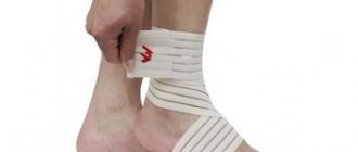
It is very important to immobilize the limb. A bandage or splint is used for this. It is better to entrust this procedure to an ambulance team. Incorrect movements can lead to complications.
General information
A radial head fracture is an intra-articular injury that can result from:
- falling on a straightened arm, elbow or hand;
- a strong blow to the elbow.
Sports injuries are usually caused by a fall on a straight arm. In everyday life, older women are often injured when they fall on their elbow. In this case, a closed fracture of the head of the left radial bone is diagnosed more often than damage to the bones of the right hand.
In patients with osteoporosis (a chronic disease characterized by a decrease in bone density), inflammatory diseases and oncological pathologies of the bones, and a severe lack of calcium and protein in the body, injury can occur even with a light force on the elbow. Such a fracture is pathological, and its root cause is a somatic disease.
Damage to the beam head can also occur in a traffic accident, as a result of an industrial accident, or after a fall from a height. In such situations, the injury is often combined with dislocations, bruises, and other fractures.