The list of the most severe and dangerous injuries includes a fracture of the femoral neck. The damage is characterized by a disruption of the integrity of the upper part of the femur, as a result of which the person may lose the ability to move and become bedridden. For older people, the injury can be fatal.
The femoral neck is the most vulnerable part of the bone, which experiences a high level of stress. With age and under the influence of a number of unfavorable factors, it triples its original strength. Therefore, even a minor impact can cause a fracture.
Given the seriousness of the problem, it is important to make a timely diagnosis and begin adequate treatment. A qualified approach ensures the restoration of motor functions and a return to a full life.
Symptoms of a hip fracture
To choose the right tactics for helping a patient, it is important to determine in a timely manner whether the femoral neck is broken or not. The presence of pathology is indicated by various signs, based on the analysis of which the presence of pathology can be suspected.
Typical symptoms of a hip fracture include:
- functional impairment: inability to move and walk;
- pain localized in the groin area when moving the leg and axial load (pressing the heel);
- rotation of the limb: at rest, the injured leg turns with the foot outward, while even with effort it is impossible to turn it inward;
- change in limb parameters: one leg becomes visibly shorter than the other;
- the appearance of a hematoma in the area of joint damage (the symptom appears after some time).
Initially, after receiving an injury, the patient may maintain motor activity, but gradually it is lost. The patient urgently requires medical attention, since without diagnosis and treatment, the likely outcome will be increased pain and the appearance of secondary displacement. A natural fight in this and a number of other cases becomes impossible, especially when it comes to hip fractures in older people.
Pain relief for amputation
In our clinic, hip amputation surgery is most often performed under epidural anesthesia. A thin catheter is inserted into the back through which a painkiller is injected. Epidural anesthesia will not only provide pain relief during the operation, but will also help provide adequate postoperative pain relief so that the patient does not have severe pain.
The operation is performed under general anesthesia in very rare cases when the general condition or characteristics of the spine do not allow spinal or epidural anesthesia.
It is mandatory to monitor the vital functions of the body during surgery and the postoperative period. To do this, monitoring equipment is connected to the patient, and a urinary catheter is installed.
Femoral neck fracture treatment
Modern treatment of a hip fracture improves favorable prognoses and increases the chances of recovery. The main task facing doctors is to restore the integrity of the bone. Important criteria for choosing treatment tactics are:
- patient’s age: a young body recovers faster and more efficiently than the body of old people;
- severity of injury and location of the fracture: the closer to the head, the more difficult;
- speed of seeking medical help;
- the presence of blood supply to damaged areas of bone tissue.
The practice of treating the femoral neck involves the use of conservative therapy and surgical intervention.
- Conservative treatment. The choice in its favor is possible in the presence of significant contraindications to surgery, as well as in case of a fracture of the femoral neck without displacement (mild form). As a rule, therapy requires long-term immobilization and rehabilitation treatment. In the case of elderly patients, conservative treatment tactics may be unsuccessful and lead to disability. This is due to the fact that with age, bone healing occurs extremely slowly (or does not occur at all).
- Surgical intervention. The operation helps the patient return to his usual life as quickly as possible. Surgical treatment includes:
- carrying out osteosynthesis: bone fragments are fastened together using metal pins and structures;
- endoprosthetics: complete replacement of a joint with a prosthesis with cemented or cementless implant fixation.
In both the first and second cases, the patient requires rehabilitation after treatment of the femoral neck. Its volume and duration are determined in accordance with the type of intervention, the severity of the body’s condition and other criteria that experts focus on.
Observation and early prosthetics
Our clinic actively prepares patients for prosthetics after amputation in our rehabilitation department. The main goal of rehabilitation measures is early activation of patients, lifting onto walkers and crutches, the fastest possible prosthetics and learning to walk on a prosthesis.
In the postoperative period, even before the sutures are removed, we send the patient for a consultation with a prosthetist, who gives recommendations on preparing the stump and discusses the situation with possible early temporary prosthetics. After receiving the prosthesis, we teach the patient to walk on it and monitor the reaction of the stump to the prosthesis.
Types of hip fracture
To classify types of femoral neck fractures, experts rely on various criteria. Depending on the type of disease, the choice of treatment tactics, prognosis and assessment of associated risks are made. For classification, the location of the injury, the direction of the fracture, the presence and degree of displacement are important.
Depending on the location of the fracture, there are:
- cervical: location – femoral neck;
- non-cervical: the damage is located at the junction of the neck to the bone;
- subcapital: fracture under the head of the bone (the most difficult option, high risk of developing necrosis).
The fracture can occur in a vertical or horizontal direction. If the crack is located vertically, the risk of displacement and other complications increases.
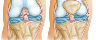
Depending on the type of displacement in a femoral neck fracture, the following can be distinguished:
- valgus fracture: the head of the bone moves upward and to the outer edge, which leads to an increase in the angle between the neck of the bone and its body;
- impacted fracture: one part of the damaged bone enters another;
- varus fracture: the head is displaced downward and inward, and the angle between the neck and the body of the bone decreases.
In older people, medial femoral neck fractures often occur, which belong to the category of intra-articular injuries. The cause of the pathology lies in the age-related decrease in blood supply to the joints and pelvic bones.
Blood and lymphatic vessels
Many vessels pass through the femoral part, each feeding specific organs and structures. The most important is the femoral artery (in Latin - a. femoralis). It continues the iliac vessel, descends along the anterior outer part of the thigh through the vascular lacuna into the popliteal cavity, where it transforms into the artery of the same name. In Scarp's triangle, the main vessel of the femur is covered only by connective tissue and skin. Other femoral arteries depart from it:
- superficial;
- deep;
- superficial epigastric;
- medial;
- lateral;
- perforating;
- external genitalia;
- descending knee
The femoral vein arises from the azygos popliteal vein and has about eight peripheral branches. One of them is the deep vein, which “works” on the back of the thigh. Also, large venous vessels pass medially and laterally and serve the corresponding parts of the upper limb. The superficial circulatory network is located directly under the skin.
In the femoral region there are large lymph nodes - superficial and deep inguinal. The first are located under the skin on a wide connective tissue element along the inguinal fold and on its anterolateral surface. You can really feel them with your fingers. The second ones are located deep in the thigh near the vein. The largest is located directly at the vascular lacuna.
Additional small lymph nodes are located singly and in groups in different femoral sections along the lymphatic vessels.
The latter also vary in depth. Superficial vessels go from the wall of the peritoneum and genital organs to the lymph nodes, and deep ones - from the lymphocapillaries of the muscles, joints, and bone structures. The lymph nodes of the femoral part connected by a vascular network form the inguinal lymphatic plexus. A complete diagram of the vessels can be seen in the photo.
What causes a femoral neck fracture?
The risk of hip fracture increases as the patient ages. In this regard, bone fragility increases, the intensity of bone tissue nutrition decreases, and joint wear occurs. That is why people over 50 years of age most often face the problem.
All it takes for a person to get injured is to turn awkwardly, trip or fall. A minimal impact is enough to cause a crack or fracture in the bone. Such a violent reaction of the body to a blow or fall is also explained by the influence of accompanying factors, which include:
- the presence of cancer pathologies in the body that cause weakness of bones and joints;
- physical inactivity and lack of physical activity;
- excess weight, causing additional stress on joints and limbs;
- deficiency of nutrients and vitamins, which develops against the background of dietary restrictions, diet, and fasting;
- menopause, which causes changes in hormonal balance and a decrease in estrogen levels;
- neurological diseases;
- rheumatoid pathologies;
- vascular diseases that interfere with adequate nutrition of bones and joints.
With age, health deteriorates, natural wear and tear takes its toll, which aggravates the severity of the injury and its consequences. Older people require more time for rehabilitation, which requires more strength and vitality from them.
Advantages of our clinic
At the Innovative Vascular Center, unique operations are performed to restore blood circulation in the limb during gangrene, so we rarely perform large amputations at the level of the upper third of the thigh. The decision to amputate can only be made by a council of doctors after a detailed study of the situation with blood circulation and the viability of the limb segment. If a limb segment cannot be saved, we restore blood flow and perform amputations at the lowest possible level.
In our clinic, the preferred surgical method is Gritty femoral amputation with preservation of the patella. Such amputations allow the patient to use lightweight prostheses and have a better quality of life. The supporting stump according to Gritty is good because the skin of the stump is not injured by bone sawdust, bedsores and trophic ulcers never occur. The skin over the patella is the strongest and densest. The patient can lean on the Gritty stump without the risk of damage. The quality of this stump allows for maximum rehabilitation and mobility of the patient. In other clinics, such amputation is used very rarely due to the reluctance to complicate the operation itself, or due to the lack of experience in reconstructive plastic surgery.
What is the danger of a hip fracture?
A bone injury is a serious test for a person who is deprived of motor activity for a long period, suffers pain and is forced to depend on outside help. But complications after a hip fracture can worsen the condition even more. These include:
- necrosis of the femoral head, causing tissue necrosis;
- vein thrombosis, which occurs against the background of blood stagnation with prolonged absence of physical activity;
- pneumonia caused by congestion (occurs in patients who lie for a long time and lack of preventive measures);
- the appearance of a false joint caused by the formation of a movable connection of individual pieces of bone;
- trauma from inserting a screw into the bone, blood vessels and nerves may be damaged, and a depression may appear on the leg;
- damage to the connecting structure, the appearance of a tent;
- concomitant tissue or joint infection;
- the development of degenerative changes causing pain and problems with movement.
In each individual case, the patient requires medical assistance. The volume of intervention and treatment tactics are selected taking into account the individual characteristics of the patient and the severity of the pathology.
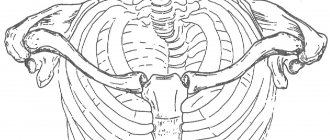
Anatomy of the human hip
The thigh, called femur in Latin, is the part of the legs located closer to the body.
It consists of bone structures, muscle masses, ligaments, and nerve branches. The tissues penetrate the vessels of the circulatory and lymphatic systems. The topographic anatomy of the human thigh includes the following areas:
- the hip joint, formed by the acetabulum of the pelvic bone and the femoral head;
- the front part of the thigh, located in front of the leg from the pubic tubercle to the patella;
- the posterior region, which starts from the transverse fold of the buttock and ends six centimeters above the knee bend;
- the area above the knee is five centimeters above the kneecap.
The internal structure of each area of the human thigh is different, but all its elements are interconnected, allowing for a variety of movements and promoting upright posture. Externally, this area of the body is protected by the skin, under which there is a layer of fatty tissue. The epidermis inside the thigh is soft and mobile, while on the outside it is elastic and dense.
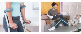
Rehabilitation after a hip fracture in a boarding house
In order to more effectively cope with the consequences of a hip fracture, restore strength and return to an active life, the patient requires competent and comprehensive rehabilitation after a hip fracture. The program of activities is aimed at accelerating the process of bone fusion, as well as restoration of motor functions.
As a rule, during the rehabilitation period the following is applied to patients:
- Massage. The purpose of the massage is to improve blood circulation in damaged tissues, as well as to activate the outflow of lymphatic fluid. With the help of regular massage it is possible to improve tissue trophism, reduce the risk of bedsores and congestive pneumonia in bedridden patients. The procedures help maintain the tone of muscle tissue, preventing its atrophy. Thanks to professional procedures, you can improve your breathing and relieve stress on your cardiovascular system.
- Physiotherapy. A set of exercises for patients after a hip fracture helps reduce the risk of complications, muscle atrophy, and osteoporosis. Subsequently, it is easier for a person to return to active movements.
- Physiotherapy. In accordance with the indications, the set of procedures also contributes to the overall strengthening and healing of the body.
In a private boarding house, care for this category of patients is provided at the highest level:
- the comfort of staying in conditions that meet the severity of the current condition is ensured;
- qualified sanitary care is provided;
- a full range of restoration measures is provided;
- psychological comfort is created.
Preparing for limb amputation
In most cases, the operation is performed for urgent indications and preoperative preparation is limited in time. An assessment of blood circulation in the limb as a whole and at the intended level of femoral truncation is always necessary. To do this, an ultrasound scan of the arteries is performed, and, if necessary, angiography.
The main goal of preoperative preparation is to diagnose the exact criteria for the need for amputation, study indicators of vital body functions (laboratory parameters of blood and urine, the state of heart and lung function, the presence of risks of thrombosis and bleeding).
Patient preparation consists of bowel cleansing, bladder catheterization, installation of an epidural catheter for pain relief during surgery and the postoperative period, and shaving the surgical field. In order to relieve emotional stress, the patient is premedicated with sedatives.
Psychological support
An injury that causes a fracture of the femoral neck seriously affects one’s well-being, deprives one of the ability to walk, and makes one dependent. All this negatively affects the psychological state. Depression, depression, anxiety and worries require contacting a psychotherapist. A professional specialist will help you overcome the problem and restore a favorable emotional background.
At the boarding house for the elderly there are conditions for providing modern psychological assistance to old people who are faced with disaster. The work of specialists is included in the complex of care activities.