Fractures of the talus
There are fractures of the neck, body, head and posterior process of the talus. Often these fractures are combined not only with each other, but also with dislocations or individual parts of the talus, or with dislocations of the foot in the subtalar joint.
- Mechanism of damage
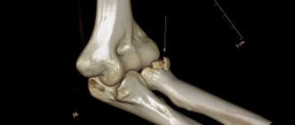
Two main mechanisms predominate in fractures of the talus. The first factor is the impact on the plantar surface of the foot. The second factor is twisting. Often both of these factors are present and complement each other. Most often, these fractures occur in motorcyclists. A fracture occurs when the motorcyclist’s foot is on the footrest - an accident, a strong blow from this particular footboard on the plantar surface of the foot. Car drivers have approximately the same mechanism, only the pedal serves as a footrest. These fractures also occur during sports, falling from a height, or an unsuccessful jump. The patient experiences severe pain. Sometimes there is a visible deformity of the foot, especially in cases of fracture-dislocations. Severe fractures are often open. X-rays and computed tomography (CT) are used for diagnosis.
- Main types of talus fractures
All, with few exceptions, fractures of the talus are intra-articular. Moreover, there are no muscles attached to the talus. This means that the nutrition of the cartilage (up to 70% of the body of the talus is covered with cartilage) largely depends on the synovial fluid in which the talus literally “floats”. Early movements are extremely important for the circulation of synovial fluid. This makes surgical treatment the main treatment for fractures of the talus. Prolonged immobilization is not acceptable here. Only surgery allows you to begin early movements, and therefore ensure normal nutrition of the talus.
There are fractures of the neck, body, head and posterior process of the talus. Often these fractures are combined not only with each other, but also with dislocations or individual parts of the talus, or with dislocations of the foot in the subtalar joint.
- Treatment of talus fractures
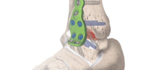
The main treatment for talus fractures is surgery. Even non-displaced fractures should be fixed with screws through skin punctures to allow early movement of the foot.
- Rehabilitation
Since the nutrition of the talar cartilage depends on the circulation of intra-articular synovial fluid, early movement is extremely important for the talus. Full rehabilitation is necessary. It’s hard to overestimate here. For each patient, rehabilitation specialists at the Ilyinskaya Hospital draw up an individual program that takes into account his needs and individual characteristics.
Medical reference books
Fractures of the talus
Epidemiology
Fractures of the talus are the second most common among fractures of the tarsal bones and occupy 2% of the total structure of fractures of the lower limb and 5-7% of injuries to the bones of the foot. Clinical anatomy (
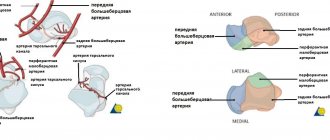
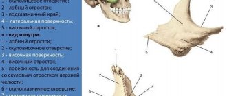
) - The body of the talus is covered on top with articular cartilage, through which the load of body weight is transmitted. — The front surface is wider than the back, which gives stability to the ankle joint. — Externally and internally along the lateral surfaces, the talus articulates with the lateral and internal malleolus, respectively. — The inferior surface forms an articulation with the posterior articular facet of the calcaneus. — The neck of the talus deviates medially by 15-25°. This is the most vulnerable place of the bone for fracture. — The head of the talus is covered by an articular surface for connection with the scaphoid. The tendons extend from it downwards. From the sustentaculum tali - in the posteroinferior direction, the deltoid ligament - medially. — Anatomically, 2 processes are distinguished: lateral (wedge-shaped, articulates with the posterior facet of the lower internal surface, and with the external ankle - the upper external surface), posterior (has 2 tubercles - internal and external, separated by a groove for the long flexor tendon of the 1st toe) cm . . — The triangular bone as an independent formation is present in 50%. It is formed by separating the center of ossification of the lateral tubercle of the posterior process of the talus. — About 60% of the surface of the talus is covered with articular cartilage. There are no attachment points for muscle tendons. Blood supply depends on connective tissue structures that are located close to the bone. Therefore, damage to the capsule can lead to avascular necrosis of the bone. — Blood supply is carried out with the help of: 1) arteries of the tarsal sinus (a. Peroneal, a. dorsalis pedis); 2) arteries of the tarsal canal (a. tibialis posterior); 3) deltoid artery (a. tibialis posterior); 4) feeding vessels of capsuloligamentous structures (see.
). Consequently, this arrangement of the supply arteries causes a high risk of aseptic necrosis of the talus when it is damaged in the neck area, especially with concomitant subluxation in the subtalar (50%) and ankle joint (95%).
1) – Medial projection of the talus (the zones presented below are marked with letters: A, B and C). A 1 – Perforating calcaneal artery; 2 – Lateral metatarsal artery; 3 – Dorsal artery of the foot; 4 – Anastomosing arteries of the deltoid branch of the dorsalis pedis artery; 5 – Arteries of the talus sinus. B 1 – Branches of the dorsal artery of the foot; 2 – Branches of the talus sinus; 3 – Arteries of the metatarsal canal of the foot; 4 – Deltoid branches of the dorsal artery of the foot. C 1 – Branches of the talus sinus; 2 – Arteries of the metatarsal canal of the foot; 3 – Deltoid branches of the dorsal artery of the foot; 4 – Posterior tibial artery.
Mechanogenesis of damage
— Very often, injuries occur as a result of a motorcycle injury or a fall from a height with excessive dorsiflexion of the foot at the ankle joint. — Fractures of the neck of the talus occur as a result of depression of the anterior edge of the distal metaepiphysis of the tibia.
Diagnostics
Clinical diagnosis
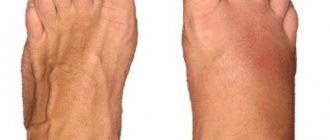
— The patient complains of pain in the foot. — Movements in the foot are painful, and crepitus may be detected. - Widespread swelling in the foot area. — Often combined injuries of the ankle and foot are detected.
X-ray diagnostics
Anteroposterior, three-quarter, lateral projections of the ankle joint are made, as well as anteroposterior, lateral and oblique projections of the foot according to indications.
The Canale view is performed to visualize the neck of the talus. It is performed in the position of maximum equinus of the foot with the anteroposterior direction of the beam and pronation of the foot 15°. In this case, the beam from the X-ray tube is directed cranially, with a deviation of 15° from the vertical. Computed tomography
is an additional imaging method. Produced in axial, frontal and sagittal projections. Helps visualize the features of fragment displacement in complex injuries of articular surfaces.
Magnetic resonance imaging and osteoscintigraphy
These studies are used to visualize possible hidden injuries to the talus.
Classification of fractures
Anatomical classification
— Fracture of the external process. — Fracture of the posterior process. — Fracture of the head of the talus. — Fracture of the body of the talus. — Fracture of the neck of the talus.
Classification of talus injuries according to Marti and Weber
(cm.
) - Type 1 - peripheral fracture (scale type).
— Type 2 – fracture of the central part without displacement. — Type 3 – fracture of the central part with displacement. — Type 4 – additional dislocation of the talus. Classification of injuries to the neck of the talus according to Hawkins
(see.
) — Type 1 – non-displaced fractures. — Type 2 – fractures with concomitant subluxation in the subtalar joint or with displacement. — Type 3 – fractures with concomitant subluxation in the subtalar joint and displacement. — Type 4 – type 3 injuries with additional subluxation in the talonavicular joint or with displacement.
Treatment
Fractures of the head and neck of the talus Non-displaced fractures (type 1)
Fractures that may appear to be non-displaced fractures or not detectable at all after analysis of plain radiographs may turn out to be comminuted or intra-articular according to CT examination.
Fractures with confirmed absence of displacement of the fragments and no signs of incongruity in the subtalar joint are classified as type 1. Treatment includes fixation in a short plaster cast for 8-12 weeks, with the absence of axial load on the limb for 6 weeks until clinical and radiological signs of union appear. Displaced Fractures (Types 2-4)
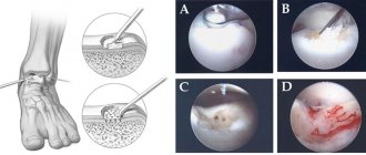
Closed reduction should be attempted with immediate open reduction and internal fixation for all open injuries or unreduced fractures. If anatomical reduction is achieved, which is confirmed by CT examination, further patient management is carried out as for non-displaced fractures. With open reduction, all major fragments must be preserved.
Accesses
- Anterointernal.
This approach can be extended from a dosed capsulotomy to a wide approach with an ankle osteotomy. Produced somewhat medially from the tendon m. tibialis anterior. Allows visualization of the neck and head of the talus. Implies careful surgical technique, because there is a risk of iatrogenic damage n. and v. saphenus and deltoid artery. — Posterior external – for access to the posterior process and body of the bone. Produced between m. peroneus brevis and m. flexor hallucis longus. It is important to bypass n. suralis. — Anterolateral - access allows you to visualize the sinus tarsi, the outer part of the neck of the bone and the subtalar joint. This may cause damage to the tarsal sinus artery. — The combination of anterior-external and anterior-internal access allows maximum visualization of the neck of the talus. Internal fixation of the fragments is carried out using 2 lag screws or special “headless” screws drawn perpendicular to the fracture line. The screws can be inserted either antegrade or retrograde. Insertion of the screw in the posterior-anterior direction is more justified from a biomechanical point of view. In areas with a bone tissue defect, bone grafting should be performed, and if there are a large number of fragments, in addition to bone grafting, a support plate should be installed. Postoperative management includes fixation with a short plaster cast for 8-12 weeks, with no axial load on the limb. Hawkins sign.
The presence of subchondrol osteopenia of the talus on anteroposterior radiography at 6-8 weeks is a sign of the viability of the talus.
Fracture of the external process
is an intra-articular injury to the subtalar and ankle joint.
Formed by excessive dorsiflexion and inversion of the foot. Posterior process fracture
This injury involves more than 25% of the articular surface, including the internal and external tuberosities.
This injury results from foot inversion or forced flexion and direct trauma. Fracture of the head of the talus
The injury results from plantar flexion and longitudinal compression along the axis of the forefoot.
It is often comminuted in nature and is accompanied by damage to the scaphoid and talonavicular joint. Dislocations in the subtalar joint
(see.
,
) With these injuries, the calcaneus, cuboid, navicular bones and the entire forefoot are displaced in relation to the talus. Dislocations are divided into medial (most common), lateral, anterior and posterior. Reduction of the dislocation is carried out with adequate pain relief in the position of flexion of the knee joint and with traction along the axis of the foot. It is especially important to achieve unblocking of the heel bone. In internal dislocations, the head of the talus is captured by the capsule of the talonavicular joint, the retinaculum extensorum and the short extensors of the toes. With external dislocations, the tendon of the tibialis posterior muscle may be inserted between the bones. In this case, closed reduction will not be successful; open reduction is indicated.
Dislocation of the talus
Complete dislocation of the talus is a fairly rare injury. Basically, dislocation is accompanied by damage to the skin. Therefore, treatment is aimed at restoring the integumentary soft tissues with open reposition of the talus. There remains a high risk of complications in the form of infectious contamination of the wound, aseptic necrosis, and post-traumatic arthritis.
Anatomy and significance of the talus
The talus is not one of those bones that can be removed from the skeleton, used to create, for example, Eve and continue to live a completely comfortable life.
The importance of this small bone, 70% covered with cartilage and not attached to any muscle, cannot be overestimated.
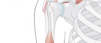
Located between the tibia and ankle, it practically takes on all the static and dynamic load from the body, redistributing it to the feet.
In the talus there are such functional zones as:
- Body;
- Head;
- Neck;
- Posterior process.
More often than others, a fracture of the neck or body is diagnosed; a fracture of the posterior process of the talus occurs much less often.
The head of the talus comes into contact with the scaphoid bone, its lower part connects to the calcaneus, and the body is clasped on both sides by the tibia and fibula. On the posterior process, two tubercles protrude - lateral and medial, separated by a tendon.
Contrary to the opinion of some experts, the talus is quite well supplied with blood, thanks to three arteries: the posterior tibial, anterior tibial and peroneal.
Mechanisms and causes of fractures
The cause of such a serious injury is excessive sudden loads in the ankle area, which are provoked by:
- Awkward movement during sports, ballet and similar exercises.
- Falling from a height;
- Impact of a heavy object on the lower leg.
A strong arch of the foot in the instep (as in a ballet position on the toes) can lead to a fracture of the neck, and reverse bending, exceeding the safety margin, can lead to a fracture of the posterior process. If such a bend is accompanied by a turn, then a fracture of the outer process is possible.
With a vertical impact, the bone is pinched between the tibia and calcaneus, and a compression fracture of the talus occurs with the formation of fragments.
In addition, severe dorsiextension plus axial loading or severe plantar flexion often results in dislocation or displacement of the body of the talus.
Treatment methods
If you suspect a fracture of the talus, you should immediately contact an orthopedist or surgeon.
He will conduct an examination, make a diagnosis and prescribe appropriate treatment. Depending on the nature of the fracture, the following treatment methods may be used:
Immobilization
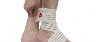
When the talus bone of the foot has received a mild fracture, without displacement, a polymer or plaster cast with an instep support in the sole of the “boot” is used as treatment, which remains on the patient’s foot for at least 6 weeks, without axial load, followed by exercise therapy.
In this case, it is necessary to take painkillers and ensure that the injured leg is in an elevated position to avoid the development of edema. The effectiveness of conservative treatment is 45-50%. Upon completion of the rehabilitation program, full functionality of the joint is restored starting from the third month, depending on age and other individual characteristics of the body.
Closed reduction
This is a medical manipulation of matching parts of a broken bone without disturbing the soft tissues surrounding the joint. The procedure is very painful and is performed only after intraosseous anesthesia.
The victim is placed on his stomach. The orthopedist bends the patient's leg at the knee joint, stretching the heel with one hand, bending the foot with the other to close the displacement, and then applies an immobilizing bandage. After seven weeks, the plaster cast is changed to a new one when the foot is bent at an angle of 90º.
The patient wears the bandage for up to four months from the date of application. After this period, the plaster is removed, and if control images are satisfactory, a rehabilitation program is prescribed.
Open reduction and osteosynthesis
Surgical intervention to detect and eliminate traumatic changes in bones and soft tissues.
First aid
Emergency care for a suspected fracture of the talus is no different from the same measures for any other fracture:
- Give the victim pain relief: Analgin, Ibuprofen, Ketorol, Nimesil (tablets or injections).
- Lay or sit so that the injured limb is not subjected to further stress.
- Call an ambulance.
- If the nature of the injury allows, remove shoes, socks and apply a fixing bandage.
- If open wounds are found, treat them with an antiseptic before applying a bandage.
- Apply cold to the injured area, controlling the time (if it is ice crushed in a plastic bag, remove it for 2-3 minutes every 10 minutes) to avoid frostbite.
- If necessary, independently, as quickly and carefully as possible, deliver the patient to the nearest medical facility.
You should know that if you delay contacting a doctor for a fracture of the talus, the consequences can be very undesirable, from prolonged treatment and rehabilitation to amputation of part of the leg.
Recovery period
The postoperative period can be burdened by various complications that develop as a result of:
- Poor quality of closed reduction;
- Poor quality of osteosynthesis;
- Injury to systems and tissues during access formation;
- Serious disturbances of innervation and blood circulation inside the joint;
- Inconsistency of loads and untimeliness of recovery programs.
The following have a great therapeutic effect:
- Exercise therapy complexes;
- Aquatherapy;
- Massotherapy;
- Various physiotherapy procedures.
However, an individual set of procedures for each individual case is drawn up by the attending physician, taking into account medical history, age, gender characteristics and other nuances.
During the rehabilitation period, you should not force an increase in loads, increasing them gradually in a safe manner. Also, do not forget about dispensary registration and control x-rays at least once a month.
Symptoms and other diagnostic methods
Symptoms of talus fractures are often similar to other injuries in this area, such as sprains:
- Swelling and pain in the ankle area;
- Severe pain when trying to stand up or moving your thumb;
- If there is displacement, visual deformation of the ankle;
- Jamming and crepitation (crunching) of fragments upon palpation.