The zygomatic bone is one of the paired elements of the skull structure, consisting of dense spongy-type plates. The fragility of these areas causes a high probability of fracture under active mechanical stress, the consequences of which depend on the complexity of the deformation. Simple cases are characterized by the absence of displacement of the fragments, as well as the preservation of the bone structure against the background of damage to the arch itself. However, any damage of this kind requires medical intervention, excluding negative consequences for the health of patients.
Skeletal structure of the cheekbone
It conventionally divides the skull into the brain and facial sections. Its shape resembles an irregular quadrangle with three planes and two processes. Due to its anatomical location, the cheekbone is bordered by four bones. These include the frontal, temporal, sphenoid, and upper jaw.
The frontal process of the zygomatic bone connects with the zygomatic branch, which is localized on the frontal bone. When they are connected, a rough seam with teeth located on it is formed.
The temporal process is located posteriorly, forming the zygomatic arch with the bone of the same name.
The structure of this department is distinguished by the presence of three planes, which include the lateral, orbital and temporal. The orbital region has a smooth surface and is part of the orbit. The orbital-zygomatic foramen is located on it. The temporal plane is involved in the formation of the fossa of the same name; on it you can find the zygomatic - temporal foramen.
The lateral surface of this bone has a convex shape.
The zygomatic bone performs several important functions, such as:
- Redistribution of workload from other departments.
- Participation in the formation of the orbit, which helps hold the eyeball in the desired position.
- Formation of the external shape of the face.
Facial bone fractures
The facial part of the skull has a complex structure. It consists of the frontal bone, zygomatic, orbital bones, nasal, maxillary and mandibular and other bones. Some of them are located deeper in the facial structure. Muscles that support the processes of chewing, swallowing and speech are attached to these bones.
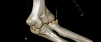
One of the most common facial fractures is a nasal fracture. Injury to other bones may also occur. One or several bones may be broken. Multiple fractures most often occur as a result of a car or other accident. Fractures can be unilateral (occur on one side of the face) or bilateral (occur on both sides of the face). Below you can see in the photo the fractures of the facial bones.
Classification and signs of a cheekbone fracture
In clinical practice, there are several classifications of cheekbone fractures. For the division, the localization of bone damage is taken into account, as well as the duration of its onset and accompanying manifestations.
A fracture of the cheek bone is divided into damage:
- With signs of displacement.
- No displacement of fragments.
- With a violation of the integrity of the sinus in the upper jaw.
A fracture in the area of the zygomatic arch is divided into:
- Damage with displacement.
- Damage without displacement.
Depending on the time when the injury occurred, the fracture may be:
- Fresh. In this case, no more than 10 days pass from the incident.
- Obsolete. From the moment of fracture it takes from 10 days to a month.
- Incorrectly fused. More than a month has passed since the impact of the injury.
According to the mechanism of bone damage, it can be:
- Open.
- Closed.
- Linear.
- Splintered.
Symptoms of a cheekbone fracture vary widely, depending on the severity of the injury and the involvement of nearby organs. Common symptoms include:
- Development of unbearable pain that occurs at rest and intensifies when trying to open the mouth.
- Impaired jaw mobility.
- External changes in the skull associated with displacement of fragments and damage to soft tissues. The face loses its asymmetry, tissue swelling increases, and abrasions with bleeding are noted.
- Impaired skin sensitivity in the area of the lower eyelid, cheekbone, and nasal wing. These symptoms are caused by damage to the infraorbital nerve. Sensitivity may decrease or disappear completely.
- Bleeding from the nasal cavity.
- Double vision.
- Feeling the bony protrusion during palpation of the cheekbone.
- The appearance of Purcher's Syndrome, which develops two days after the injury. The disease is accompanied by rapid visual impairment caused by retinal damage in the form of detachment or optic nerve atrophy.
- Growth of hematoma in the area of the eye sockets, cheeks, and lower jaw. With continued bleeding, the hematoma grows and after a few days can spread to the neck.
Rehabilitation
How long it will take for a fracture to heal - this period depends on its nature and severity. With conservative treatment, consolidation occurs within 3 weeks; after surgery, it may take more than a month.
Throughout this period, rehabilitation is carried out, which is aimed at accelerating consolidation, restoring function and eliminating cosmetic disorders. It includes electrical procedures, phototherapy, and magnetic therapy.
After the fixation is removed, the diet is expanded; chewing gum is recommended to develop muscles. A massage is prescribed with ointments that improve microcirculation to restore blood circulation and nerve sensitivity, and eliminate facial numbness.
Diagnosis of the zygomatic bone
Diagnosis of a cheekbone fracture begins with examining the patient and clarifying complaints. The doctor clarifies the mechanism of injury, as well as the conditions of its occurrence, the dynamics of the pathological process and methods of providing assistance before the arrival of a medical professional.
During the examination, the condition of the skin is assessed, their color is determined, as well as the presence of soft tissue damage. During palpation, the presence of bone fragments is determined by crunching sounds (crepitus), as well as the sensitivity of the surrounding tissues and the degree of swelling. Additional methods used:
- X-ray of the skull. The study is performed in several projections, which makes it possible to determine the condition of the zygomatic bone and its processes. For minor damage, the method may not be informative enough, since it does not have high accuracy.
- Magnetic resonance or computed tomography. These diagnostic methods are the most accurate, as they can detect minor changes in the structure of bone tissue.
First aid for suspected fracture Fractures of the zygomatic bone and arch require emergency care in the early stages after injury.
Important! Prompt assistance significantly reduces the risk of complications and facilitates the provision of qualified assistance.
The first measures are prescribed:
- Applying cold. When using improvised cold objects, they must be wrapped in a clean cloth or bandage. Application is carried out for 10-15 minutes with a break of 10 minutes.
- Applying a sterile bandage to the damaged surface. If there are foreign objects or fragments, it is prohibited to remove fragments yourself. In order to reduce the risk of infection, apply a sterile napkin.
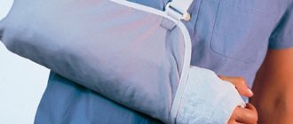
- Fixation of the jaw. In case of severe pain that occurs during conversation or movement, the lower jaw is covered with a bandage attached to the back of the head. To do this, you can use a bandage or any available means, such as belts or belts.
- Stop bleeding. Damage to blood vessels may be accompanied by heavy bleeding, which is stopped by the patient himself by squeezing the artery or vein at the site of the breach.
Classification
The classification according to the location of bone injuries is already discussed below. What other basis for dividing into types of fractures exist? By nature, open and closed fractures are distinguished. Most often among the open ones there are fractures of the bones of the dentition. The remaining local injuries are often closed in nature, unless they are injuries from a sharp object or firearm. Since the risk of injuries to the lower jaw is much higher, there is a separate classification for them:
- Fracture of the mental body;
- Angle;
- Base of the condylar process;
- Body of the condylar process;
- Bases of the coronoid process.
According to the mechanism of occurrence there are:
- Mechanical. In case of falls, impacts and other external factors.
- Firearms. The wounds are deep, open, with a high degree of severity and an increased risk of complications.
Treatment
Treatment of a cheekbone fracture is carried out using two main methods. These are surgical and conservative methods. Their selection is carried out depending on the type of fracture site, as well as what symptoms are identified and whether neighboring organs are involved in the process. It also takes into account when the injury was received and the dynamics of well-being.
If the injury is mild and there are no signs of displacement of fragments, conservative methods are prescribed.
Surgical treatment is indicated in cases where there is displacement of bone fragments or neighboring areas are involved in the pathological process. Currently, a large number of operational access techniques are used.
Types of fractures depending on the type of damage
This classification helps to assess the global scale of injuries, develop diagnostic and treatment strategies, and also helps determine the duration of recovery and possible risks. What types are distinguished:
- Complete and incomplete. Incomplete ones are otherwise called cracks and are easier to treat.
- According to the multiplicity of manifestations. Singles, doubles and so on. This parameter reflects the amount of damage inflicted on one bone.
- Single-sided and double-sided. The lateral surface of the face on which the damage is localized. If the injury is within two parts of the face, then it is a bilateral fracture.
- The direction is oblique and longitudinal.
- By offset. There are fractures with displacement, without it, and with the formation of fragments, which also tend to shift.
Methods of surgical treatment
It is necessary to treat with the help of operations a zygomaticomaxillary fracture or a fracture of a displaced bone with damage to the walls of the maxillary sinus. Surgical treatment is carried out after the patient has been fully prepared and the required scope of examination has been completed. This type of treatment is performed in a hospital setting in the department of maxillofacial surgery.
Malarchuk Khodorovich method
A similar technique is used in surgical practice in the treatment of recent fractures, as well as long-term injuries. The procedure involves the application of a special fixation device, which is fixed at the point of support on the parietal bone. After it is completed, there is no need to use more complex surgical techniques, which involve realigning the zygomatic bone and its arches with subsequent application of a bone suture. The positive effect after using the Malarchuk Khodorovich technique is achieved in more than 90%.
Keen method
The method of surgical intervention is one of the most technically complex. It is indicated for the treatment of patients with a fracture and separation of the cheekbone from the upper jaw. And also for injuries in the area of the temporal and frontal bones. The technique consists of dissecting the mucous membrane in the area where the transitional fold to the alveolar ridge is formed. After the bones are exposed, they are pulled upward and outward using a Karapetyan elevator, thereby fixing the fragments.
Dubov method
This type of surgery is indicated for fractures that result in damage to the maxillary sinus. The technique consists of making an incision in the area of the upper areolar fold of the mouth, followed by a maxillary sinusotomy. After opening the sinus, its cavity is leveled, washed and tamponed with cotton wool.
Cotton wool is moistened with iodoform solution. It must be administered through the nasal passage. After two weeks, the turunda is removed, as the fusion of fragments begins.
Kazanyan method
The extraoral approach technique is used in cases where the patient is diagnosed with a complex fracture. With this type of injury, the bone cannot support itself. An incision is made under the eye socket, as well as in the area of the cheek bone. After exposing the bones, several holes are made through which the wire is subsequently pulled. Subsequently, the wire is pulled and the bone fragments are connected. The wire is brought out and fixed with a loop and hook. These elements are attached to the rod of the headband-cap.
Limberg method
This method is indicated for trauma to the cheekbone with minor damage to the sinuses. In clinical practice, this technique is often used. To perform it, the patient must be laid on a flat surface on the side opposite to the fracture. A single-tooth hook is used to puncture the skin, after which it is inserted in a horizontal position under the displaced part of the cheekbone. After fixing the fragments, a characteristic click is heard, while the doctor performs movements in the opposite direction from the shift. Due to the fact that the hook is inserted through the skin, antibiotics are required.
Duchant method
A similar method of fixation is used for minor fractures. To perform this technique, forceps with cheeks with sharp teeth are used. The bone is fixed by making a puncture in the skin and picking up the displaced bone with forceps. It is returned to its place maintaining its natural position.
Nasal bone fractures
This type is the most common. The nasal bone consists of two thin bones. Breaking the nasal bones requires less force than breaking other bones because they are quite thin. When a fracture occurs, the nose usually looks deformed and pain appears. Swelling may make it difficult to assess the injury. Nosebleeds and bruising around the nose are common symptoms with this injury.
Conservative treatment
Conservative treatment methods are needed in cases where a cheekbone fracture is detected without displacement or damage to adjacent bone structures. In addition, treatment is prescribed in accordance with the patient’s well-being, the presence of concomitant injuries or somatic diseases.
The patient is advised to remain in bed. In this case, nutrition should be gentle with the exception of eating solid food. Food must be prepared in crushed form, which will avoid subsequent tissue trauma.
Medicines used:
- Non-steroidal anti-inflammatory drugs. They reduce the severity of swelling and inflammation of tissues, and also reduce pain. The most commonly used are Ibuprofen or Ketorol.
- Narcotic analgesics. This group of drugs is indicated for severe pain.
- Antibiotics. Agents with antibacterial activity are indicated for the prevention of subsequent suppurative processes. The average duration of treatment is 7-14 days. In this case, it is preferable to use drugs with a broad spectrum of antimicrobial action.
First aid
In a situation with a serious injury, the people around him must provide first aid to the victim. Medical recommendations exclude independent adjustment of individual parts of the skull or contact with open wounds. The main tasks are to create conditions of complete rest, eliminate re-injury, try to stop the bleeding and call an ambulance team.
In case of acute pain, an injection of an anesthetic is allowed. To stop continuous bleeding, it is necessary to press down the artery. In cases where the wound is small, you can treat the area with hydrogen peroxide, as well as ensure careful application of cold.
How long does it take to grow together and healing time?
The duration of tissue healing in a fracture of the zygomatic bone is strictly individual. This is due to the severity of the fracture, concomitant pathologies, duration of first aid, as well as complications. The age of the patient must also be taken into account. This is due to the fact that at a young age the rate of fusion is faster due to the composition of the bone tissue.
The average healing time for a non-displaced zygomatic bone fracture is 3 weeks.
If fractures of the cheekbone and arch with displacement of bone fragments are detected, when the patient requires medical assistance with surgical intervention, the healing time increases. It can be two months.
Causes
The causes of injury can be countless. All of them can be divided into the following types:
- Sports and home;
- Production;
- Transport (accidents).
Most often, a fracture of the zygomatic bone occurs as a result of a strong blow to this area.
This can be a deliberate slap in the face with a fist or a hard object, as well as an unfortunate fall of the head onto a hard surface.
Possible consequences
The consequences of a cheekbone fracture may be associated with untimely provision of medical care, severe traumatic effects, as well as damage to neighboring areas or the appearance of multiple fragments.
In clinical practice, complications are quite rare.
The most common consequences include:
- External deformation of the facial skull caused by changes in the zygomatic bone.
- Development of the inflammatory process. Infection can occur when the integrity of soft tissues is violated, odontogenic spread of microbes, and also due to damage to the sinus. The process is accompanied by tissue swelling, hyperemia, and pain on palpation. The transition to suppuration is manifested by the formation of a purulent plaque with an unpleasant odor.
- Chronic sinusitis.
- Contracture of the lower jaw. The patient's speech becomes slurred due to the inability to fully open the mouth.
Frontal bone fractures
The frontal bone is the main bone in the forehead area. The fracture most often occurs in the middle of the forehead. This is where the bones are thinnest and weakest. The damage may cause the bone to be pressed inward. It takes significant force to break the frontal bone, so this injury can often be accompanied by other facial, cranial, or neurological injuries. This can cause liquorrhea (leakage of cerebrospinal fluid), eye injuries, and damage to the nasal canal.
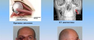
Orbital fractures
There are three main types of such injuries:
- Fracture of the orbital rim (outer rim), the thickest part of the eye socket. It will take a lot of force to break this bone. Such a fracture may be accompanied by damage to the optic nerve.
- Fracture of the rim extending to the inferior margin and floor of the orbit. In this case, there is a fracture of the facial bone under the eye.
- Fracture of the thinnest, lower part of the orbit. In this case, the orbital rim remains intact. The eye muscles and other structures may be injured. With such an injury, the mobility of the eyeball may be limited.
Fractures of the midface
With blunt trauma, fractures often occur along three lines running along the joints of bones, in the thinnest and weakest places, as well as where physiological openings are located. According to Le Fort's classification, there are three main types of fractures, but variations can also occur:
— Le Fort I fracture. With such an injury, the zygomatic bone and upper jaw are broken, they are completely separated from the other bones of the skull. Often accompanied by a fracture of the base of the skull.
— Le Fort II fracture. The fracture line runs from the bottom of one cheek, under the eye, through the nose and to the bottom of the other cheek.
— Le Fort III fracture. In this case, the alveolar process breaks off. The fracture line passes through the nasal floor and maxillary sinuses. With such an injury, the maxillary nerve ganglion is damaged.