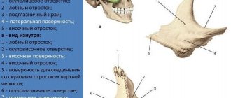
In humans, the scapula is a flat, paired, triangular bone. It is located on the back surface of the chest on the side of the spinal column. A fracture of the scapula is observed relatively rarely; among all fractures, this damage accounts for 1 to 1.5%. Why exactly this happens can be found out by understanding the anatomical features and reasons.
Blade structure
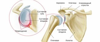
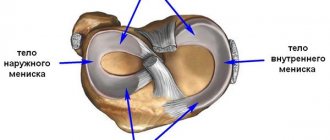
The main function of the scapula is to connect the clavicle and humerus into a single movable complex. The name of the bone comes from the fact that it has a triangular shape with three edges and corners and resembles a digging tool that everyone is used to using in the country or in the garden. The scapula consists of the body, the coracoid process, the spine of the scapula, which passes into the acromion process. The outer angle continues into the neck, followed by the articular surface or glenoid cavity, which in appearance resembles a small saucer. It is due to the articular surface and the head of the humerus that the joint is formed.
The main formations are located on the posterior surface, which has an uneven relief; the anterior surface is smooth and has traces of ribs. The muscles responsible for various movements, including rotation of the shoulder (rotator cuff), are attached to the shoulder blade. Numerous vessels and nerves pass nearby. A photo gives a clear idea of the anatomy of the scapula.
Causes of scapula injuries
The comparative rarity of the occurrence of damage lies in the fact that the bone is located in the thickness of the muscles and has greater mobility; the shape is of no small importance. The most common reasons are:
- falling on your back;
- blow to the shoulder blade;
- falling on hand;
- road accident;
- industrial injuries.
Causes
Scapula fractures are caused by direct trauma with a large vector of force. As a rule, in 80% of cases, scapula fractures are accompanied by injuries to the chest, lungs and shoulder. Therefore, in the presence of a scapula fracture, a thorough examination is necessary for the presence of concomitant injuries. The main causes of a scapula fracture are as follows:
- Road traffic accidents
- Falls with direct shoulder injury
- Falling on an outstretched arm
- Direct blow, such as with a baseball bat or hammer
Damage classification
Damage to the scapula can be open or closed, when there is no damage to the skin. They can also have one fragment or several, which complicates the treatment process. Scapula fractures are generally classified according to the location of injury:
- axis area;
- damage to the glenoid cavity;
- neck fractures;
- injuries of the coracoid process;
- damage to the acromion process;
- fracture in the area of the upper and (or) lower corner;
- longitudinal, transverse, comminuted;
- perforated ones are distinguished separately (in case of a bullet wound or impact with a sharp object).
The glenoid cavity and acromion are most often damaged. Such situations require special treatment, after which pain may persist for some period. Fractures of the neck of the scapula are often the cause of serious consequences and complications.
Radiation diagnostics
Radiography
Linear radiolucent areas passing through the scapula, displacement of bone tissue fragments. Other radiological symptoms depend on concomitant injuries.
CT semiotics
Linear sections of fractures, in comparison with radiography, better determine the relationship of fragments and the degree of their displacement.
MRI semiotics
T1-VI: Linear areas of decreased signal with signs of trabecular edema.
T2 FS: Linear areas of decreased signal due to trabecular edema, increased signal from adjacent soft tissues.
It is possible to visualize damage to other structures of the articular region (ligaments, muscles or damage to the brachial plexus).
Symptoms of damage
Damage to the upper edge of the scapula
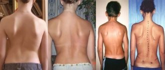
There are characteristic signs of a scapula fracture that make it possible to establish the correct diagnosis. Immediately after the injury, the victim notices pain in the area of the shoulder, shoulder girdle and scapula, which intensifies when attempting to move. The back surface of the shoulder girdle swells; at the site of injury after injury, an abrasion or wound attracts attention.
The tissues become saturated with blood, resulting in a bruise that is clearly visible under the skin. After some time, the bruise goes down the shoulder. On palpation, pathological mobility may be observed, if the victim has a crack, a similar symptom will be absent.
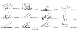
The previously described swelling completely follows the shape of the bone, having a triangular shape. This phenomenon is called the “Comolli triangle”. When trying to palpate the damaged area, the pain increases if the fracture is displaced and crepitus of the fragments is clearly audible.
Sometimes after a fracture the arm rises along with the shoulder blade, this indicates damage near the joint. Blood accumulates in the joint cavity, causing it to increase in size. When the neck of the scapula on the affected side is damaged, the shoulder hangs down, the acromion process protrudes forward, and the coracoid process deepens.
With an open fracture, attention is drawn to the wound, at the bottom of which fragments are visible. If the nerve is damaged, sensation in the upper limb may be impaired. The victim experiences pain when taking a deep breath.
Possible complications
Breaking a shoulder blade is harmless. This injury often damages the cartilage of the shoulder joint, which can subsequently lead to arthritis in the area. In the event of a displaced fracture of the scapula, a dangerous curvature of the triangular bone occurs. In addition, the triangular bone can move freely between the ribs, which is usually accompanied by an unpleasant cracking sound and pain.
As for the operation, it can provoke:
- muscular dystrophy;
- chronic dislocations;
- intercostal neuralgia;
- limitation of shoulder movements.
However, if time does not allow the operation, the person may completely lose his ability to work and become disabled. It is extremely important to diagnose this fracture in time, and then the negative consequences will be minimal.
Providing first aid to the victim
Immediately after an injury, it is important to provide first aid to the victim, this will reduce pain and swelling. To begin with, the victim is given a painkiller tablet. Until the doctors arrive, a cushion is placed in the armpit. A headscarf is made from available means and placed on the injured upper limb. The limb can be immobilized using a Krammer wire splint. It must be modeled based on the healthy upper limb of the victim.
If materials for applying a bandage are not at hand, you can use an undershirt, T-shirt, or shirt held up. With proper immobilization of the hand, the pain becomes less, and the risk of damage to blood vessels and nerves, muscles and skin during transportation is reduced.
When there is a break in the skin, it is covered with a bandage using sterile bandages. Also, first aid for a fractured scapula is to be sure to apply cold, you can take it in the freezer or refrigerator, after wrapping the object in cloth. The duration of cooling is 20 minutes, after which a break is taken for 10 and the procedure is repeated at least three times in a row.
The victim must be transported to a medical facility in a sitting position. Upon arrival, doctors can give the victim a pain-relieving injection.
Diagnostics
The diagnosis of a scapula fracture is established based on examination and analysis of instrumental research methods.
- An X-ray of the shoulder and chest is performed.
- CT scan of the abdominal cavity and chest to identify possible injuries to other organs and systems.
- A CT scan of the shoulder is necessary if it is necessary to exclude an intra-articular fracture.
Sometimes scapula fractures are diagnosed during examination associated with more serious injuries (falls, traffic accidents).
Danger of scapula fracture
The threat of a scapula fracture can be in three main ways. In the first situation, when the glenoid cavity of the scapula, covered with cartilage, is damaged and comes into contact with the head of the humerus. The fracture is called intra-articular because it communicates with the joint cavity. The result can be joint instability and dislocation of the humerus. Subsequently, arthrosis of the shoulder joint develops, which can be caused by even a slight displacement. Even a small step makes it difficult for the head to slide, and movements are accompanied by restrictions or pain.
Orientation of the glenoid cavity relative to the axis
In a healthy person, the glenoid cavity has a certain orientation to the scapular axis. Normally, the inclination varies from 12 degrees posterior to 14 anterior. The vertical tilt is approximately 92 degrees. Such orientation in space allows for the normal functioning of the muscles, primarily the rotator cuff. Damage to the neck leads not only to pain, but also to limited mobility; sometimes the rotator cuff tendons are damaged by fragments. A change in the axis of the articular surface can cause dislocations and instability in the future.
When moving the upper limb, the scapula slides along the surface of the ribs, the same thing happens when breathing. Damage to the body leads to disruption of the smoothness of the surface, which cannot slide normally along the ribs. When the fracture heals with displacement, constant pain and crunching noises occur when moving.
Osteosynthesis for fractures of the upper limbs
Osteosynthesis for humerus fractures
For humerus fractures, immersion or external fixation is used.
The external fixator is optimal for temporary use. It can be quickly installed in any operating room and can be adjusted, if necessary, after surgery. In what cases is it better to use external fixation of the humerus:
- Serious injuries in a patient with multiple injuries to various bones of the extremities.
- Severe injuries to the humerus, including extensive soft tissue damage.
In other cases, various immersion clamps are used. Acute and pathological fractures are best fixed with nails.
A plate is used to align multiple fragments. For fractures with a markedly displaced proximal portion, it is critical to achieve anatomical reduction, especially if the patient is elderly and at risk for nonunion and loss of arm function. To achieve this, the plate must provide sufficient fixation points at the proximal humerus/humeral head. It should also be long enough.
For fractures in which bone fragments may be compressed, a compression plate is used.
Depending on the morphology of the fracture, it is sometimes advisable to obtain stable fixation with lag screws and then secure it with a neutralizing plate. Alternatively, it may be better to not intervene in the fracture area and simply apply a bridging plate.
Osteosynthesis for fractures of the lower (distal) part of the shoulder
In emergency cases and for complex multiple fractures, external fixation is used.
In osteoporotic bone, the distal articular fragment may be very short, and varying degrees of metaphyseal comminution may be present in the ulnar or radial column.
The most difficult task is to stabilize a very short articular fragment when the quality of the bone leaves much to be desired. This can be achieved using standard methods, preferably with a pre-adjusted corner stable plate.
Osteosynthesis for forearm fractures
In transverse or short oblique fractures of the radial neck, fixation of the head to the diaphysis is achieved through T-shape and plating (restoration of the retaining ligament over the plate).
Proximal metaphyseal fractures of the ulna are usually part of a more complex injury involving subluxation or dislocation of the proximal radius of the elbow joint. In such cases, external fixation is used (for complex fractures) or a plate (as a splint for displacement).
Transverse olecranon fractures without fragmentation can be treated with a wire tension band or plate. This operation is suitable only for young active people.
Complex multiple fractures of the upper forearm are treated with external (usually hinge) fixation only.
A similar fixation technique is used for fractures of the distal forearm.
Regarding fractures of the radius and ulna. Open fractures requiring surgical treatment are fixed with an external percutaneous module. In other cases, use a plate, adding thread locks if necessary.
Osteosynthesis for wrist fractures
Scaphoid fractures are the most common wrist fractures. In most cases, the fracture is fixed with a screw, sometimes with a percutaneous screw. For displacements with a screw in combination with spokes.
Perilunate dislocations are pure ligament injuries. They occur as a result of high-energy trauma. They can cause severe disruption of wrist anatomy, resulting in profound changes in wrist biomechanics.
Rupture of the scapholunal ligament is the first event in any sequence of perilunar ligament ruptures. This is the most common cause of wrist instability. It is treated by fixation with knitting needles and suturing the ligaments.
Perilunar fracture-dislocation is treated and treated in the same way. Fractures of adjacent carpal bones may occur instead of ligament tears when crushing forces are applied to the midcarpal joint.
Recognizing and restoring all osseous and soft tissue components is essential to restoring wrist stability and preventing post-traumatic degenerative joint disease.
Osteosynthesis for hand fractures
Simple fractures of the metacarpal head usually result from axial loading of the hand. They often occur from hitting a hard object such as a wall or an opponent. Fixation occurs with knitting needles.
Fractures of the metacarpal head can be simple, but often consist of several fragments. In such cases, screws are used.
If the metacarpal bone itself is fractured, a plate is applied. For an open or very oblique fracture, several screws are used.
A common site for fractures of the base of the metacarpal bone is the fifth metacarpal bone. Most of these fractures are comminuted and depressed, and are often associated with dislocation fractures of the carpometacarpal bone. These fractures are usually fixed with plates or wires in the case of small fragments.
Unstable fractures of the thumb and phalangeal bones are fixed with pins, screws, or plates, depending on the location and complexity of the fracture.
Consult with our specialists or request a second medical opinion from an orthopedic traumatologist on the recommendations already received.
Treatment of scapula injuries
The treatment strategy for any scapula fracture depends on the nature of the injury. Basically, conservative treatment is carried out, which requires immobilization. The operation is performed when there is displacement, when it is necessary to fix the fragments using metal structures. Fixation of the fracture is carried out within a month - one and a half, after which the victim is shown gymnastics.
Surgical treatment
It is carried out if there are indications:
- Intra-articular fracture with a step of more than 5 millimeters. Surgery is also indicated when the damage affects more than a quarter of the circumference of the articular surface.
- With subluxation of the head of the humerus, which is more likely when the angle of inclination of the articular surface changes.
- Damage to the superior supporting complex of the scapula.
Tactics for damage to the articular surface
Variants of intra-articular fractures
There are many options for damage to the articular surface of the scapula. In some cases, only the edge is affected on one side or along the circumference; in other cases, the line of damage is longer. With an intra-articular fracture, the function of the upper limb may be impaired, especially if displaced. To ensure that the function of the limb does not suffer, the fracture is fastened with a metal structure.
When surgery is performed to fracture the scapula, the displacement is eliminated, after which a metal structure is installed. The intervention is called metalosteosynthesis, and screws are used to carry it out.
Damage before and after surgery
After the intervention, the limb is fixed with a sling with mandatory abduction of the limb. A person walks in this position for 3 to 4 weeks, after which the fragments begin to heal and the risk of displacement passes. The limb must be abducted in any situation; in a pressed position, the risk of developing deforming osteoarthritis increases. After the fixation is removed, exercise therapy begins.
Sling and diverter bandage
Damage to the neck of the scapula
When this anatomical area is damaged, displacement is almost always observed. In a situation where the displacement exceeds 10 millimeters or when the inclination of the glenoid cavity changes by more than 40 degrees (around any axis), restoration of the normal position with the help of a metal structure is required. If the misalignment is not corrected, the person may suffer from joint instability in the future, resulting in dislocation and rotator cuff dysfunction.
Rehabilitation period
Recovery from scapula injuries begins from the first days, regardless of the choice of treatment method. The main activity is aimed at preventing muscle atrophy of the upper limb .
How long does recovery take?
The rehabilitation period, regardless of the type of treatment, takes from 4 to 6 weeks . During this time, the bone tissue has time to completely grow together and mineralize. At the same time, both immobilization and skeletal traction do not take more than 3–4 weeks.
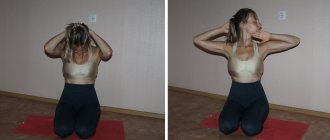
Photo 2. To restore limb mobility, you need to perform a set of exercises. Source: Flickr (kenga86)
Exercise therapy for recovery
Already from the first days of treatment, you should begin exercising the muscles of the hand and forearm. At the same time, all sorts of small movements are performed with the fingers, rotation, flexion and extension in the wrist joint. If the situation allows, the elbow joint is also involved.
After completing skeletal traction or immobilization, it is necessary to carry out a standard set of warm-up exercises for the shoulder girdle and upper extremities every day.
It is important! Any strength exercises (lifting weights, bench press, squats with a barbell, rows of the upper or lower block, pull-ups, etc.) should be avoided for a period of 2 to 3 months from the date of injury.
Rehabilitation treatment
During the recovery period, the main role is played by gymnastics, the task of which is to restore the function of the upper limb. Gymnastics is prescribed by a rehabilitation specialist or attending physician. An approximate set of exercises is described below.
You can start by lying on your back and spreading your shoulders 45 degrees to the sides, they are located at level with the body, and take the rod in your hands. You need to raise your hands up and behind your head, freeze for 3 seconds and return to the starting position. Afterwards, without changing the starting position, the arms are moved to the sides, the amplitude is individual.
Then you need to stand up and lean forward, lean on the ball with outstretched arms, the weight is directed as much as possible onto your hands. Swings are performed, the ball moves up and down, and then to the right and left. At the initial stage, gymnastics is performed with the condition of bending at 90 degrees, and after that you can lean on the arm on the side of the injury. In a similar position, rest your healthy arm on the table, with the limb hanging freely on the side of the injury. The body is swayed in different directions, while the hanging limb moves at the same time.
In a vertical position, a towel roll is placed in the armpit. The arm is bent at a right angle, and a fixed, tensioned elastic band is compressed in it. The forearm is abducted and adducted inward, resistance is applied to the band. The torso remains motionless during the exercise.
When the previous exercise is completed, you need to lie on the couch, raise your arms up and move them to the sides, fixing them for a short time. An outsider or doctor tries to push the elbow in different directions in order to move the arm. Over time, the force of the shocks increases. It is useful to sit on a chair and hold a rope in your hands, which is passed through your waist. It is necessary to slowly raise your arms up alternately.
After completing the previous exercise, it is useful to lie on the couch and clench the limb on the side of the fracture into a fist, place a rolled towel under the elbow, and move the shoulder to the side 40 degrees. After the injured hand rises up, it is allowed to help the healthy one. Without changing the position, the straightened arm extends upward; it must be raised as high as possible.
After completing this, you need to stand in front of a wall and bend your arms at the elbow joint at a right angle. The ends of the elastic band are clamped in the hands without changing the position of the limbs, the band is stretched, and the shoulder blades are brought together.
In a position on the side, the arms are bent at a right angle and are in a relaxed state. A pillow is placed between the hands, and the doctor moves the shoulder blade sideways and inward, down and up. The victim resists the effects. It is useful to perform gymnastics in water that reaches the neck. In a straight position, the arms are parallel to the body. The limbs are raised upward in a "V" shape until the thumbs touch the surface of the water.
What position to sleep in
Damage to the scapula can disrupt the victim’s proper sleep. Without adequate sleep, a person can only endure so much, but the pain can disrupt all plans to get enough sleep. First of all, it is better to sleep on your healthy side and put a soft pillow under your back that will prevent you from turning onto your back. It is better to sleep on a hard surface so that the body does not sink. You can also sleep in a position on your stomach; in this position, the load on the shoulder blade is minimal. The limbs can be placed parallel or in any comfortable position that does not cause pain.
The pillow should be low to minimize the load on the neck and prevent tension in the muscles attached to the shoulder blade. It is better to get out of bed from the healthy side, without relying on the limb on the injured side.
Types of fractures
A scapula fracture can be divided into nonspecific types (common to all fractures) and specific types, which can only be attributed to the scapula.
The first include:
- open (with damage to skin and muscles),
- closed,
- with offset,
- without displacement,
- complicated (attachment of bacterial flora or lysis of damaged bone),
- combined (injuries to other bones or organs).
Specific injuries include:
- bodies,
- awns,
- cervix,
- acromion,
- coracoid process.
All scapula fractures can be divided into stable and unstable. The first include injuries to the body and spine, all others disrupt the motor function of the upper limb girdle and are called unstable.