Knee replacement is done, of course, in compliance with the rules of asepsis and antisepsis. At the same time, not only doctors, but also patients themselves do everything necessary to avoid complications. But even preparation for surgery and rehabilitation do not always protect a person from unforeseen consequences.
Surgeons are increasingly wearing such attire so as not to become a source of infectious complications themselves.
Complications after endoprosthetics can be early and late. The former develop due to infection, improper installation of the implant, or a blood clotting disorder in response to surgery. The cause of early postoperative complications may also be the patient’s insufficient physical activity or failure to comply with the doctor’s recommendations.
In a later period (after a year or more), complications arise due to the gradual destruction of bones due to osteolysis. Less commonly, patients develop allergic reactions to metals in endoprostheses.
Knee replacement in the Czech Republic: guarantees, prices, rehabilitation, reviews and statistics.
Find out more
Pain as a sign of complications
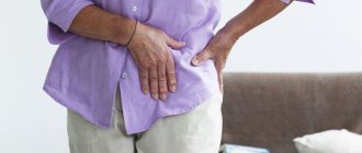
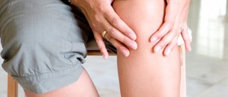
Knee replacement is performed to relieve chronic pain and improve a person's quality of life. After surgery, people are able to move normally and stop taking pain medications (unless they have severe osteoporosis in other joints). However, sometimes, after endoprosthetics, the patient develops alarming symptoms. His temperature rises, severe pain, swelling, crunching in the knee and other unpleasant phenomena appear.
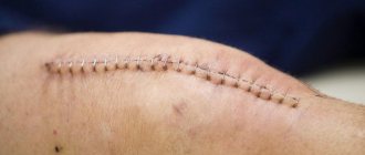
This is what a “quiet” seam looks like.
Painful sensations may indicate the development of infectious complications, contractures, synovitis, joint instability or other dangerous consequences.
With purulent-inflammatory complications, fever, chills, headache and general weakness appear. The knee also hurts, and the surrounding skin becomes red and hot to the touch. The pain is bursting in nature, and pain relief with ointments and tablets has no effect. Why is my knee hot? This is explained by the accumulation of pus inside the joint and the development of an acute inflammatory process.
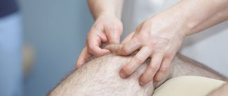
This is what a knee looks like and should be further examined for complications.
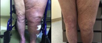
With thrombophlebitis of the veins of the lower extremities, the leg swells and a feeling of fullness appears in the lower extremities.
What to do if pain in the knee appears after endoprosthetics? You need to go to the doctor immediately. In the worst case, he will recommend repeated surgery and revision of the joint cavity; at best, he will advise what ointment or medications to use. It is possible that knee pain is caused by nerve irritation and will go away within 2-3 months.
Treatment
Treatment of knee instability is carried out taking into account the degree of damage to the ligamentous apparatus. For mild to moderate injuries, conservative techniques can be used, while severe ruptures require surgical intervention.
The choice of treatment tactics is based on the advantages of each component in the complex treatment of knee ligament injuries. The most common conservative methods are:
- Immobilization of the knee with a plaster cast or orthosis.
- Drug treatment.
- Physiotherapy.
- Massage and exercise therapy.
- Puncture of the knee joint (for hemarthrosis).
After receiving an injury, it is necessary to provide first aid to minimize tissue damage and gain time to see a doctor. There are simple recommendations that everyone should know. Self-help activities include:
- Ensure rest of the injured limb.
- Apply cold to your knee.
- Raise your leg above the horizontal plane.
- Secure the knee with a bandage (gauze or elastic).
- Take painkillers if necessary.
In the future, you cannot postpone a visit to the doctor, since the speed of recovery and the absence of unwanted risks depend on this.
Drug treatment
Taking medications in the acute stage of injury can reduce pain, relieve inflammation and swelling. In addition, medications improve tissue healing and create favorable conditions for faster restoration of joint function. The doctor prescribes the following medications:
- Non-steroidal anti-inflammatory drugs (meloxicam, diclofenac, nimesulide).
- Decongestants (L-lysine escinate).
- Chondroprotectors (glucosamine and chondroitin sulfate, hyaluronic acid).
- Improves blood circulation (pentoxifylline).
- B vitamins (neurorubin, milgamma).
In the acute period, the use of drugs by injection is justified, and as the symptoms subside, you can switch to taking tablet forms. There are a large number of topical medications (ointments, gels) that can be used for knee ligament tears. Of these, we can highlight Dolobene, Nicoflex, Menovazin, Apizartron.
However, their use is limited by the need to immobilize the joint. But after the cast is removed, rubbing medication into your knee will help speed up recovery.
You can take medications yourself only as prescribed by a doctor - ignoring the recommendations can cause adverse consequences.
Physiotherapy
Physiotherapy is of great importance in the complex of conservative measures and as a component of rehabilitation after surgery. Some methods work well with medications used immediately after injury.
Others are applicable only after the elimination of swelling and inflammation. However, all have a positive effect on soft tissues, improving biochemical processes and microcirculation, thereby promoting healing. In case of ligament ruptures, it is recommended to undergo a course of treatment with the following procedures:
- Electrophoresis of drugs.
- Cryotherapy.
- Laser treatment.
- Magnetotherapy.
- UHF therapy.
- Paraffin and mud therapy.
- Electromyostimulation.
- Balneotherapy.
Physical impact on damaged tissue enhances the effect of drug treatment and speeds up recovery from injury. To obtain the maximum result from the procedures, it is necessary to follow all the recommendations of the physiotherapist, who will select the optimal methods taking into account the characteristics of the patient’s body.
Massage and exercise therapy
Among rehabilitation measures, a special place is given to therapeutic exercises and massage. In this case, a gradual impact is necessary so as not to harm the damaged joint. You can start training after eliminating the acute consequences of the injury, even during the period of immobilization.
At this stage, perform gymnastics for the unaffected limb, as well as exercises in the ankle and hip joints on the affected side. Massage of the free areas of the thigh and lower leg is also indicated.
It will be possible to work on the injured knee joint no earlier than after 3–6 weeks, which depends on the severity of the damage to the ligamentous apparatus. At first the exercises are passive, and then they move on to active exercises. Massage of the periarticular area can also be done after removing the plaster cast.
Early activation of the motor function of the lower limb is a prerequisite for successful treatment of ligament ruptures. This helps prevent muscle wasting and the development of joint stiffness.
Infectious complications after knee replacement
The frequency of infectious complications after surgery is 0.2-4.5% for primary prosthetics and 4.5-12% for revision (repeated) prosthetics.
The development of purulent-inflammatory complications in the first 12 months is due to microbial contamination during surgery. Pathogenic microorganisms can penetrate the knee joint aerogenously or by contact. They are carried into the wound with dirty air, the surgeon's hands or surgical instruments.
Things are bad.
The risk of postoperative complications is especially high in elderly patients with diabetes mellitus, obesity, rheumatoid arthritis, immunodeficiency conditions and in patients taking corticosteroids. The prognosis is also worsened by the limited experience of the operating surgeon, large blood loss, the duration of the operation over 3 hours and the use of bone cement without an antibiotic in the composition.
In a later period (a year or more after surgery), inflammatory complications are a consequence of the hematogenous spread of microorganisms. Pathogenic microbes penetrate into the joint cavity through the bloodstream from foci of chronic infection.
Possible sources of hematogenous dissemination:
- skin;
- organs of the genitourinary system;
- Airways;
- oropharynx;
- lower parts of the gastrointestinal tract.
Often before operations the oral cavity is not even examined, which as a result causes severe complications.
The severity of clinical manifestations in a patient depends on the source of infection, the virulence of the pathogen and the time of development of the pathology. The classic bright picture of purulent inflammation (fever, swelling and hyperemia of the knee, fistula formation) is observed in less than half of the patients. Others may be bothered by constant pain in the knee joint, which intensifies with movement.
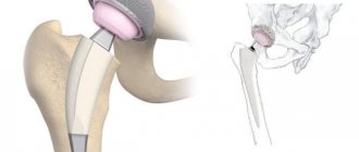
To successfully combat paraprosthetic infection, an integrated approach is required. The most effective is open sanitation of the joint cavity with complete removal of all components of the prosthesis (preservation of any of the components of the implant may ultimately lead to repeated surgery). At the same time, the patient is prescribed local antibiotic therapy by creating a depot in the inflammatory focus.
On the left is the implant after the primary operation, on the right after the revision surgery, it is significantly larger in size for strong fixation.
Conservative treatment of infectious complications is possible only with early diagnosis, low virulence of the pathogen and the presence of contraindications to surgery.
Implant dislocations and ways to solve the problem
Dislocations after knee replacement are very rare. The hip and shoulder joints are less favorable in this regard - their endoprostheses are displaced much more often.
This may be due to inappropriate design of the prosthesis, incorrect installation, or incorrect behavior of the patient during rehabilitation. The components of the implant may become dislodged in the early postoperative period when the patient first attempts to move the leg. Dislocations occur more often after revision arthroplasty than after primary arthroplasty.
Most often this is the result of a fall or other injury.
Displacement of the implant parts causes severe pain in the patient and leads to impaired joint mobility. The patient cannot move normally. A dislocated part of the endoprosthesis injures nearby tissues.
Implant dislocations can be treated using several methods. The simplest and cheapest of them is closed reduction. After it, relapses often occur. In case of repeated dislocation, the patient is recommended to undergo primary total arthroplasty or open revision prosthetics.
Contraindications for radiography
Pregnancy
For women during pregnancy, almost all diagnostic procedures except ultrasound are contraindicated. X-rays are contraindicated in the first place, since their radiation can negatively affect the development of the fetus and provoke the appearance of serious pathologies in it.
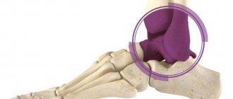
Metal prostheses and bolts in the knee
The metal screens X-rays and prevents them from reaching the film on which the image is created. As a result, the image turns out unclear and unsuitable for further analysis and diagnosis.
Severe obesity distorts the image
Large fat deposits complicate the passage of X-rays, and the image may turn out to be uninformative and unsuitable for analysis. In this case, the patient will still receive a dose of negative radiation.
Schizophrenia
Since it is difficult to predict how X-ray radiation will affect the condition of a patient suffering from schizophrenia, the use of this diagnostic method is not recommended. In addition, such a patient, not fully in control of himself, may not be able to remain still during the scan, which will negatively affect the quality of the image.
Contracture - the knee does not bend
Contracture is a restriction of mobility of the knee joint, which is accompanied by aching pain and difficulty walking. The operated leg may be in a forced, incorrect position.
It is impossible to fully straighten the leg.
The reason for the development of contracture is prolonged inactivity of the limb. Muscles weaken and their functionality is impaired. When a person begins to move the operated leg, a reflex muscle contraction occurs. Due to the spasm, the patient cannot bend and straighten the knee freely. Temporary contractures soon pass without any consequences.
If for any reason the patient requires long-term immobilization of the joint, there is a high risk of developing persistent contracture. It occurs after three weeks of inactivity of the limb. Persistent contracture is much more difficult to treat than temporary contracture.
In advanced cases, surgical treatment of contracture is used.
The most effective methods of combating this pathology are adequate physical activity and physical therapy. Exercises help develop muscles and return them to normal functional activity. Treatment includes physiotherapy and massage.
Congenital patellar displacement
Congenital dislocation is extremely rare. Usually directed outward. Can be one- or two-sided. There are three degrees of the disease:
- there may be no complaints, the knee is abnormally mobile;
- there is instability when walking with the patella turning outward;
- periodic blockades are observed that prevent flexion; the calyx is in an unnatural position with a pathological lateral deviation of the tibia.
Diagnosing congenital displacement of the patella becomes possible after the small patient begins to walk. Therefore, early diagnosis of pathology is difficult.
Conservative therapy is usually prescribed, aimed at strengthening muscles and ligaments:
- electromyostimulation;
- massage;
- exercise therapy complex.
If congenital displacement becomes habitual, surgery is indicated.
Venous thromboembolic complications
Deep vein thrombosis of the lower extremities develops in 40-60% of patients who have undergone major orthopedic surgery. An analysis of a number of clinical studies has shown that after knee replacement, this complication occurs in 85% of patients. In 0.1-2% of cases, thrombosis leads to fatal pulmonary embolism (PE).
Scheme of thrombosis formation.
Risk factors for thromboembolic complications:
- obesity ІІ-ІІІ degree;
- age over 75 years;
- chronic heart failure;
- varicose veins;
- diabetes;
- oncopathology;
- previous heart attack;
- long-term immobilization;
- taking steroids and hormonal contraceptives;
- chronic nonspecific lung diseases (CNLD).
During the operation, the human body begins to release substances that increase blood clotting. Thrombosis begins during surgery, so they occur in the early postoperative period. In 50% of cases they appear on the first day, in 75% - in the first 48 hours after surgery.
These compression cuffs are used for the first hours and days.
In orthopedics, mechanical and medicinal methods are used to prevent postoperative thrombosis. The first include compression, electrical neurostimulation and physical therapy. As for medications, unfractionated and low molecular weight heparins, vitamin K antagonists, and coagulation factor X inhibitors are used for prophylactic purposes. In recent years, doctors increasingly prefer oral anticoagulants (Apixaban, Rivaroxaban, Dabigatran etexilate).
The minimum duration of taking prophylactic doses of anticoagulants is 10-14 days, the recommended is 35 days. Premature withdrawal of medications can lead to spontaneous development of thrombosis. Therefore, doctors recommend long-term courses of prevention to patients. If thromboembolic complications nevertheless occur, the doses of anticoagulants are increased to therapeutic levels.
Knee replacement in the Czech Republic: guarantees, prices, rehabilitation, reviews and statistics.
Find out more
Causes of dislocations
Dislocation can be caused by many different factors and their complex combinations. Among the most commonly diagnosed causes of dislocations, the following points should be noted:
- various types of injuries;
- careless jumps, strong sharp turns at the knee;
- excessive loads on the ligaments, causing them to rupture;
- low leg muscle strength;
- pathology of the structure of the lower extremities.
If any of the above provoking factors is present, the patient should pay increased attention to preventive measures (will be described in one of the following sections) and try to protect the knees from any kind of damage with all their might.
Allergy to metal
According to foreign studies, allergic reactions may occur in 10%. According to statistics, allergies are the cause of 5% of unsuccessful joint replacements. Allergens include chromium, cobalt and nickel.
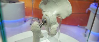
Knee implant.
Metal-containing endoprostheses are widely used in orthopedics. According to statistics, 99% of implanted prostheses contain metals or their alloys. Their contact with biological fluids causes corrosion of implants, which leads to the entry of metal salts into the human blood and the development of a delayed-type hypersensitivity reaction.
An allergy to the metal components of the endoprosthesis usually manifests itself as pain, redness of the skin and itching in the knee joint.
People who have had allergic reactions to any metals throughout their lives undergo patch testing before endoprosthetics. Skin patch tests determine microelement intolerance. This avoids the implantation of a prosthesis, which will ultimately cause allergies.
For patients who have already undergone endoprosthetics, patch tests are performed to confirm the diagnosis of allergy. The combination of a positive skin test and characteristic symptoms of an allergic reaction is an indication for reoperation. The patient's old prosthesis is removed and a new one is placed in its place.
Examination by an orthopedist, tests and diagnosis
The diagnosis is made based on:
- typical patient complaints;
- anamnestic data indicating the fact and mechanism of injury;
- results of an objective examination;
- data from instrumental research methods:
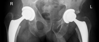
- radiography (of both joints in a standing position in the infero-anterior and lateral projections);
- Ultrasound (to verify soft tissue damage);
- CT scan (can be done with the joint flexed);
- MRI (the most accurate method, allows you to identify damage to tendons and muscles);
- results of biochemical studies indicating an inflammatory process in the joint area:
- examination of the joint fluid (articular puncture is performed);
- biochemical and general blood tests.
Instability at the patellofemoral joint
One of the complications of total arthroplasty is instability of the knee joint in the area of the patella. The reason is a violation of the normal sliding of the latter in the frontal plane due to incorrect orientation of the implant. During the first year after surgery, instability in the patellofemoral junction is detected in 1.5% of patients.
The incidence of complications does NOT depend on the type of prosthesis and the experience of the surgeon who performs the operation.
An example of incorrect installation of the femoral component and the consequences in the form of increased wear.
To eliminate patellar instability, the patient undergoes revision arthroplasty. During the intervention, surgeons eliminate errors in the orientation of parts of the implanted implant. At the same time, superficial prosthetics of the patella is performed.
Less common complications of the femoral-patellar joint:
- damage to the patellar prosthesis;
- aseptic loosening;
- patella fractures;
- rupture of the patellar ligament;
- snapping patella syndrome.
Displacement classification
Pathological changes in the position of the patella based on pathogenesis factors can be:
- habitual – with regular changes in the position of the patella, accompanied by a pronounced pain symptom complex;
- partial – with an unstable position of the patella, prone to displacement with minor impacts on the knee joint;
- congenital - due to joint injuries received at birth.
Depending on the scale, the displacement is classified into:
- partial – provoked by a sharp turn of the leg;
- complete – represents a dislocation of the patella with displacement forward or backward due to a strong blow.
© designua — stock.adobe.com
Osteolysis and aseptic loosening of components
Osteolysis is a pathological process that leads to bone destruction at the site of fixation of the endoprosthesis. The main reason for this phenomenon is the prevalence of resorption processes over bone formation processes. Over time, osteolysis causes aseptic (non-infectious) loosening of the endoprosthesis parts.
Pathological mobility of the implant can also be a consequence of the destruction of the cement that was used to fix it. Due to the disruption of the strong connection between the surfaces of the bones and the endoprosthesis, the latter loses support. This leads to its loosening. The patient may experience knee pain, discomfort, and difficulty walking.
There should be no areas of distinct color or outline between the implant and the bone. This may indicate loosening and instability.
Aseptic loosening of the endoprosthesis occurs at a later stage. According to statistics, in the first ten years after surgery it develops in 10-15% of operated people. Instability of the knee joint is an indication for revision arthroplasty late after surgery. The patient is given an implant with longer legs. Such an endoprosthesis provides reconstruction of lost bone tissue and allows for durable fixation.
A number of medications are used to prevent aseptic instability and implant loosening. These include bisphosphonates, calcium supplements and vitamin D. In addition, the patient is recommended to eat a calcium-rich diet. The intake of osteolysis inhibitors, vitamins and minerals into the body slows down the development of osteoporosis. The bones of the lower extremities stop deteriorating. This allows you to avoid pathological loosening of the endoprosthesis or delay the occurrence of unpleasant complications.
Factors in the development of pathology
Displacement of the kneecap can be caused by:
- injuries (impacts and falls);
- high loads (weightlifting or triathlon);
- damage to the menisci, tendons and ligaments, increasing the vulnerability of the patella;
- hypotrophy of the leg muscles (quadriceps femoris) due to a sedentary lifestyle;
- abnormalities in the development of the legs, including their X-shaped deformity;
- dysplasia of the femoral condyles;
- abnormally high localization of the patella;
- tumors of the knee joint;
- chronic lesions of the knee joints (brucellosis), leading to their instability.
Typically, trauma-induced dislocation is accompanied by ruptures of the collateral ligaments. With torsional horizontal displacement, the quadriceps tendon with the patella ligament apparatus is damaged.
Congenital pathologies predisposing to habitual displacement of the patella include:
- valgus joint deformity;
- patellar hypermobility;
- hyperextension of the leg;
- hypoplasia of the femur.
The horizontal and habitual patellar displacements described above are treated surgically, followed by a rehabilitation period of up to six months.
Prevention of complications in the first month
In the first 5-6 days after, the patient is in the hospital, where he receives all the necessary medications and procedures. To prevent thromboembolic complications, the patient is given anticoagulants to avoid infection - antibiotics. In the early stages after surgery, the patient is recommended to wear compression stockings.
The day after endoprosthetics, doctors remove drainage from the wound, which served to drain fluid. Afterwards, walking with elbow crutches or walkers is allowed. The medical staff helps the patient get up and explains how best to walk and what exercises to do. Early physical activity and exercises help avoid the development of contracture.
Arthromot provides passive development of the joint.
If the patient does not experience any complications, he is soon discharged from the hospital. Working people are given sick leave. The length of sick leave can range from 1.5 to 3 months. Upon discharge, doctors give the patient a number of recommendations for caring for the postoperative wound. After two weeks, the patient will need to come to the hospital to have the stitches removed.
For 5-6 weeks the patient is forced to walk with crutches. After this period ends, he will no longer need them. However, you can return to long walks, swimming and other vigorous activities in 5-6 months. The question of playing sports is decided together with the attending physician. Orthopedists prohibit heavy physical activity.
This is what the suture should look like 3-4 months after surgery.
A follow-up examination of the patient is usually carried out 2-3 months after surgery. If, during the examination, doctors identify signs of instability, infection, thromboembolic or other complications, the patient is given the necessary assistance. After the first scheduled examination, a person visits a doctor once every 2-3 years.
Prevention of late complications of endoprosthetics
At the end of the rehabilitation period, it is worth continuing to do the exercises for several more years. Therapeutic exercise is necessary to maintain normal muscle tone and functional muscle activity. Exercise helps a person maintain weight. Since obesity accelerates the development of osteoporosis, preventing it helps prevent loosening and instability of the knee joint.
The ligamentous and muscular system around the implant must be strengthened with regular training.
Endoprosthetics are often performed on older people prone to osteoporosis. Surgery further traumatizes the bones, which contributes to their further destruction. Therefore, to prevent osteolysis, the patient needs to take drugs that normalize metabolic processes. Medicines must be taken daily.
Drugs used for preventive purposes:
- Alendronate;
- Recostin;
- Fosamax;
- Alendronic acid;
- Bonviva;
- Rizendros;
- Zolerix.
The listed drugs inhibit proteolysis, that is, they slow down the destruction of bone tissue. Along with them, doctors recommend that patients take calcium, phosphorus and vitamin D supplements. They saturate bone tissue with essential minerals, thereby strengthening them. Particularly effective in this regard are medications containing hydroxyapatite (Calcimax, Ossein-Hydroxyapatite). This substance is very well absorbed and acts much more effectively than calcium carbonate or calcium citrate.
And even 20 years after primary endoprosthetics, the patient needs to consult a doctor to monitor the need for revision surgery. The specialist must carefully examine and examine the person. If the patient shows signs of instability of the knee joint, re-endoprosthetics may be necessary. After revision surgery, the incidence of complications is higher than after primary surgery.
Rehabilitation after a knee fracture.
Rehabilitation after a knee fracture includes several basic rehabilitation and control measures. In particular, control radiographs are taken throughout the entire period of restoration of joint function after a knee injury. Also, rehabilitation after such a displaced fracture includes periodic injections to suck out the blood accumulated in the joint. When performing exercises aimed at recovery after such a fracture, the doctor and the patient work together, since the intensity of the exercise is set according to the principle of maximum tolerance - that is, you need to step on the leg or bend it with the effort that is acceptable due to pain.
Rehabilitation after a fracture of the patella contains exercises such as flexion and extension of the sore leg using the healthy leg or arms; also, as the joint recovers after fractures of the patella (kneecap), new exercises are added. These are bending, walking, running and jumping. When performing exercises, the performance of the diseased and healthy leg is compared, thus drawing conclusions about whether rehabilitation after a fracture is effective.
In addition to exercises, rehabilitation after fractures involves taking special restorative medications, including vitamins, as well as calcium, iron and other minerals. For each patient, rehabilitation after a fracture requires its own therapeutic nutrition regimen.
After the knee regains its motor function, the attending physician evaluates the success of rehabilitation and decides whether to extend it or discharge the patient.