A Bankart lesion is an injury to the anterior labrum of the shoulder joint. This injury is associated with repeated anterior subluxation/dislocation of the shoulder. Anterior subluxation of the shoulder joint can also damage the connection between the labrum and the joint capsule. This is usually due to the absence or abnormality of the medial glenohumeral ligament. Bankart damage is typical for athletes who play volleyball, tennis, handball, as well as people whose professional activities involve overhead movements of the arms.
Epidemiology/Etiology
The glenohumeral joint is the most mobile joint in the human body. At the same time, its mobility is achieved at the expense of stability. Due to poor bony congruence and capsular laxity, the glenohumeral joint is a very unstable joint, making it very vulnerable to dislocation. Its stability is ensured by dynamic stabilizers and neuromuscular control. Anterior shoulder instability is the most common traumatic type of instability, representing approximately 95% of all injuries resulting from shoulder instability. Shoulder dislocations are primarily caused by abduction, extension, and external rotation of the shoulder.
Friends, Georgy Temichev’s seminar “Diagnostics and treatment of shoulder joint problems” will take place very soon. Find out more...
In many cases, patients with anterior shoulder dislocation have a Bankart injury. A reverse Bankart lesion can occur with a posterior dislocation.
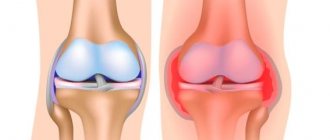
Bankart tear of the shoulder joint
A Bankart tear of the shoulder is another type of injury. With it, there is no violation of the integrity of bone tissue. Rupture of the cartilage tissue of the labrum can occur due to excessive physical activity or a dislocation of the shoulder.
A Bankart tear can only be treated with surgery. After the operation, a long period of rehabilitation is required. If it is not carried out, then a relapse of the pathology is possible in the near future, since in order to fully restore cartilage tissue it is necessary to strengthen the surrounding muscles of the upper shoulder girdle and restore impaired microcirculation of blood and lymphatic fluid.
Diagnostic procedures
Many patients who suffer a shoulder dislocation have a Bankart injury. Although this type of injury is common in patients with shoulder dislocation, it is difficult to detect on physical examination.
Thus, magnetic resonance imaging can be used to identify a Bankart lesion. It can also be used to quantify associated lesions, including the inferior glenohumeral ligament.
You can read about subacromial pain syndrome here.
According to some studies, a Bankart lesion can be diagnosed if there is a contrast agent between the glenoid cavity and the capsulolabial complex.
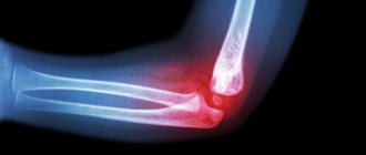
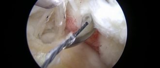
Bankart soft tissue lesions can be seen on arthroscopy and magnetic resonance arthrography as a fragment of the labrum attached to the anterior bundle of the inferior glenoid glenohumeral ligament and a rupture of the periosteum of the scapula. A bony Bankart lesion can also be detected by x-ray.
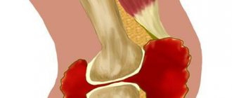
Bone Bankart lesion of the shoulder joint
Bone Bankart is the most severe type of injury. It is preceded by several stages in the development of articular lip pathology:
- primary degeneration with dehydration and loss of elasticity due to impaired diffuse exchange of fluids with surrounding muscles;
- reduction in the height of the articular labrum and cracking of its surface;
- primary dislocation with disruption of the integrity of cartilage tissue;
- formation of gross scar deformation at the site of rupture;
- repeated dislocation and formation of extensive scar deformation of the articular labrum;
- dysfunction of the articular endplate and its partial sclerosis;
- osteoarthritis and bone deformation in the area of the glenoid cavity of the scapula;
- partial separation of bone tissue at the moment of excessive tension of the muscles and the entire shoulder joint as a whole.
A Bankart bone lesion causes severe pain and limited mobility of the upper limb. Immediate medical attention is required. It is best to visit a traumatologist immediately after an injury. This doctor will order an x-ray to rule out a violation of the integrity of the bone tissue.
If a Bankart-type injury is detected, primary treatment is prescribed to stop bleeding and inflammation. Then a planned endoscopic operation is performed using arthroscopy. During surgery, the integrity of the bone tissue and articular cartilaginous labrum is restored.
Bone damage to the Bankart shoulder joint without timely treatment leads to disability, as hemarthrosis, ankylosis and complete contracture of the joint are formed. In the absence of movement, complete degeneration of the cartilage tissue of the joint is observed. Then the joint capsule begins to collapse. An excess amount of scar tissue forms inside it. As a result of this, the shoulder joint completely loses its mobility.
Inspection
When it is necessary to examine a patient with a shoulder joint problem, you can use a special test that allows you to evaluate the amount of resistance to internal rotation. This is a reliable test that allows you to differentiate intra-articular pathology from impingement syndrome of the shoulder joint (sensitivity and specificity of the test are 88% and 96%, respectively). In any case, this is evidenced by a study in which 110 patients participated (they were divided into 2 groups: patients with internal impingement of the shoulder joint and patients with superior thoracic outlet).
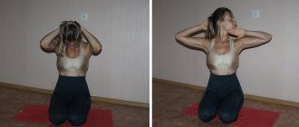
Bankart damage testing
How is this test performed? You stand behind a patient who is sitting. His arm is in 90 degrees of abduction and 90 degrees of external rotation. You should ask the patient to resist external rotation as much as possible, followed by internal rotation. If the patient demonstrates good strength in external rotation and weakness in internal rotation, the test is considered positive.
The idea is that if this test is positive, then with the arm in a fixed position, the internal rotators of the shoulder will anteriorly displace the head of the humerus against the anterior part of the labrum.
A positive test is predictive of internal impingement, which is often associated with Bankart injury. A negative test suggests the presence of superior outlet syndrome.
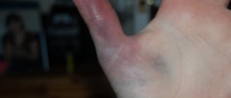
Symptoms
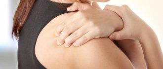
You can check for bankart damage by external signs and based on examination of the patient:
- there is severe pain arising due to the inflammatory process in the joint ligaments;
- when moving the arm back or lifting it upward, the head of the bone slips, which accompanies a click;
- swelling and hematoma form at the site of injury;
- The range of movement is significantly limited due to swelling, dislocation and severe pain when trying to raise or move the arm to the side.
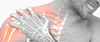
Diagnosis is carried out using X-rays, MRI and CT. The methods make it possible to establish the location and extent of a bankart tear.
Assessing the extent of the injury is critical to determining effective treatment options. Based on the results of instrumental diagnostics, the traumatologist makes a decision on the advisability of surgical intervention.
Treatment
Arthroscopic repair with suture anchors is an effective surgical treatment for isolated Bankart lesions. Identification of risk factors for relapse allows for appropriate patient counseling and consideration of open stabilization. Risk factors include age under 25 years, ligament laxity, and large (>250 mm3) Hill-Sachs lesion. After arthroscopic surgery, at least one third of patients experience at least one dislocation within 8 to 10 years. Naturally, this negatively affects shoulder function.
After failed arthroscopic Bankart repair, open surgery demonstrates positive results, including low recurrence rates and the most complete restoration of function (unless there is loss of the external rotation component after open surgery).
Physical therapy
There are several options for intervention when Bankart is damaged. First of all, we can differentiate between operative and non-operative interventions.
Possible surgical interventions include arthroscopic repair and open surgery. With arthroscopic repair, muscle strength is restored more quickly, but the relapse rate after open repair is significantly lower (Level of Evidence: 4). Several studies have shown that the recurrence rate after surgical treatment is significantly lower compared with non-operative treatment (evidence level 2B). Of course, after surgery, rehabilitation is necessary, which may be slightly comparable to the program for conservative treatment (evidence level 4).
Conservative nonoperative treatment for Bankart lesions had a significantly worse outcome, with recurrence rates of 17–96% in patients under 30 years of age (LE: 2B).
List of used literature and sources
- Diagnostic Imaging: Musculoskeletal: Trauma / DG Blankenbaker, KW Davis. 2nd edition. ELSEVIER. Philadelphia, 2016. – P.1122. ISBN: 978-0-323-39253-2.
- Differential Diagnosis in Musculoskeletal MRI / GM Hollenberg, EP Weinberg, SP Meyers. Thieme Medical Publishers, Inc. New York, 2015. – P.657. ISBN 978-1-60406-683-8.
- MRI of the Musculoskeletal System/TH Berquist. 6th edition. Lippincott Williams & Wilkin. Philadelphia, 2013. – P. 173. ISBN 978-1-4511-0918-4.
- Musculoskeletal Imaging / BJ Manaster, DA May, DG Disler. 4th edition. ELSEVIER. Philadelphia, 2013. – P.590. ISBN: 978-0-323-08177-1
- Musculoskeletal Imaging / Th.L. Pope, Jr. at all. 2nd edition. ELSEVIER. Philadelphia, 2015. – P.1291. ISBN: 978-1-4557-0813-0.
Rehabilitation
There are 7 key factors to consider when rehabilitating an unstable shoulder.
- Onset of pathology (traumatic, chronic).
- Degree of instability.
- Relapse rate.
- Direction of instability (anterior, posterior, multidirectional).
- Concomitant pathology.
- Ultimate range of neuromuscular control.
- Level of premorbid activity (level of evidence 1A).
In addition to these key factors, the physical therapist must also keep in mind that each patient is different and that we must never forget about personalizing the rehabilitation program.
The primary focus should be on increasing dynamic stability, scapular positioning, proprioception, and improving neuromuscular control, as there are no specific exercises to directly restore/strengthen the labrum (Evidence Level 1A). Typically, the rehabilitation program is divided into 3 stages. Non-operative treatment and post-operative rehabilitation programs are very similar.
The first phase of rehabilitation involves the use of a limited active range of motion (ROM) brace for 0 to 4 weeks, which allows the arm to move through a range of 20 degrees of abduction and 40 degrees of internal rotation (Evidence Level 1B). This ensures an earlier return to functional activity. External rotation immobilization reduces the risk of recurrent shoulder dislocations (Level of Evidence: 4). After 14 days, passive movement in a pain-free zone is acceptable. Strengthening exercises begin as isometric contractions to initiate activation of the rotator cuff muscles. These are mostly closed chain exercises (such as pressing the shoulder against a wall in external rotation). The goal is to reduce pain and protect the healing soft tissue.
In the second phase, progression in passive movements is important, as well as active-assistive exercises aimed at increasing the range of motion (evidence level 1A). Strengthening the rotator cuff muscles begins with dynamic exercises performed in an open kinematic chain. Examples of exercises include shoulder movements performed with resistance bands or dumbbells (Level of Evidence 3A). Rehabilitation should include both closed and open chain exercises. An example of a closed chain exercise is a quadruple exercise that involves prostration of the scapula followed by a trilimb movement.
The third phase is marked by the restoration of the full range of passive movements. The rehabilitation program is aimed at restoring the full range of active movements. This phase emphasizes the gradual increase in resistance during dynamic exercises. This is necessary to restore strength to carry out normal activities of daily life. In fact, this is the goal of the rehabilitation program in this phase.
First aid.
If you suspect a dislocation, you should contact a traumatologist as soon as possible. Self-reduction of the shoulder joint can lead to negative consequences and lead to serious complications. Only an experienced specialist can correctly assess the damage and prescribe the type of treatment necessary in a particular case.
The sooner after the injury the patient gets to the doctor, the easier it will be to solve the problem, and the less likely complications will arise - after just one day, muscle contraction will occur, as a result of which it will be more difficult to influence the affected area.
Before visiting a doctor and during transportation of the patient, it is recommended to fix the arm in the position in which the injury occurred. If the patient cannot return the arm to the correct position, this should not be done. Cold compresses can be applied to reduce pain.
When communicating with a traumatologist, you need to describe to the doctor as accurately as possible exactly how the injury occurred, as well as talk about all the symptoms and sensations. This will help to quickly diagnose the type of disorder.
Types of deviation
The type of pathology depends on the intensity and type of displacement. The joint can deviate in the vertical or horizontal plane (there is also a combined displacement).
There are 3 types of pathology that appear for the following reasons:
- Severe injuries - dislocations or subluxations due to blows and bruises. Displacement occurs, causing rotator cuff instability. Because of this, functionality is impaired and mobility is reduced. Scar tissue gradually forms, which increasingly impairs functionality and leads to the pathology becoming chronic.
- Sprains of the joint capsule - appear due to increased mobility or regular physical activity. This leads to a decrease in the stability of the joint capsule, which in the future will provoke dislocation or subluxation.
- Instability in the horizontal or vertical plane - appears in childhood and represents hyperelasticity. It manifests itself in the form of discomfort during physical activity, as the shoulder joint is greatly displaced.