If arthrosis progresses, doctors often prescribe surgery - arthroscopy. A special endoscopic instrument equipped with a video camera is inserted into the joint and displays an image with multiple magnification on the monitor. The procedure is performed under anesthesia, gives the patient a good prognosis, but requires a serious, very responsible attitude during the recovery period. What recommendations must be followed to avoid complications in the form of adhesions, contractures and relapses?
Knee arthroscopy is not as scary as rehabilitation after it
What Happens During the Early Recovery Period
After the end of the operation, 2-3 days pass before the drainage tube is removed. This is the early recovery period, during which:
- painkillers and antibacterial drugs are administered;
- an aseptic bandage is applied to the incision area;
- apply an ice pack;
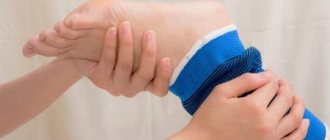
- It is recommended to use a special bandage or elastic bandage for immobilization to prevent swelling.
Any load on the knee is contraindicated. At this stage of treatment for arthrosis or osteoarthritis, it is important to observe bed rest. The bed should be such that the operated leg is in a stable position. Loads on the limb must be strictly dosed - according to the recommendation of the attending physician.
Orthosis and crutches are mandatory companions for the patient in the early recovery period
Exercises to restore knee joint function
Already in the early recovery period and after its completion, the patient is prescribed exercises to strengthen the muscles around the operated joint. They should be performed under the supervision of an instructor, gradually increasing the intensity and duration.
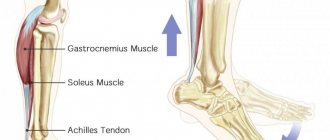
On the second day after surgery, light rotation of the ankle is allowed. A little later - in agreement with the doctor and exercise therapy instructor (usually within 3-7 days after surgery) - the patient performs the following exercises:
- Bend your knees while lying on your back so that the action does not cause pain.
- While lying on your stomach, press down with your foot the cushion placed under the foot.
- Raise the straightened leg 30 cm and hold it suspended for 5 seconds.
- Bend your knees while lying on your back with tension in your gluteal muscles.
- In the position of standing sideways against the wall, lift the straightened limb 45° above the floor surface (then a similar action, but with the foot turned outward).
A week after arthroscopy, a different, more intense set of exercises is recommended, where most of the actions are performed in a standing position. If the condition of the joint allows, you can connect an exercise bike or treadmill.
At different periods after knee arthroscopy, different exercises are performed.
In the first week after surgery:
From the first to the fourth week:
From the fifth week to the end of the second month:
Carrying out arthroscopy of the meniscus.
Surgical instruments used during arthroscopic surgery.
First, appropriate anesthesia is administered. The patient is then positioned for easy access. The surgeon makes 2 or 3 small skin incisions to insert surgical instruments: an arthroscope (a narrow tube with a camera at the end), a cannula (tubes) for washing the joint cavity, and an instrument to improve visibility. After diagnosis and obtaining a complete picture of the damaged area, the necessary manipulations are performed. If it is possible to restore the integrity of the meniscus, the torn edges are sutured. If parts are torn off or there is prolonged compression of the cartilaginous layer, resection is performed. At the same time, modern surgical technologies make it possible not to affect and preserve healthy tissue as much as possible.
Suturing
With a fresh injury in an area with sufficient blood circulation, as well as in relatively young patients, complete restoration of its integrity is most possible. This is also done surgically by suturing the tears during arthroscopy. If you follow the recommendations of specialists, rehabilitation occurs very quickly.
Meniscus resection
If all tissues of the damaged meniscus cannot be restored to their previous functionality, then they are partially removed. All viable parts are preserved and reconstructed as much as possible, trying to give the most anatomical shape. Completing a rehabilitation course after resection under the supervision of orthopedic traumatologists and rehabilitation specialists is the key to a successful and speedy return to knee functionality.
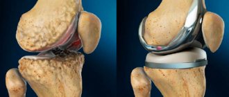
Complete removal of the meniscus (meniscectomy)
Meniscectomy is performed for more severe pathologies, when other treatment methods are excluded. Modern medicine has sufficient experience in the subsequent management of such patients. Observation in such a situation is necessary in order to avoid various complications in the future, for example, arthrosis.
Drug treatment of arthrosis after arthroscopy
Arthroscopy does not lead to immediate recovery, so medication assistance is necessary during the recovery period. The patient is prescribed non-steroidal anti-inflammatory drugs to prevent complications. You also cannot do without taking chondroprotectors, for example the drug Structum, Dona or Elbona.
Two weeks after arthroscopy you can recover on a treadmill
Treatment of damage and injury to the meniscus - treatment of meniscopathy
Treatment of meniscopathy , that is, treatment of meniscus injury , depends on the degree and nature of its damage.
Speaking without going into purely medical details, there are 3 fundamentally different types of meniscus damage : pinched meniscus, its partial rupture (tear) and tear.
When torn, the meniscus is either completely separated from its attachment site in the joint, or a piece of the meniscus itself is completely “broken off” and, figuratively speaking, “dangles” freely inside the joint cavity. This is the most severe form of meniscus injury. Such damage is almost impossible to cure without surgery: a completely torn meniscus does not “take root” and must be removed on the operating table. Or you have to remove the torn part of the meniscus (when part of it is torn off).
Fortunately, this form of meniscal injury is the least common, accounting for approximately 10–15% of all meniscal injuries.
Much more common is a pinched meniscus (approximately 40% of cases) or a tear (partial tear) - up to 50% of cases. When pinched, the meniscus usually only “gets wedged” between the cartilage of the knee, and when it is torn, as you understand, the meniscus is also torn, but does not come off completely. This means that in most cases he is able to recover. That is, such damage should be tried to be cured with therapeutic methods, without surgery.
Note from Dr. Evdokimenko. Removing the meniscus (or part of it) is a radical, but most often controversial decision, regardless of whether the meniscus was removed during a routine operation, or it was removed using the now “fashionable” arthroscopy. Indeed, although the operation leads to a rapid restoration of the functions of the damaged joint, in the future, as already mentioned, the absence of a meniscus (or part of it) in the knee contributes to the development of gonarthrosis.
I have seen how, after surgery to remove the menisci, gonarthrosis developed even in 30-35-year-old young people who should not have arthrosis at this age. And it is much more difficult to treat knee joints that have ever previously been operated on for meniscopathy than knees that have not been operated on. Undoubtedly, there are situations when such an operation is necessary (for example, when a meniscus is torn or when the same meniscus is pinched 2-3 times), but I believe that in most cases the primary meniscus injury should be treated with therapeutic methods.
If the meniscus is pinched (or torn), you should first try to release the meniscus that is pinched between the cartilages of the knee (regardless of whether it is torn or not). That is, it is necessary to reposition (reduce) the joint using manual manipulation . A good orthopedist, traumatologist or chiropractor can, in the vast majority of cases, eliminate a pinched meniscus in one, two, three or four sessions. If you do not take into account surgical treatment, it is usually not possible to unblock the joint so quickly in any other way.
It takes much longer for a pinched meniscus to be eliminated using hardware traction of the knee (that is, using hardware traction of the joint), if the doctor for some reason cannot or does not know how to reduce the meniscus. Hardware traction requires more time and more treatment sessions. But this procedure still does its job - the meniscus is gradually released and removed “from under the attack.”
The paradox, however, is that in most of our medical institutions they try to treat pinched meniscus not with manual manipulation or traction, but with medications and physical therapy. This treatment usually aims to relieve pain and swelling of the joint. But swelling, as already mentioned, can be protective. And before you fight swelling and swelling of the knee, you need to eliminate the root cause of this phenomenon!
Only after eliminating the root cause (jamming, pinched meniscus) with the help of manual therapy or hardware traction, can you proceed to physiotherapeutic restorative treatment. After the joint has been reduced (reduced), its recovery can be accelerated using laser, ultrasound with hydrocortisone and magnetic therapy.
For long-term persistent (after reposition) edema, intra-articular injections of corticosteroids (diprospan, kenalgon, hydrocortisone, etc.), as well as non-steroidal anti-inflammatory drugs (voltaren, movalis, nimulide, ibuprofen, etc.) will help us.
And you need to consolidate the success of the above therapeutic measures with the help of therapeutic exercises, taking chondroprotectors and, possibly, two or three injections of hyaluronic acid preparations into the joint (drugs Ostenil, Fermatron, Giastat, Synvisk).
Chondroprotectors and therapeutic exercises will be discussed in this article: Chondroprotectors - glucosamine and chondroitin sulfate *
Glucosamine and chondroitin sulfate belong to the group of chondroprotectors - substances that nourish cartilage tissue and restore the structure of damaged joint cartilage.
The use of glucosamine and chondroitin sulfate helps restore the cartilaginous surfaces of the knee joint , improve the production of joint fluid and normalize its “lubricating” properties. In addition, chondroprotectors restore the cartilage tissue of the menisci well.
The duration of treatment with chondroprotectors may vary, but most often I suggest that my patients with meniscus damage take chondroprotectors daily for 3-4 months.
And, of course, when treating meniscus damage, do not forget about therapeutic exercises. In this case, you can’t do without it!
A video of gymnastics for the treatment of menisci can be viewed here *
READ MORE:
- Causes of meniscus damage
- Symptoms of meniscus damage
- Diagnosis of meniscus damage
- Exercises to restore the knee with meniscal injuries
Chapters from other books by Dr. Evdokimenko
How to improve your diet
After surgical treatment of osteoarthritis of the knee, it is very important to receive substances from food aimed at strengthening the operated tissues. The body should not be deficient in protein, Omega-3 fatty acids, sulfur and selenium. Therefore, during this period it is recommended to include in the diet:
- cottage cheese and fermented milk products;
- cheeses;
- condensed milk;
- egg yolk;
- dried fruits;
- seaweed, shrimp, mussels, seafood;
- broth, aspic, jellied meat;
- compotes, fruit drinks, fruit jellies and marmalades;
- mineral water.
The right diet will help you lose weight
Is it possible to avoid all this?
Knee arthroscopy is a difficult surgical procedure that requires the patient to be patient and carefully follow all recommendations. It is often practiced in the second and third stages of arthrosis, while the problem can be dealt with in another, minimally invasive way.
Intra-articular injections of Noltrex help restore mobility in the knee joint and relieve pain for 9-24 months. The product is a synthetic substitute for synovial fluid. The course includes 2-5 Noltrex injections depending on the extent of the lesion and does not require the patient to undergo a difficult recovery period, as after arthroscopy. The therapeutic effect is noticeable after the second procedure; there are no risks compared to surgery.
Arthroscopic procedure
Many people are hesitant to undergo surgery because they do not know that manipulation of the meniscus of the knee joint
not that scary.
Partial or complete resection is done in the most gentle way: without pain, cuts and blood, using a safe and effective regional anesthetic. Patients are concerned about whether it is possible to run after removal of the meniscus
of the knee joint (watch our video on the topic) and engage in physical exercise?
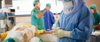
In the operating room.
Let's just say that people are allowed to lead their usual lifestyle, where running, jumping, cycling, and so on are not contraindicated, but only after a thorough restoration of the operated area. There are many real examples where even football players return to the game after such an intervention, and actively take part in competitions, playing at a professional sports level.
Exercise not on an unstable platform.
It is very important for the patient’s life to undergo rehabilitation so that after surgery the meniscus of the knee joint does not change for the worse. It will be as dynamic and rich as before. Well, we already discussed the dangers of inaction at the very beginning of the article. Now, actually, let's talk about the surgical procedure itself.
Modern medical technologies allow injured cartilaginous structures to be restored minimally invasively. Thanks to this, recovery after surgery, usually a suture is placed on the meniscus, is quick and without any difficulties. For therapeutic and restorative purposes, the arthroscopy method is used. The procedure is performed using a fiber-optic endoscopic device equipped with a video device called an arthroscope.
- The optical device looks like a thin probe. The probe is inserted through a puncture in the skin (diameter 5 mm) inside the joint and brought directly to the damaged object, which is visualized on the operating screen in enlarged sizes. The minimally invasive technique can also be used as a diagnostic tool if traditional diagnostic methods turn out to be insufficiently informative.
- Having decided on the treatment tactics, which will depend on the type and severity of the damage, the surgeon, using an additional puncture and special instruments, begins to perform the main tasks of eliminating defects on the meniscal body. The doctor, as far as possible, will try to preserve the cartilage tissue as much as possible, sparingly removing only clearly non-viable areas.
- There are two most common correction techniques: suturing the linear tear or excision of the marginal dislocated areas. In the first case, a suture will be applied using medical threads using a special technology. In the second, microsurgical instruments will be used to resect the flaps in the peripheral part, and then polish the edges of the meniscus. If free fragments are detected, the specialist removes them from the joint space.
- At the end of the session, the surgical field is washed. Small skin incisions are sutured, treated with antiseptic agents and covered with a sterile dressing. No plaster needed.
In case of generalized crushing of structures or an excessively large gap, the cartilaginous layer will most likely be completely removed. In practice, such a clinic is extremely rare. As for implantation or transplantation, today the methods of implanting artificial implants and donor cartilage grafts of the knee are at an experimental stage. Therefore, such high-tech methods in modern meniscus surgery have not yet become widespread.