Trauma to the bones of the hand is one of the most common problems when visiting emergency rooms. This is due to the fact that the upper girdle of the limbs is mobile and is involved in most of the actions performed by a person. Displaced arm fractures are a more serious injury than simple ones. This is due to the formation of bone fragments, which, when displaced, damage soft tissues, muscles, nearby nerves and large vessels. The consequences become deeper and require long-term rehabilitation. Read more about displaced arm fractures (what they are, treatment tactics, emergency care for the victim and rehabilitation methods) further in this article.
A little about anatomy
The anatomical structure of the bony skeleton of the hand is varied. Therefore, the resulting injuries with a violation of its integrity can be of a different nature. It is necessary to have an understanding of the structure of the limb in order to understand the specifics of its fractures. The arms can move in different trajectories; they are very flexible and mobile due to the large number of articular connections between conventionally designated sections:
- Shoulder girdle. This section includes paired bones - the scapula and collarbone. They are a transitional place from the torso to the limbs. These are not all their functions. With their help, a person can make rotational movements in the shoulder joint and raise their arms in different projections.
- Shoulder. Includes the direct attachment of the humerus to the scapula by the acromion process (which is considered an integral part of the shoulder). As well as a large joint connecting the shoulder girdle and shoulder.
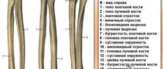
- Forearm. This section is represented by two main bones: the radius and the ulna. It also connects the elbow joint to the humerus. They are unpaired, but are next to each other. At the elbow joint, their epiphyses are connected to form a strong contact with the shoulder. They participate in the organization of rotational movements and help maintain the mobility and functionality of the arm as a whole.
- Brush. A complex and amazing education. Five fingers, each of which is formed by three phalanges. The connection between the hand and forearm is provided by many ligaments (radial collateral, pisiform-macula, semilunar, etc.). At the base of the hand lies a complex formation of eight bones, with the help of which rotation ability is maintained.
Interesting fact! Nature has protected such an important tool for humans as hands. She placed the muscle and tendon structures in a special protective “film” (fascia). With their help, the elements of the hand became more protected from mechanical influences.
Classification
There are general principles for classifying all bone fractures. This includes open and closed. By the way, a displaced fracture of the arm is most often accompanied by a violation of the integrity of the skin. Therefore, open injuries are more typical for him. There are also intra-articular fractures. They are characterized by damage to the bone of the arm in the area of the articular capsule. The difficulty lies in the high probability of complications in the form of filling the joint cavity with blood. The consequences impair the functionality of the joints, which will further affect the function of the hand as a whole.
A narrow classification has emerged based on the external characteristics of the displaced fracture and the type and location of the injury. So this includes:
- Incomplete. It is characterized by a partial fracture of the bone with the formation of a fragment, while maintaining the integrity of a separate area.
- Wedge-shaped. It is characterized by the formation of a large bone fragment as a result of a double fracture of one bone.
- Multi-fragmentary. Complex, with multiple formation of fragments of different sizes. It is often a common characteristic of a wedge-shaped fracture.
- Torsion. Basis for classification based on the method of mechanical action. One piece of bone is held in place by one mechanical force, while the opposite piece is twisted and traumatized by another.
- Helical. Characterized by an oblique, twisting fracture line.
- Compression. Another type depending on the mechanical impact. Such arm fractures are formed from strong pressure exerted on the bone, under which it cannot maintain its integrity.
- Longitudinal. It runs along the axis of the bone and is characterized by elongation, but a simpler treatment regimen. Most often it is a closed fracture.
- Transverse. Runs perpendicular to the axis.
Interesting fact! The hand is an important verbal element. With the help of gestures, a person is able to verbalize his emotions, feelings and show intentions.
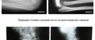
This is what happens even with simple fractures!
1. Immediately after injury, the displacement is 15 degrees.
2. Reduction, comparison and fixation with plaster, everything seemed fine.
3. A month later, the bones shifted again and fused with displacement.
It is not too late to perform the operation, but it will be more difficult than osteosynthesis performed in the first 2-3 weeks after the injury, and the result may not be as good.
It is necessary to perform an osteotomy, eliminate the deformity, replace the defect with artificial or your own bone, and fix it with a plate. For strong fixation, the screws must lock into the threads of the plate, creating a single structure with it. The radiograph on the left shows the Chronos block in the defect of the radius after correction of its shortening and deformation. The plate is precisely molded to the shape of the bone. Thanks to stable fixation, empty spaces are quickly filled with bone regenerate.
7 months after the operation, the bone looks monolithic, the regenerate has filled the empty spaces, the shape of the bone and the function of the hand have become normal. The plate does not need to be removed.
Diagnostics
When a patient with a suspected fracture comes to the doctor, he first performs a visual examination and palpates the damaged area. A fracture of the arm with displacement of fragments often manifests itself already at this stage of diagnosis, since fragments penetrate into the soft tissues and are palpated. Afterwards, radiography in different projections is required. This measure provides a complete understanding of the fracture pattern and allows us to develop a strategy for treatment and further rehabilitation. In addition to these measures, it is possible to use additional diagnostic tools:
- General tests. They allow you to assess the patient’s current condition and help make predictions for the process of bone fusion.
- Imaging methods - CT (computed tomography) or MRI (magnetic resonance imaging). Used when complications arise from other organ systems (nervous, circulatory, muscle groups, etc.)
- Puncture. Used to diagnose intra-articular fractures. Determines the presence of blood and the need to extract it and stop bleeding.
Causes of displaced fracture
The following reasons are identified that lead to displacement of bone fragments:
- Primary displacement. Formed under the influence of physical force.
- Secondary displacement. Occurs due to contraction of the muscles that are attached to the broken bone.
- Tertiary displacement. Occurs after the fracture itself due to external factors. For example, improper transportation of the patient or careless handling of the injured limb.
When displaced between the bone fragments, muscles or tendons can become wedged. This complicates the fracture and affects its treatment.
First aid
Correctly provided first aid will speed up the healing process and eliminate the risk of negative consequences. Following the algorithm in the correct sequence will help reduce the patient’s suffering. What is this algorithm:
- Anesthesia. Severe pain is the first symptom of an injury. To remove it, it is necessary to give the patient an anesthetic. This can be any painkiller that you have on hand. It is worth giving it according to the dosage, without exceeding it. It is better if it is possible to administer the drug intramuscularly. This will speed up the effect of the medicine.
- Immobilization. It is necessary to apply a splint from available materials or at least apply a fixing bandage. The simplest option is considered to be a scarf bandage, for which you only need fabric whose width corresponds to the length of the forearm. If the fracture is localized in the hand, then it is necessary to fix all its elements so that it is impossible to carry out any actions.
- Providing peace. After taking measures in case of a closed fracture, it is worth giving the victim a rest until the ambulance arrives.
- Disinfection. An open fracture must be disinfected. Iodine, brilliant green, hydrogen peroxide, chlorhexidine digluconate, and alcohol are suitable for this.
- Application of a tourniquet. If the bleeding is intense, then you need to apply a tourniquet 10-15 cm above the source of blood.
Important! When providing emergency care, do not try to dislodge fragments if they are palpable under the skin or visible in an open wound. This can cause damage to associated structures and cause additional consequences for the victim. It will be more difficult for doctors to correct the consequences and cure a broken arm.
Treatment
Depending on the severity of the injury, treatment tactics are chosen and the expected time frame for bone fusion is put forward. It is fair to note that when treating displaced fractures, the surgical method is most often chosen so that bone restoration proceeds safely, without pathological processes.
Conservative
Includes wearing fixing items that provide limb immobilization. Such items include:
- Gypsum. The most famous and no less effective method among other options. It consists of applying bandages soaked in plaster to the damaged area to fix the hand. The wearing period of the cast is within 6 weeks.
- Orthosis. A modern fixation method that is individually selected for the patient. It is made of medical plastic and is used to treat hand fractures.
- Splint. For displaced fractures, it is used as a temporary option until it becomes possible to place a full-fledged fixator.
- Bandage. Used to treat the bones of the forearm and shoulder.
Fixation is not the last stage of conservative treatment. While wearing a cast or other brace, the patient is prescribed a healthy diet with a high content of foods rich in vitamin D, C and calcium. Vitamin D helps absorb calcium and strengthen bone tissue. Vitamin C is a participant in metabolic, energy and regenerative processes. Medicines are also used - anti-inflammatory, painkillers and chondoprotectors.
Operational
Osteosynthesis operations are performed for complex hand injuries. These include:
- Multiple formations of fragments.
- Fractures of several arm bones at once.
- Damage to muscles, blood vessels, nerves.
The operation involves combining the fragments and fixing them using plates, screws and knitting needles. So how long does it take for a fracture to heal? Complex, with displacement, it is restored after only six months of active treatment and rehabilitation. The degree of consolidation is monitored over time using x-rays. Once the bone has healed, a second operation is performed to remove the fixing instruments from the healed bone.
Immobilization period
A cold compress immediately after an injury will reduce pain, swelling and inhibit inflammation.
Here's how to treat a broken arm after a cast has been applied:
- Drug therapy:
- paracetamol, analgesic or NSAID;
- calcium preparations (as indicated and under control of its level in the blood);
- D3 and B vitamins;
- Tablets with mumiyo, propolis, calamus root extract.
- Therapeutic exercise – special gymnastics for a plastered arm (6-8 times a day, 15-20 minutes each) + available general developmental exercises. A set of exercises and instructions for their implementation are given individually by a doctor or exercise therapy instructor.
- Self-massage below and above the plastered area.
- Physiotherapeutic procedures available with plaster and/or metal wires.
- Dietary food - fatty fish, walnuts, buckwheat porridge, chicken breast, fermented milk products and drinks, fresh vegetables and fruits, drinking or mineral water (2.5 l/day), limiting salt to 2-3 g per day.
- Quitting smoking and alcohol.
On a note. How to treat a hand after a fracture if there is pain? For mild or moderate pain - Nimesulide, Meloxicam, and for severe pain - Ketorolac, Diclofenac, Parecoxib. Nizilat is suitable for patients with stomach diseases.
Rehabilitation and how to develop a broken arm
The bone grows together within 1-1.5 months. The next 4.5 - 5 months are devoted to restoring the functions of the hand and strengthening the bones. Several methods are traditionally used for this:
- Exercise therapy (physical therapy)
- Physiotherapy.
- Massage.
- Proper nutrition, daily routine, etc.
Massage
Massage begins after the fixation is removed and after partial or complete healing of the wound. What are the goals of performing a massage:
- Prevention of atrophy. During a long stay in a state of immobilization, the strength of the arm muscles decreases. Massage helps restore muscle strength and gradually return to its original state before injury.
- Acceleration of blood circulation. Vibrations, claps, and pinches help increase blood flow in the area being massaged. Increased blood circulation helps accelerate the regeneration of damaged tissues and reduces the time spent on recovery.
- Elimination of puffiness. Fluid that accumulates in soft tissues as a result of injury interferes with proper blood flow. Stroking and gentle vibrations help reduce excess fluid.
Example of massage technique:
- Stroke areas of the arm just above and just below the injury.
- After lightly patting, move in the direction from the shoulder to the hand.
- Then apply vibrations - these are low-amplitude movements towards the injury.
- The massage must be completed with stroking again.
Attention! All techniques, duration and intensity are determined by the attending physician. Self-indulgence in this matter can lead to irreversible consequences.
Physiotherapy methods
Such methods rely on the ability of bone tissue to be a dielectric and conduct electrical impulses. In addition, the effects of UHF therapy and magnetic therapy are used. Let's look at each of them in more detail:
- UHF. Allows you to reduce pain and remove excess fluid from the surrounding soft tissues.
- Magnetotherapy. Accelerates metabolic and regenerative processes and also fights pain.
- Electrophoresis. Performed with local anesthesia for patients with persistent pain. Helps start regeneration processes.
- Ultrasound. One of the few methods to improve the condition of joints after damage.
- Laser. Used to stimulate metabolic functions.
Exercise therapy classes
The right hand is the dominant one for most people, so restoring fine motor skills is extremely necessary for a full return to normal life. Exercises are used in writing, drawing and other operations with small objects that are made at multiple magnification. Gradually, as the functions of the right hand are restored, pens and pencils are reduced, and abilities are improved. If the left arm of a left-hander is broken, then similar exercises are performed with it.
What tasks does exercise therapy perform:
- Strengthening muscles. After prolonged immobilization, muscles lose their volume, endurance and strength. Exercise therapy exercises help you gradually return to your previous and even better state.
- Restoring motor function of the hand. Each exercise is based on performing actions that help develop joints, restore muscle response to brain signals, and improve motor functions.
- Acceleration of blood circulation. Any activity generates increased blood circulation, which in turn helps reduce rehabilitation time.
- Launching metabolism.
Fractures of the radius of the arm in a typical location
Fractures of the distal radius of the arm are the most common fractures of the forearm and account for about 16% of all skeletal fractures. Typically caused by a fall on an outstretched arm. The description and classification of these fractures is based on the presence of fragments, fracture line, displacement of fragments, intra-articular or extra-articular nature, and the presence of a concomitant fracture of the ulna of the forearm. Incorrect fusion of the distal radius after untreated or secondary displaced fractures reaches 89% and is accompanied by angular and rotational deformation of the wrist joint and shortening of the radius. Classification of radius fractures:
• Intra-articular fracture: A fracture of the radius in which the fracture line extends into the wrist joint.
• Extra-articular fracture: A fracture that does not extend to the articular surface. • Open fracture: When there is a break in the skin. Damage to the skin can be from the outside to the bone (primarily open fracture), or damage to the bone from the inside (secondary open fracture). These types of fractures require immediate medical attention due to the risk of infection and serious problems with wound healing and fracture healing. • Comminuted fracture. When a bone is broken into 3 or more fragments. Causes of radius fractures:
• The most common cause of distal radius fractures is a fall on an outstretched arm.
• Osteoporosis (a disease in which bones become brittle and more likely to break under significant stress or impact) can contribute to a fracture from a minor fall on the hand. Therefore, these fractures most often occur in people over 60 years of age. • A fracture of the radius can certainly occur in healthy, young people if the force of impact is sufficiently great. For example, a car accident, falling from a bicycle, work injuries. Symptoms of radius fractures:
• Immediate pain;
• Hemorrhage; • Edema; • Crepitation of fragments (crunching); • Numbness of fingers (rare); • In many cases, it is accompanied by displacement of fragments, and as a consequence, deformation in the area of the wrist joint. Diagnosis of fractures
Most fractures of the distal radial part are diagnosed by conventional radiography in 2 projections.
Computed tomography (CT) is necessary for intra-articular fractures. Treatment of fractures of the radius:
1) Conservative: Fractures of the radius in a typical location without displacement are usually fixed with a plaster or polymer bandage to avoid displacement.
If the radius fracture is displaced, then the fragments must be returned to their correct anatomical position and fixed until the fracture heals. 2) Surgical treatment of radius fractures Sometimes the displacement is so critical and unstable that it cannot be corrected or kept in the correct position in a cast. In this case, percutaneous fixation with pins or surgery may be required: open reduction, external osteosynthesis with a plate and screws. During this operation, the displacement of the fragments is eliminated and the bone is fastened with a metal structure, the choice of which is determined by the nature of the fracture. Complications
One of the most common complications in fractures of the radius is Sudeck's syndrome.
Sudeck's syndrome (Sudeck's atrophy) is a pain syndrome that occurs after injury to the limbs, accompanied by long-term vasomotor, trophic disorders and osteoporosis. Clinically, constant intense pain is noted, which intensifies with the slightest movement or touch. In this regard, there is a sharp limitation of them. After immobilization, the pain does not disappear. Diffuse dense edema develops quickly. The skin is initially warm and hyperemic, then cold and cyanotic (so-called marbled skin). Gradually, the skin becomes thinner, becomes shiny, and loses its folded structure. Fragility, thinning and brittleness of nails and hair, atrophy of muscles and subcutaneous tissue are noted. Hair growth on the affected part of the body increases. X-rays show patchy osteoporosis of the bones. Treatment is usually conservative. In the initial stage, drug therapy is carried out (analgesic and vasodilator drugs, muscle relaxants, B vitamins, anabolic hormones, etc.), which is combined with hyperbaric oxygenation, acupuncture, laser therapy, etc. In order to normalize calcium metabolism, calcitrin is prescribed. If there is no effect, novocaine blockades or sympathectomy are sometimes performed. After reducing the intensity of the pain syndrome, physiotherapy, exercise therapy, and massage are prescribed. The use of intense thermal procedures and gross violent manipulations should be avoided, as this causes an exacerbation of the pathological process. The prognosis for function is not always favorable. Rehabilitation after a fracture of the radius
Most patients return to their daily activities after a fracture of the distal radius within 1.5 to 2 months. During the recovery period, the patient must be prescribed the following procedures: physiotherapy, massage, physical therapy. The duration of rehabilitation after a fracture of the radius depends on many factors: the nature of the injury, the method of treatment, and the body’s reaction to the injury.
Possible consequences
Unfortunately, bone fractures do not go away without leaving a trace. In some cases, the echoes of trauma bother a person throughout his life. For example, if fragments are improperly fixed, the bone grows together in an anatomically incorrect shape. Sometimes this is not noticeable externally, and sometimes external deformations occur in the form of pathological bends of the arm, shortening of the length of the limb, etc. In such cases, an osteotomy operation is indicated - the bone is broken and reassembled. Incorrect fusion is not the only possible consequence:
- False callus. This is a phenomenon when the bone does not fully regenerate and instead of bone tissue, a fibrous callus is formed. One of the symptoms of this complication is increased bone mobility where it should not be. It can only be treated surgically, also with a large number of consequences.
- Nerve conduction disorder. Nerve damage may be permanent. In this case, certain functions of the hand suffer, as the connection with the brain is lost. Or the sensitivity of certain areas of the skin is lost.
- Osteomyelitis. This is the name of the inflammatory process that affects bone tissue and bone marrow. With open displaced fractures, the risk of developing osteomyelitis is especially high due to the penetration of pathogenic flora into unprotected areas.
- Bleeding. Due to damage to soft tissues from fragments, injuries to large vessels (veins and arteries) are likely. Such bleeding leads to serious blood loss. This reduces the body's protective potential, increases the risk of infection and slows down regeneration processes. Therefore, in case of bleeding, it is so important to quickly stop it by applying a tourniquet and treating the open wound.
- Joint mobility disorders. In the case of intra-articular fractures, the joints suffer greatly. For their mobility, the release of a lubricating fluid is necessary. In case of injury, its synthesis decreases or stops. This consequence, if left unattended, can lead to disability.