A hip fracture is a dangerous injury, most often found in older people due to increased bone fragility. Although it can also be diagnosed in young and middle-aged people who have been involved in serious accidents, fallen from a height, etc. It accounts for about 6% of all cases of fractures.
When a femoral neck is fractured, the fragments heal poorly, so conservative therapy is ineffective and is used only if it is impossible to perform the desired type of surgery. Moreover, this injury, especially in older people, can cause the development of fatal complications that claim the lives of about 30% of victims. Therefore, it is extremely important to diagnose a fracture of the femoral neck as early as possible, carry out treatment appropriate to the situation and help the patient to properly recover from the injury.
What is a femoral neck fracture
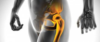
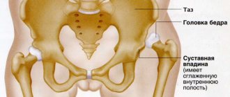
A femoral neck fracture is an injury accompanied by a violation of the integrity of varying degrees of severity of the thinnest part of the femur connecting its body to the head. The femoral head is round in shape and is part of one of the largest and most powerful joints in the human body - the hip.
The hip joints are paired hinge joints designed to withstand combined mechanical loads. Each of them is formed by the femur, which connects to the cup-shaped acetabulum of the pelvis with its head. Around the acetabulum is articular cartilage. It also surrounds the head of the femur and strengthens the hip joint. From the top of the head of the femur to the center of the acetabulum there is a thin ligament through which an arterial blood vessel passes.
The neck extending from the head of the femur is located at an angle of about 120° to the body and connects it to the body of the femur, which forms the base of the thigh. Behind it are the greater and lesser trochanters, to which the muscles and joint capsule are attached. Thus, the femoral neck is located inside the articular cavity and is covered by the articular capsule, but does not have periosteum, which in other cases provides nutrition to the bone tissue. Its nutrition is organized through arteries that penetrate the bone along the lower edge of the articular capsule and into the recesses between the trochanters.
As the body ages, the ligament and artery that supply the head of the femur become overgrown, which leads to the fact that it receives nutrition only from the vessels passing through the neck and trochanters. Due to such anatomical features, a fracture of the femoral neck leads to impaired circulation and difficulty in accessing blood to the injured part. This causes the development of aseptic necrosis. It is the most common complication of a fracture of the femoral neck, accompanied by its death and gradual resorption.
This type of injury can also lead to:
- The formation of a false joint, which is caused by non-union of fragments with the formation of a movable connective tissue joint between them. This leads to impaired leg function of varying degrees of severity.
- Vein thrombosis, which most often becomes a consequence of prolonged bed rest due to the occurrence of stagnation in the vessels.
- Congestive pneumonia, developing mainly in elderly patients undergoing conservative treatment of a hip fracture, due to stagnation of sputum in the lungs.
- Arthrosis, which is a degenerative disease of the hip joint. Its development leads to dysfunction of the joint.
A fracture of the femoral neck can occur at any part of the femur. Its specific position is of great importance for assessing the prognosis and determining treatment tactics, since the closer it is to the head, the higher the risk of developing its necrosis. Thus, depending on the level of location of the fracture, the following are distinguished:
- Basiscervical - the fracture line is located at the transition of the femoral neck into its body. They heal much better than other types of femoral neck fractures.
- Transcervical - the neck is broken in its narrowest part (central).
- Subcapital - the fracture is located on the border between the head and neck. It has the least favorable prognosis.
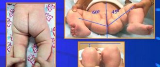
Also, the femoral neck can break at different angles, which has important prognostic implications. Moreover, the more vertical the fracture line is, the greater the risk of displacement of fragments, leading to impaired circulation. So, with an angle of up to 30°, the outcome is most favorable. If the bone is broken at an angle of 30-60°, the prognosis is less favorable, but still good. But when the deformation angle is more than 60°, there is a high risk of developing avascular necrosis.
Displacement of fragments can also occur according to different scenarios, on the basis of which they distinguish:
- varus fractures - the head of the femur is displaced downward and inward, which leads to a decrease in the angle between the body and the femoral neck;
- valgus - the head moves up and out, which leads to an increase in the angle between the neck and the body;
- impacted - accompanied by a shortening of the length of the femoral neck due to the impact of one fragment into another.
Impacted fractures are often valgus.
Drug therapy
Advertising:
Drug therapy is prescribed to elderly patients after surgery and conservative treatment. Medications help relieve pain spasms, relieve inflammation and reduce hematoma, and speed up the recovery process.
To eliminate sharp pain, a Novacoin blockade is performed.
It is not prescribed during the period of deterioration in the patient’s well-being, when blood pressure decreases, or when the patient develops a state of shock.
3-4 days after the exacerbation period, injections with non-steroidal anti-inflammatory drugs - Diclofenac or Ibuprofen - are prescribed. After 3-4 days, the injections are replaced with oral medications.
Such medications not only effectively eliminate painful spasms in the area of the injured joint, but also inhibit the further development of the inflammatory process. For complete regeneration of damaged bone elements, drugs from the group of chondroprotectors are prescribed - Dona, Alflutop.
Complivit Calcium D3, Kalcemin, Calcium D3 and other complexes that contain calcium help strengthen bone tissue and stimulate recovery processes. Additionally, diuretics may be prescribed to relieve swelling - Furosemide, Mannitol.
Causes
In more than 90% of cases, hip fractures occur in people, and 4 times more often in women over 65-70 years of age, who already have grade 2-3 osteoporosis. In such situations, a fall from one’s own height is enough to cause such a dangerous injury. Since in such cases the bones have a low degree of mineralization, and blood circulation is usually impaired due to the development of other concomitant diseases, the fractures heal very poorly.
Osteoporosis is a chronic disease accompanied by a decrease in bone mineralization, causing them to become more porous and fragile.
Also predisposing to getting a hip fracture in older people:
- presence of cancer;
- maintaining a sedentary lifestyle and pathologies of the nervous system leading to limited mobility or coordination of movements, including Parkinson’s disease, residual effects after a stroke;
- obesity;
- poor nutrition, starvation, leading to a deficiency of nutrients of different groups in the body;
- hormonal changes characteristic of menopause;
- atherosclerosis and other vascular pathologies;
- decreased visual acuity, which increases the risk of falling and injury.
At the same time, hip fractures are rare in young people, since the femurs are highly durable and resistant to various traumatic factors. In such situations, fractures are usually the result of:
- road accident;
- work injury;
- falls from great heights;
- participation in armed conflicts.
Recovery time
Recovery time depends on several factors - the age and health of the person, the severity and specificity of the fracture. On average, with strict adherence to all medical instructions, the rehabilitation period is 6 months.
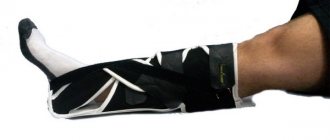
3 days after applying the plaster cast, massage sessions of the lumbar region are prescribed. After a few days, you can move on to massage the uninjured leg, and after another 7-10 days - to massage the thigh that was injured.
With normal recovery of the femur, after 2-3 weeks the plaster cast is removed, and the patient is advised to make light movements of the injured limb. This should only be done under strict medical supervision.
After 4 weeks, you can begin to perform special exercises to restore the injured hip - for example, simple flexion and extension of the limb.
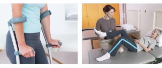
Attempts to sit down can be made only after 8-10 weeks and only under the supervision of a doctor. After 12-15 weeks, the patient is recommended to begin to stand up and move independently, relying on crutches and a healthy leg.
Gradually, the load on the injured limb increases; with a normal recovery process, after 6-8 months the person can return to a full, active life.
Symptoms of a femoral neck fracture
If a femoral neck fracture occurs, victims may encounter the following clinical manifestations:
- violations of the supporting function of the leg, accompanied by the patient’s inability to stand on the injured limb and walk, a violation of the configuration of the hip joint;
- minor pain in the groin area that occurs when trying to move the injured leg and passes at rest;
- turning the injured leg outward, which can be determined by the rotation of the knee and foot, with the inability to turn the leg inward;
- pain when pressing or tapping on the heel with the leg straightened;
- shortening of the leg (typical of varus fractures of the femoral neck), which cannot always be noticed independently, since the amount of shortening is insignificant;
- hematoma in the groin area, most often occurring a couple of days after injury due to the depth of the bone structures in the thigh.
If there is a suspicion of a femoral neck fracture, first aid consists of immobilizing the injured limb in the exact position in which it is located. It is necessary to immediately call an ambulance, and while waiting for its arrival, you need to try to calm the victim and convince him to remain still and not try to get up. Specialists fix the injured leg and pelvis with a special splint. In the absence of such, improvised means are used, for example, long sticks. If there is bleeding, a tourniquet is applied.
It is important to choose professionals
By contacting our clinic for hip replacement surgery, each patient can be confident in the high professional qualities of our specialists.
Modern equipment, the introduction of the best world achievements and many years of experience of doctors allow operations to be performed with minimal risks of complications.
The level of interventions performed in our clinic is in no way inferior to those of the world's best clinics in terms of effectiveness and safety. This is confirmed by years of practice and reviews from grateful patients.
Diagnostics
Victims suspected of having a femoral neck fracture are hospitalized in the traumatology department. An orthopedic traumatologist examines the patient and questions him about the circumstances of the injury.
To confirm the diagnosis, radiography in two projections is required. Using the obtained images, the type and angle of the fracture line are determined.
Fractures without displacement and signs of impaction in elderly people are difficult to diagnose using X-rays immediately after injury. Therefore, such patients are often mistakenly diagnosed with arthrosis of the hip joint and prescribed appropriate treatment, which does not produce results. In such situations, it is important to perform a CT scan or repeat an x-ray of the affected joint 2 weeks after the fall and the start of treatment.
But delaying treatment for a fracture of the femoral neck is fraught with aseptic necrosis of its head. Therefore, it is important to diagnose the injury as soon as possible after it occurs. Therefore, to obtain a complete picture of the condition of the hip joint, not only x-rays are often performed, but also CT, MRI, and scintigraphy.
Economic efficiency
Strict requirements are put forward for the design of prostheses. Two are of greatest importance:
- Inertness of materials for the prevention of rejection.
- Maximum reliability and durability.
Rigidity is provided by a titanium alloy, and the mobility of the articulated joint when choosing total prosthetics is achieved thanks to modified polyethylene or ceramic-based material.
A polymer liner is considered standard. For this purpose, polyethylene is additionally processed using compaction technology. The material becomes resistant to abrasion and retains shock-absorbing properties.
Ceramic components resist wear best, but they are characterized by some fragility and require special care during prosthetic surgery.
Thanks to the developed medical equipment market, every person has the opportunity to choose the best option. A single-pole prosthesis is relatively inexpensive, but has narrow indications.
The operation of total prosthetics is progressive, but also more expensive. The most modern designs with a ceramic liner and head are the most physiological. But their cost is also more significant.
Treatment of femoral neck fracture
Treatment of injuries of this kind is carried out by orthopedists. Conservative therapy, i.e. non-surgical treatment, is indicated only if there are serious contraindications to surgical intervention. This is due to the fact that most patients tolerate surgery much easier than long-term and ineffective conservative therapy, which requires prolonged bed rest.
In addition, for older patients, who account for more than 90% of all hip fractures, early intervention is extremely important. Otherwise, there is a serious risk of complications, including bedsores and congestive pneumonia. But severe inflammatory processes in the lungs can be fatal for older people. In young patients, prolonged immobility often causes muscle atrophy and post-traumatic contractures of the knee and hip joint. Therefore, whenever possible, preference is given to surgery for a femoral neck fracture.
The only case where effective treatment of a femoral neck fracture is possible is an impacted fracture with a horizontal line. In such situations, for young patients, the entire hip joint is fixed with a plaster splint, which reaches all the way to the knee, for 3-4 months. At this time, patients can walk, relying on crutches and without putting stress on the injured leg. Elderly patients have to be hospitalized and remain in skeletal traction for 1.5-2 months. From the first days of treatment, exercise therapy is carried out.
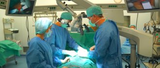
Today, surgical treatment of injuries of this kind is carried out by internal osteosynthesis or hip replacement. Young patients, as a rule, are offered osteosynthesis, while elderly people are almost always recommended to undergo endoprosthetics, which is due to circulatory disorders in the hip joint, slow regeneration of bone tissue and a low rate of fusion of fragments.
Carrying out osteosynthesis for elderly people sharply increases the incidence of complications, including due to the fact that the implantation of metal implants into the bone sharply increases their tendency to resorption. In addition, osteosynthesis for older people is fraught with the formation of a false joint, which causes disability.
In all cases, surgery should be performed as soon as possible after the injury. If this possibility is not available for one reason or another, then skeletal traction is performed before the operation.
Osteosynthesis
The essence of the internal osteosynthesis technique is the correct placement of fragments formed as a result of a fracture and their reliable fixation using pins, screws, knitting needles, plates and other means made of biocompatible materials. Most manufacturers use titanium, molybdenum-chromium-nickel and other alloys for their manufacture, since they have shown themselves to be the most resistant to oxidation in body tissues.
But for osteosynthesis to be successful, several conditions must be met:
- absence of severe osteoporosis;
- maintaining normal levels of bone mineral density;
- maintaining normal nutrition of the bones of the hip joint;
- the ability to accurately and firmly compare the fragments formed as a result of a femoral neck fracture;
- no need to traumatize periarticular tissues during surgery;
- compatibility of implant and bone.
Thus, osteosynthesis is not always possible even in young victims. If the above conditions are not met, there is a high probability of fracture nonunion. Therefore, in such situations, in order to reduce risks, preference is given to endoprosthetics. But even if the patient’s condition fully complies with the requirements for osteosynthesis, it does not always lead to the expected result. Thus, in 10–30% of cases, nonunion of a femoral neck fracture is observed, and in 10–40% of patients, aseptic necrosis of the femoral head develops. In such cases, it is also necessary to subsequently undergo total hip replacement.
Today there are several different techniques for performing internal osteosynthesis:
- Osteosynthesis using 3-blade Smith-Petersen nails. These nails are very thick and, thanks to the presence of 3 blades, are able to reliably hold fragments of the femur. During the operation, a nail is inserted into the femoral neck from the side of the femoral trochanters using a special hammer.
- Osteosynthesis using 3 screws. This method of surgical treatment of a fracture is more reliable and consists of screwing a number of thin knitting needles into the neck with a drill from the side of the trochanters. Then X-rays are taken and the most successfully installed knitting needles are selected, and the rest are removed. They are used as conductors for further screwing in special screws in the form of a hollow tube with an external thread.
- Osteosynthesis using a dynamic DNS hip screw. The method involves introducing a special metal structure with several screws into the femur. It requires special professionalism from the surgeon, and the design itself is quite bulky, although it provides reliable fixation.
The surgeon chooses a specific technique based on the individual characteristics of the patient’s femur structure, the type of fracture, its direction and some other factors. But always after internal osteosynthesis, patients undergo early activation, since this largely determines the success of the operation.
After internal osteosynthesis, recovery takes at least 12 months; joint replacement is associated with a shorter rehabilitation period - 5-6 months. Regardless of the type of surgical intervention performed, all patients in the early postoperative period are prescribed antibiotic therapy and drugs to prevent the occurrence of thromboembolic complications.
After osteosynthesis, patients can get out of bed and move their legs after 3-5 days. Subsequently, they must be assigned:
- massage;
- physiotherapeutic procedures;
- exercise therapy;
- swimming in the pool.
Metal plates, pins or other devices installed during surgery are removed only after the femoral neck fracture has completely healed and normal functional activity has been restored. This usually occurs 1-1.5 years after osteosynthesis. But if the operation was performed on older people, metal structures may not be removed.
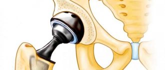
Endoprosthetics
Hip replacement is the main method of treating femoral neck fractures in elderly patients and young people with severe injuries. Carrying out this operation meets all the requirements for preventing the development of complications in people of the older age group, since it allows for early activation. After its completion, getting out of bed and physical activity are possible within 2-3 days, provided there are no complications, and after the end of the rehabilitation period, the person is able to move independently absolutely normally.
The essence of endoprosthetics is to replace the hip joint or only part of it with an artificial implant that completely repeats the functions and structure of the deformed part. This operation is shown:
- all patients over 70 years of age with a hip fracture;
- in the presence of a false joint;
- all patients with aseptic necrosis of the femoral head;
- for severe fractures, the formation of several bone fragments, bone fragmentation, etc.
In addition, hip replacement is performed in cases of grade 3 deforming coxarthrosis and the presence of tumors in the hip joint.
Depending on the severity of the situation, in case of injury with aseptic necrosis of the femoral head, total (complete) arthroplasty with replacement of the entire hip joint, as well as only partial arthroplasty using monopolar or bipolar femoral heads, can be performed. In the first case, the head and neck of the femur, as well as the acetabulum, will be replaced with an artificial prosthesis. Its elements are firmly fixed on the surface of bones, including those affected by osteoporosis, through the use of special cement, which is especially important for elderly patients.
In young patients, a cementless method of fixing a total endoprosthesis is more often used. In this case, a special spongy layer is located between the surface of the implant and the bone. Over time, bone grows through it, which ensures strong fixation of the prosthesis.
Total arthroplasty for hip fractures has a favorable outcome in more than 90% of cases and leads to a complete recovery. But after 15-25 years they may need to be replaced.
Sometimes patients may be offered the installation of a monopolar femoral head prosthesis, which replaces only the neck and head of the femur, i.e. the socket is not replaced. The disadvantage of such devices is the rapid wear of the articular cartilage due to the constant friction of the synthetic head against the glenoid cavity. Bipolar femoral head prostheses do not have this disadvantage. For devices of this kind, the head is enclosed in a special capsule, which, after installation, is adjacent to the acetabulum. Therefore, the main friction of the endoprosthesis elements occurs within itself, which reduces wear of the joint.
But, despite all the advantages, endoprosthetics cannot always be considered due to the presence of concomitant diseases that increase intraoperative risks. The operation is contraindicated for:
- severe heart failure;
- chronic respiratory failure grade 2-3;
- inflammatory process in the hip joint or other organs;
- previous history of sepsis;
- the patient’s inability to move independently as a result of other injuries or diseases;
- absence of a medullary canal in the femur.
Thus, some contraindications are temporary and after they are eliminated, the operation can be performed. In other situations, patients are shown conservative treatment, which, although it does not lead to fusion of bone fragments, allows saving life. It consists of performing skeletal traction for 5-10 days, after which the patient is allowed to turn on his side, sit up in bed and hang his legs from it. Painkillers are used all this time. After 3 weeks from the start of conservative therapy, the patient is allowed to walk on crutches, without which he will not be able to move independently.
After endoprosthetics, rehabilitation takes much less time and is easier. Already on the second day after surgery, the patient can get out of bed, but only with the help of medical personnel. Soon he is allowed to move independently using a walker or crutches. These aids should be used for 2-3 months, after which, if recovery proceeds properly, they can be abandoned. Patients are also prescribed massage, physiotherapy and exercise therapy, and if all the doctor’s recommendations are strictly followed, they can return to their normal lifestyle within 6 months after the operation.
Ostiosynthesis is the joining of fragments with metal structures.
Osteosynthesis is usually used in relatively young patients, without serious concomitant diseases, with simple fractures, when there is a high chance that the bone will heal. The fragments are fixed with screws, sometimes they are connected to a plate that is attached to the femur. Recovery usually occurs within 4 months, after which the injured leg can be supported. You can walk on crutches almost the next day after surgery. However, osteosynthesis does not always help. There is no guarantee that the fragments will heal successfully. Sometimes after surgery, avascular necrosis develops due to impaired blood supply to the head. Sometimes the shape of the head is disrupted, and the joint ceases to adequately perform its functions; over time, arthrosis forms.
Akhov Andemir Olegovich
Traumatologist-orthopedist
Careful planning of the intervention, assessment of all the pros and cons, and the correct choice of prosthesis help to minimize the risk of complications. Of course, a lot depends on the skill and experience of the surgeon.
Rehabilitation
All patients after a particular operation are indicated for rehabilitation. It implies approximately the same set of measures, which are introduced at different times after surgery.
A massage is required, which helps to activate blood and lymph circulation in the thigh, helps reduce the risk of blood clots and bedsores, as well as pneumonia. In addition, therapeutic massage helps to avoid muscle atrophy. But its duration and intensity for each patient is determined individually by a specialist.
Also, all patients are prescribed exercise therapy in accordance with the patient’s physical capabilities, the speed of his recovery and the characteristics of the operation performed. Therapeutic exercise helps reduce the likelihood of complications, avoid a decrease in muscle tone and achieve the restoration of normal motor activity. Initially, breathing exercises and exercises are performed in the supine position, using the muscles of the arms, neck, abdominals, and healthy leg. Gradually, the exercises become more complicated, but their implementation, especially at first, is allowed only under the supervision of a specialist, and only after complete mastery of the technique are independent exercise therapy sessions allowed.
It is also important to provide adequate nutrition to patients, although a decrease in appetite is often observed after a hip fracture. Therefore, it is necessary that the portions correspond to human needs, but the menu includes sufficient quantities of fruits and vegetables rich in fiber, as well as milk and dairy products. It is better to limit meat food.
Thus, although a hip fracture is not a common injury, almost every elderly person can experience it. Moreover, it often causes the development of life-threatening complications leading to death. Therefore, after falls and other traumatic factors on the hip area, especially if symptoms of a femoral neck fracture occur, you should contact an orthopedist and, when diagnosing an injury, do not be afraid of surgery. Modern methods of surgical treatment allow us to count on a favorable outcome and restoration of normal motor activity without complications.
Prevention
No one is immune from injury: neither a young nor an elderly person. But you can still reduce the risk of fractures, in particular such dangerous ones as a hip fracture. For this it is recommended:
- organize your daily diet in such a way that it provides the body with all the necessary nutrients, in particular calcium;
- if necessary, use complex vitamin and mineral supplements to improve metabolic processes and reduce the likelihood of developing osteoporosis;
- Move as much as possible, for example, swimming in the pool and special gymnastics are very useful and safe for older people.
Of course, you should also try to avoid traumatic situations, be careful on the road, etc.
Thus, the most favorable outcome of injury is during hip replacement. In such situations, older people manage to fully recover and return to a more or less normal lifestyle. In other cases, no doctor will give any guarantee for strong fusion of the femoral neck and restoration of limb mobility. Moreover, the effect of treatment largely depends on how timely it was started, as well as on the general health of the patient and his psychological mood.
Indications and contraindications
In the structure of injuries of the proximal zone of the femur by type of fracture, as much as 55% are lesions of the neck, and they occur mainly against the background of age-related osteoporosis. For comparison, in approximately 40% of cases the integrity of the trochanteric region is affected and in only 5% the subtrochanteric region is affected. PSB is intra-articular and therefore requires urgent early treatment. In 99.99% of cases, it cannot heal on its own, without high-precision reposition, which almost always serves as a reason for surgical intervention. The absolute indications for surgical reposition are:
- identified displacements of bone fragments of the femur, even minor ones;
- the presence of a comminuted type of fracture (more than 2 fragments are determined);
- vertical line fault;
- combined form of injury, for example, in combination with dislocation;
- improper bone fusion, developing pseudarthrosis after unsuccessful conservative or surgical therapy.
Bilateral osteosynthesis.
Like any orthopedic operation, osteosynthesis has contraindications. This is a complex and traumatic intervention, often involving opening of the joint and extensive blood loss. It is contraindicated in conditions such as:
- poor general health of the patient;
- coma, shock;
- severe diabetes mellitus;
- active form of tuberculosis;
- acute infectious pathologies;
- infections, suppuration, inflammation of the skin, soft and bone tissues in the affected area;
- severe diseases of the respiratory system, heart;
- severe thrombophlebitis of the lower extremities;
- intolerance to general anesthesia;
- serious mental disorders;
- the function of the hematopoietic system is severely impaired, coagulopathy;
- patient age >70 years (requires endoprosthetics);
- subcapital fracture, that is, the lesion is localized near the head (similarly, installation of an endoprosthesis is required).
Extramedullary method
Extramedullary technology is the application of plates with screws, cerclage sutures made of wire material or rings on the bone. The fixing element will be located outside the medullary canal. Models of modern designs are represented by a varied assortment (L-shaped plates, plates with a three-blade nail, etc.). Bony-type implants, if we are not talking about wire sutures and the installation of rings, for the most part do not require the use of additional external fixation (plastering) of the limb.
Extramedullary.
To create a “ligament” with the extramedullary plate, the affected segment is first exposed. The neck fragments are then placed in the correct position, ensuring that the adjacent ends match exactly. Afterwards, the plate is tried on, and then it is placed on top of the bone and pressed with a bone holder. Through its holes, screws are screwed into the cortical layer of each fragment one by one. The threaded part is screwed into the end point only after bone compression has been performed using a special device.
Preoperative preparation and anesthesia
The preparatory stage before the procedure includes a number of laboratory and hardware tests and medical consultations that will allow the specialist to:
- choose the optimal surgical tactics, determine the type and dose of anesthesia;
- diagnose concomitant pathologies and at the same time take all necessary preventive measures (if there are absolute contraindications for the patient, another treatment benefit is selected);
- think in advance about a rehabilitation treatment strategy, taking into account the nature of the injury and the chosen operation, the person’s health status and age characteristics;
- find out whether the patient is psychologically ready to undergo a complex procedure and an equally simple postoperative recovery.
The period of preparation for this pathology should take place according to an accelerated program. It is necessary to operate on the patient as quickly as possible, while the injury is still “fresh”. Preoperative examination:
- X-ray of the affected area in several projections (CT, MRI is required in exceptional cases);
- biochemical blood test (including group and Rh factor, sugar level);
- hemostasiogram (blood clotting is checked);
- general clinical analysis of urine, feces;
- tests for anesthetic drugs;
- electrocardiography;
- fluorography (if not done during the current year);
- digital densitometry (a procedure to determine the degree of osteoporosis)
- examination by some highly specialized doctors (generalist, cardiologist, anesthesiologist, etc.).
Before the surgical session, the intestines are thoroughly cleansed, an antibiotic is injected and taken to the operating room. The patient is put under anesthesia on the operating table. The procedure mainly takes place under general endotracheal anesthesia with the use of muscle relaxants and artificial ventilation. The use of epidural anesthesia is quite acceptable, but it is rarely used. Conducting catheterization of the epidural space in elderly people, whose spine is significantly degenerated due to age, is very problematic and unsafe.
Intramedullary method
With the intramedullary technique, the distal and proximal fragments are connected with special rods or pins passed through the medullary canal of each of them. Today, the advantage is on the side of blocking type endosystems. At their opposite ends there are ready-made holes for screws or peculiar bends that increase the degree of stabilization of the system. Intramedullary devices, which can be rigid or semi-elastic, can even fix multiple fragments.
Intramedullary.
The introduction of a rod/pin into the diaphysis can be done either after preliminary preparation of the canal by drilling it out, or without perforation. All intraosseous structures must be removed, which means another intervention after complete bone fusion. The repeated procedure for removing the fixators in most cases does not involve intra- and postoperative difficulties.
Intraoperative process
Let us briefly consider the features of open surgery. The entire surgical session takes place under operational X-ray control in the axial and anteroposterior projection.
- A tourniquet is applied to the limb to avoid excessive blood loss. The skin is extensively treated with an antiseptic solution.
- In the projection of the injured area, a linear incision of the soft tissue structures is made with a scalpel. The skin and tendon-muscle tissues entering the field of surgical access are dissected layer by layer. The length of the cut is approximately 10-15 cm.
- The dissected soft tissues are separated and pulled apart, and the fracture site is exposed. The fragments are released and tightly aligned with each other in a perfectly aligned position.
- The next stage is the correct installation of the selected retainer according to the planned implementation technique.
- At the last stage of the session, the operated field is thoroughly disinfected, the wound is drained and sutured layer by layer.
- If the implemented tactics of metal osteosynthesis include plaster immobilization, a plaster cast is applied to the leg.
An intensive course of antibiotic therapy must be prescribed immediately after surgery, since biological structures have been subjected to large-scale and deep invasion. The doctor also prescribes medications for pain and to improve the venous-vascular system. It is necessary to activate the patient at the earliest postoperative stages in order to avoid the development of serious congestion and atrophic phenomena in the body. Surgical sutures are removed after approximately 12 days.