Home Articles How to care for a patient with a hip fracture
For older people, a common injury is a hip fracture. Older women have more fragile bones than men, so they are more likely to experience fractures. For 20% of such patients, the injury can be fatal if proper treatment is not provided and proper patient care is not provided by a professional nurse.
Lateral or lateral fractures are typical when falling on the side. If the patient is cared for properly at home, middle-aged people will regain their ability to work within 5 months.
Rules of care for a hip fracture
For effective recovery of the patient it is necessary:
- Record information about nutrition, physiological functions, medication intake, pulse, temperature and pressure, and hygiene care.
- Support the patient morally: do not limit yourself only to care and medical manipulations, talk to the person, read to him.
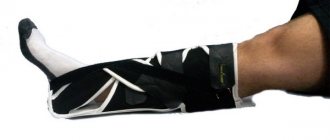
- Use a Lada bedpan with a small side for bowel movements to reduce pain in the leg. For urination, you can use a standard 0.8 liter jar or a special vessel.
- Record information about the dynamics of pain, adaptation, and independent exercise.
- Provide plenty of fluids (at least 1.5 liters), including soup, to avoid bedsores and constipation.
- Install a Balkan frame above the bed to facilitate movement in the patient’s bed when it is necessary to change linen, place a bedpan or treat bedsores. It also helps you start doing physical exercise.
- Add more vegetables and fiber to the menu.
When is a fixation bandage needed?
The difficulty with a hip fracture is that it never heals.
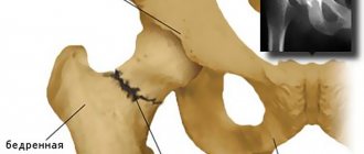
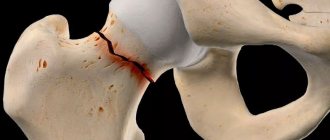
The fracture of the bone tissue goes along the acetabulum. During an injury, the arteries that supply the head of the joint are torn. Due to the lack of supply of nutrients, this part is absorbed 3-6 months after injury. It is for this reason that doctors insist on hip replacement surgery. Only endoprosthetics can return a person to a full life. Very old people who have received such an injury often refuse surgical intervention, given the risks.
Often receiving such an injury can be fatal due to its negative consequences. The patient finds himself bedridden. Blood circulation throughout the body is disrupted, and bedsores appear. There is a high probability of developing congestive pneumonia. When the patient lies down all the time, fluid accumulates in the lungs. It is an ideal environment for the proliferation of pathogenic bacteria. This type of pneumonia is difficult to treat.
The main task of doctors is to provide a patient with a serious injury with at least partial mobility. If the patient can get up and move slowly around the apartment or house, then most of the negative consequences of the injury will not overtake him. It is for these purposes that an orthosis is used for a femoral neck fracture.
Physical activity
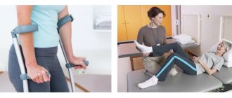
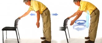
It is necessary to explain to the patient from the very beginning how important early physical activity is for him. To prevent pneumonia, breathing exercises are prescribed. Loading is necessary to maintain muscle tone. You should train the joints of the lower leg and knee, try to strain your thigh muscles, and lift yourself up using the bar.
With a healthy limb, they perform circular movements, bend the fingers, the sole of the foot, flex and extend the limb at the hip joint.
It is important to remember that excessively early stress on an injured limb can be dangerous, so activity should only begin after consultation with your doctor.
Description of the disease
A hip fracture is a serious injury that requires surgery to completely or partially replace the hip joint. But in medical practice there are situations when endoprosthetics is contraindicated. Old age, concomitant complex diseases, weak heart - in all these cases, there is a high probability that the patient simply will not survive the operation. Doctors choose palliative treatment tactics. Their main task is to relieve pain and at least partially restore the patient’s mobility. The patient will be able to move around his home, but will have to wear a hip brace constantly.
The choice of such devices is made by the doctor. He gives recommendations to patients and talks about the advantages of specific types of retainers. You can purchase them on order; regular pharmacies do not offer a wide range of such devices.
Types of bandages
Hip bandages for hip fractures can be divided into two broad categories - hard and soft. Each of these varieties has its own classification.
Rigid orthoses are divided into:
- One-sided;
- Double sided;
- With hinge;
- No hinge.
Such bandages are made of dense materials. The upper part is attached to the waist, the lower part is attached to the hip. They are connected using a hinge, which performs the functions of the damaged joint. The maximum bending angle can be adjusted.
Such devices are prescribed immediately after an injury, when the hip is very painful, and any movement is accompanied by a lot of unpleasant sensations.
Soft thigh bandages are also common. They are used during the rehabilitation period, reduce tissue swelling, provide blood flow to the damaged area, warm it and nourish it. Recovery is faster this way.
All bandages are left-handed and right-handed. Some manufacturers produce designs for both legs. They are used by patients whose integrity of the hip joints is compromised not due to injury, but due to dystrophic changes and autoimmune diseases.
Wearing a brace after joint replacement surgery
Opinions about the need to wear a hip orthosis after hip replacement surgery vary among doctors. Some argue that this is necessary because the use of a bandage reduces the risk of dislocation of the endoprosthesis in the first weeks after surgery. The latter argue for the refusal of the fixator by the fact that the muscles with its constant use relax, do not perform a supporting function, and the recovery process is delayed. Both sides are right, they make sound arguments.
If the operation went without complications, the recovery period goes on as usual, then there is no urgent need to use an orthosis. If there is severe swelling of the tissues and pain in the incision area, then in the first 2-3 weeks you can wear a soft retainer, but not on an ongoing basis, but for several hours a day.
The patient must independently get used to certain leg positions while walking, sitting, and sleeping. Every movement must be brought to automaticity. Therefore, using the orthosis on a permanent basis is not recommended.
Rules for selection and use
In order for the bandage to perform all its functions, it must be chosen correctly. The type of design, the presence or absence of a hinge - all these details are discussed with the doctor. Only an experienced traumatologist can assess the need to use an orthosis and give recommendations regarding its choice.
It is important to choose the correct size according to the waist, hip, and height of the patient. There are retainers designed for patients of different ages. This criterion must also be used. The patient's weight is of paramount importance in the rehabilitation process.
Obese patients have a more difficult time recovering. They are recommended to use only a rigid orthosis, which takes over all the functions of the hip joint. In this way, the load on damaged tissues is significantly reduced.
Department of Traumatology and Orthopedics
article in PDF format
QUOTE LINK:
A. Y. ZAROV, V. R. GUDKOV
GBOU HPE First Moscow State Medical University named after. THEM. Sechenov, Moscow
ANO Central Clinical Hospital of St. Alexis of the Moscow Patriarchate, Moscow
Information about the authors:
Zarov Alexey Yuryevich – GBOU HPE First Moscow State Medical University named after I.M. Sechenov. Department of Traumatology, Orthopedics and Disaster Surgery, assistant department of the Central Clinical Hospital of St. Alexius of the Moscow Patriarchate
Gudkov Vitaly Robertovich – Central Clinical Hospital of St. Alexius of the Moscow Patriarchate
Currently, the opinion about the advisability of organ-preserving operations for fractures of the femoral neck is becoming increasingly widespread. Features of the anatomical structure and blood supply of this area lead to special tactics for treating intra-articular fractures of the femoral neck. This article provides an overview of modern osteosynthesis options, including osteosynthesis with cancellous screws, dynamic hip screws, and the Targon FN system.
Key words: cancellous screws, dynamic hip screw, osteosynthesis, femoral neck fracture.
Introduction
Currently, the opinion about the advisability of organ-preserving operations for fractures of the femoral neck is becoming increasingly widespread. Features of the anatomical structure and blood supply of this area lead to special tactics for treating intra-articular fractures of the femoral neck. This article provides an overview of modern osteosynthesis options, including osteosynthesis with cancellous screws, dynamic hip screws, and the Targon FN system.
The incidence of fractures of the proximal femur, according to various estimates, ranges from 27 to 80 cases per 100 thousand population, with about 50% being intra-articular fractures of the femoral neck (Parker MJ, white A., Boyle A. 2008). In Russia, this figure is 61 cases per 100 thousand people and increases with the age of patients. Thus, 230 out of 100 thousand people over 75 years old experience fractures in this area (Shersternya N.A. 2005). An increase in the risk of hip fracture with age is associated with a progressive decrease in bone density - osteopenia (Babhulkar S., Tanna DD 2013). Osteopenic processes under the age of 60 are more pronounced in women, which is associated with hormonal changes in the postmenopausal period, however, in older patients, gender does not have a significant effect on the incidence of the disease (Vakulenko V.M. 2010).
Mortality during the first year after a hip fracture can be 16–28% and during the second year increases to 32.9% (Kurtinaitis J. et al. 2012). It is also important to note the significant functional impairment that develops in patients after a fracture (Johnell O., Kanis JA 2004). The quality of life of patients and mortality largely depend on whether surgical treatment was performed (Lesnyak O. et al. 2007). In recent decades, hip replacement has been considered the leading treatment method for femoral neck fractures. However, at present, some authors note the advantages of organ-preserving operations, mainly intramedullary osteosynthesis (Karev D.B. 2010; Sachse D. et al. 2014). Osteosynthesis operations take less time, cause fewer complications and are associated with less mortality (Parker MJ, white A., Boyle A. 2008). In this regard, more and more new metal structures are appearing for osteosynthesis of femoral neck fractures, which require study, improvement, and the formulation of selection criteria.
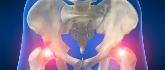
Anatomical aspects of femoral neck fractures
The proximal femur includes the head, neck, intertrochanteric and subtrochanteric regions of the femur. The neck of the bone is bounded by the boundaries of the head superiorly, the intertrochanteric line anteriorly, and the intertrochanteric ridge posteriorly (Babhulkar S., Tanna DD 2013). The plates of the spongy substance of the head and neck of the femur form a special system of trabeculae, the direction of the bundles of which corresponds to the lines of force. The intersections of the arcuate fascicle with the trochanteric and cephalic fascicles form two arches that can withstand heavy loads. In this case, the area between the two arches is the weakest and most susceptible to osteoporosis. Fractures of the femoral neck most often pass through this area (Kapandzhi A.I. 2010).
With a femoral neck fracture, there is also a disruption in the blood supply to the femoral head due to damage to the arteries and compression of the vessels by the intracapsular hematoma, while the greatest risk occurs with intra-articular femoral neck fractures. In subcapital fractures, when the fracture line passes distal to the entry into the head of the feeding vessels, only 8% of the blood flow is retained from a small number of vessels in the subcapital area and the vessels of the round ligament, which can lead to aseptic necrosis of the femoral head (Zhilyaev R.A., Tyazhelov A. .A., Zaritsky A.B. 2009).
It has been shown that different types of femoral neck fractures are accompanied by varying degrees of nutritional loss. Thus, subcapital abduction fractures without displacement are characterized by impaired blood supply within 10.2%, which provides a good prognosis in such patients and allows treatment with intramedullary osteosynthesis to be considered. With subcapital fractures with displacement, the most significant disturbance in the nutrition of the femoral head is observed (loss of up to 54.4%), which, taking into account age and general condition, may be an indication for endoprosthetics of the damaged joint (Litvinov A.A. 2002).
To take into account possible disturbances in the blood supply to the femoral head when choosing treatment tactics, the Garden classification is used. This division is based on the degree of valgus dislocation of the femoral head. When displacement occurs, the likelihood of necrosis of the femoral head increases due to disruption of its blood supply (Yang JJ et al. 2013). Therefore, non-displaced fractures of type I, II according to Garden are most favorable for treatment (Murphy DK, Randell T. 2013). In patients under 65 years of age with Garden I and II femoral neck fractures, osteosynthesis is preferred (Parker MJ, white A., Boyle A 2008).
Thus, due to its structural characteristics and blood supply, the femoral neck is one of the most vulnerable sections, which emphasizes the relevance of the problem of choosing treatment tactics depending on the specific clinical case. Determining the type of fracture according to different anatomical classifications makes it possible to predict the outcome of treatment and select the most appropriate treatment method for a given clinical case. However, difficulties in treatment can be caused not only by the location of the fracture and the presence of displacement, but also by other features of this area.
Possibilities of osteosynthesis in the treatment of fractures of the proximal femur
The proximal femur has a complex biomechanical structure that protects the area from fracture under axial loads. When implanting structures for osteosynthesis, a change in the biomechanics of the system occurs, which causes stress remodeling - a change in the composition, structure, volume and properties of bone tissue. If mechanical stresses exceed the tensile strength of bone tissue, then its destruction occurs, which must be taken into account when choosing a structure for osteosynthesis (Be'eryLipperman M., Gefen A. 2006).
It is obvious that the biomechanical features and characteristics of the blood supply to the proximal femur largely determine the healing process, which must be taken into account both in the conservative and surgical treatment of these fractures. To achieve a good treatment result, the following principles must be observed: anatomically accurate reposition of fragments, their reliable fixation, early activation of patients, gentle surgical intervention (Gilfanov S.I. 2010).
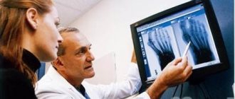
The results of osteosynthesis of the femoral neck vary significantly. The success of treatment is largely determined by an individual approach to each patient, careful selection of osteosynthesis techniques, competent technical execution, as well as attentive postoperative care. Errors that lead to a decrease in the rates of positive outcomes of osteosynthesis include: inadequate assessment of the severity of osteoporosis, incorrect interpretation of radiographs and characteristics of the fracture, incorrect selection and location of the fixator, insufficient adaptation of fragments, premature loading on the operated leg (Karev D.B., Boltrukevich S.I., Karev B.A. 2009).
Despite existing errors, treatment success, according to most authors, is observed in more than 70% (Zhang NN et al. 2013). The main percentage of negative results of treatment of intra-articular femoral neck fractures is mainly due to the frequent development of complications. These indicators reflect the need for a comparative analysis of the results of using various osteosynthesis techniques in the treatment of intra-articular fractures of the femoral neck.
Modern methods of osteosynthesis of femoral neck fractures
The most common methods of treating the femoral neck today include osteosynthesis with cancellous screws.
The main disadvantage of the method is the lack of stability due to the lack of fixation of the neck-diaphyseal angle. This leads to varus deformation in the fracture area due to the pressure of the screws on the lateral cortical bone (Parker MJ, Raghavan R., Gurusamy K. 2007). It is also highly likely to develop shortening of the femoral neck, which changes the biomechanics of the hip joint and negatively affects the functional result (Liu Y. et al. 2013). Another disadvantage of the method is the risk of migration of metal structures due to the movement of screws to the lateral side as the fracture consolidates. A retrospective analysis of 116 intra-articular femoral neck fractures stabilized with cancellous screws showed lack of consolidation and avascular necrosis of the femoral head in 14.7% of cases (Lee KB, Howe TS, Chang HC 2004). In another study, the formation of a false joint was found in 19.4% of cases (Basov A.V. 2012). It was shown that the majority of cases of lack of consolidation occurred in displaced fractures (Lu QH, Yu FP 2012).
Osteosynthesis with a dynamic hip screw (DHS) is widely used in the treatment of femoral neck fractures. The peculiarity of this screw is its ability to “spring”. The so-called monoaxial dynamization along the axis of the femoral neck causes dynamic compression of the fracture line necessary for bone consolidation. The screw is secured using an extramedullary plate located on the outer surface of the femur, which ensures fixation of the neck-diaphyseal angle (Ananko A.A., Babko A.N. 2007).
However, when using a dynamic hip screw in patients with intra-articular femoral neck fractures, a positive result was achieved only in 73.4% of cases (Majernicek M. et al. 2009). It should be noted that in addition to standard complications, DHS osteosynthesis in some cases may be accompanied by migration of the screw and displacement of bone fragments (Hrubina M., Skotak M., Behounek J. 2010). There is also a very strong possibility of rotational misalignment given the lack of a derotation component in the design. Although the DHS system is widely used in patients with trochanteric fractures, there are insufficient studies on the treatment of intra-articular neck fractures to confirm its effectiveness in these patients.
The obvious disadvantage of this technique is the relatively high invasiveness of the operation.
The least traumatic methods of treating femoral neck fractures include osteosynthesis with a bundle of wires. The method is minimally invasive and minimally damages bone tissue. However, it is noted that osteosynthesis with wires is not cruel enough and creates a high risk of infection spreading along the parts of the wires protruding above the skin (Karev D.B. et al. 2009). One of the options for osteosynthesis with a bundle of wires is osteosynthesis with bundles of V-shaped wires. When inserting bundles of V-shaped wires, the bone beams are moved apart without their destruction along the perimeter of the fixator, which distinguishes the method from osteosynthesis using large structures. In combination with dynamic tension in the system, this creates optimal conditions for healing even against the background of osteoporosis.
However, when using this method, non-union of the fracture was observed in 17.6% of cases, and in the long term a large percentage of unsatisfactory results were noted, mainly associated with shortening of the femoral neck (Ardashev I.P. 2012). There also appears to be a high risk of varus deformity due to the lack of fixation of the neck-diaphyseal angle.
The undoubted advantage of this technique is its unprecedented low cost.
The Targon FN (TFN) fixation system was developed taking into account all the shortcomings of previously used methods. The system includes a plate with four holes for insertion of cancellous and bicortical screws. Telescopic cancellous screws allow you to control the consolidation of the fracture, while preventing the migration of structures into the soft tissue. The presence of a femoral plate for securing the screws ensures fixation of the neck-shaft angle and rotational stability.
The surgical technique makes it possible to perform manipulations through mini-approaches and avoid complications such as bending of the guide pin and sinking of the pin into the pelvis (MJ Parker, R. Raghavan, Gurusamy K. 2007). A large study of this system included 320 patients. Of the 112 non-displaced fractures, in three cases (2.7%) the fracture did not heal or secondary displacement occurred, and in five cases (4.5%) necrosis of the bone head occurred. Among 208 patients with displaced fractures, consolidation could not be achieved in 32 (15.4%), and necrosis developed in 23 (11.1%). In addition, the work revealed cases of secondary fractures in the area of the structure (1.9%) (Parker M., Cawley S., Palial V. 2013).
R. Biber proposed the following tactics for managing patients with intra-articular fractures of the femoral neck. Patients under 60 years of age underwent osteosynthesis with a TFN construct regardless of the fracture characteristics, while elderly patients received TFN only for Garden types I and II. At the same time, the frequency of postoperative complications was 16.4% and prevailed in patients with displaced fractures (Biber R., Brem M., Bail HJ 2014). According to the authors, the main complication of osteosynthesis with this system is perforation of the metal structure, which does not agree with the data of other studies (Parker MJ, Stedtfeld Hw 2010).
TFN has proven to be a more effective method of osteosynthesis for intra-articular fractures of the femoral neck compared to DHS, cancellous screws and wires, however, it is worth noting a complication uncharacteristic of other techniques - a fracture in the area of insertion of the structure.
Discussion
In the literature one can find studies on the results of using various designs for osteosynthesis of the femoral neck, but the question of which method is the most effective remains open. E. Brandt showed that the TFN design can withstand twice the load than a dynamic hip screw and is comparable in strength to a design of three cannulated screws (Brandt E., Verdonschot N. 2011). A comparison of the results of surgical treatment of 52 patients with femoral neck fractures showed that TFN osteosynthesis is less often accompanied by migration of metal structures than DHS (Eschler A. et al. 2014).
Displaced intra-articular fractures are less treatable than others. According to a retrospective analysis of the treatment of 78 patients with such fractures, osteosynthesis with a TFN construction is accompanied by a lower incidence of nonunion compared with cancellous cannulated screws (3.2% compared to 46.8%) (ein R. et al. 2014).
The temporary disadvantage of the TFN system is associated with a specific method of installing the structure, which requires mastery and experience (Korver RJ et al. 2013). At first, this may lead to longer surgery times and higher radiation exposure compared to the widely used methods of osteosynthesis with cancellous screws and DHS.
Due to the fact that femoral neck fractures heal less well in older people, a comparison of osteosynthesis with TFN and cancellous screws in patients over 65 years old, conducted in the UK, is relevant (Gri n xL et al. 2014). It was found that both methods are equally effective for intra-articular femoral neck fractures in elderly people. At the same time, certain complications develop with the same frequency. The authors of one study draw attention to the need to evaluate factors such as the traumatic nature and duration of the operation, the amount of blood loss during surgery and the length of hospitalization when treating elderly people (Lee YS et al. 2008).
According to the data presented, the most promising method of osteosynthesis of femoral neck fractures at the moment is TFN. Although there is no difference in mortality between different techniques and in the incidence of major complications such as fracture nonunion and avascular necrosis of the bone head, TFN is able to withstand greater loads and migrates less frequently into soft tissue.
conclusions
1. Osteosynthesis is an organ-preserving operation and is therefore the preferred option for surgical treatment of the femoral neck.
2. The main criteria for determining the tactics of surgical treatment of femoral neck fractures are the type of fracture and the age of the patient.
3. Determining the type of fracture according to different anatomical classifications makes it possible to predict the outcome of treatment and choose the optimal surgical tactics. Namely, to make a choice between osteosynthesis and hip replacement.
4. The severity of the circulatory disturbance and, consequently, the frequency of development of avascular necrosis and fracture pseudarthrosis, first of all, depends on the degree of displacement in the fracture zone. In view of this, the leading classification for choosing a method of surgical treatment is the Garden classification, since it is based on the degree of fracture displacement.
5. All existing methods of osteosynthesis, such as osteosynthesis with cannulated screws, DHS, intramedullary osteosynthesis, osteosynthesis with tension wires, osteosynthesis with the Targon FN system, etc. When performed according to indications, in the vast majority of cases, fracture consolidation can be achieved.
6. The optimal fixation should combine the principle of dynamic fixation, rigid fixation of the neck-diaphyseal angle and the presence of a derotational component.
Bibliography
1. Lazarev A.F., Solod E.I. Methodological recommendations: minimally invasive osteosynthesis of femoral neck fractures using V-tensioned wires, CITO named after. N.N. Priorova.Moscow 2013
2. Ananko A.A., Babko A.N. Modern traumatological tactics for proximal femur fractures: (review of German lit.) // Ukr. honey. chasop. 2007. No. 1. P. 75-80.
3. Ardashev I.P. Experience in treating femoral neck fractures with V-shaped wire bundles // Medicine in Kuzbass. — 2012. T. 11. No. 2. pp. 18-23.
4. Basov A.V. et al. Experience in treating femoral neck fractures with cancellous screws // Bulletin of new medical technologies. 2012. T. 19. No. 2.
5. Vakulenko V.M., Vakulenko A.V., Nedelko A.A., Lapai V.V. Structure of fractures of the proximal region of the femur // Ukrainian Medical Almanac. 2010. Volume 13, No. 3. pp. 35-36.
6. Gilfanov S.I. Treatment of fractures of the proximal femur: Abstract of thesis. dis. ... doc. honey. Sciences: 14.00.18 – Moscow, 2010.
7. Zhilyaev R. A., Tyazhelov A. A., Zaritsky A. B. Variant features of the blood supply to the femur // Trauma. 2009. T. 10. No. 1. pp. 36-39.
8. Kapandzhi A.I. Lower limb // Functional anatomy. M.: Eksmo, 2010. T. 2. Page. thirty.
9. Karev D. B., Boltrukevich S. I., Karev B. A. Errors and complications in the treatment of patients with medial fractures of the femur // Bulletin of Vitebsk State Medical University. 2009. T. 8. No. 1.
10. Karev D. B. et al. Osteosynthesis with compression screws as a treatment option for patients with medial fractures of the femur // News of surgery. 2009. T. 17. No. 3. pp. 96-102.
11. Karev D. B. Valgus reconstruction in the treatment of patients with fractures of the proximal femur // Journal of GrSMU. 2010. No2 (30).
12. Lesnyak O. M., Bakhtiyarova S. A. et al. Quality of life in osteoporosis. Prospective observation of patients who suffered a fracture of the proximal femur // Osteoporosis and Osteopathy. 2007. T. 3. P. 4-8.
13. Litvinov A.A. Features of intraosseous circulation during surgical treatment of medial femoral neck fractures in adults: Abstract of thesis. ...cand. honey. Sciences: 14.00.27 / Litvinov Andrey Aleksandrovich; RGMU named after. Pavlova. Ryazan, 2002.
14. Lazarev A.F., Solod E.I. Methodological recommendations: minimally invasive osteosynthesis of femoral neck fractures using V-tensioned wires, CITO named after. N.N. Priorova. Moscow 2013
15. Shesternya N.A., Gamdi Y., Ivannikov S.V. Femoral neck fractures. M.: BINOM, 2005. 104 p.
16. Babhulkar S., Tanna DD Proximal Femoral Fractures. – JP Medical Ltd, 2013.
17. Biber R., Brem M., Bail H. J. Targon Femoral Neck for femoral neck fracture xation: lessons learned from a series of one hundred and thirty ve consecutive cases // Int Orthop. 2014. T. 38. No. 3. P. 595-9.
18. Brandt E., Verdonschot n. Biomechanical analysis of the sliding hip screw, cannulated screws and Targon1 FN in intracapsular hip fractures in cadaver femora // Injury. 2011. T. 42. No. 2. P. 183-7.
19. Eschler A., Brandt S., Gierer P., Mittlmeier T., Gradl G. Angular stable multiple screw xation (Targon FN) versus standard SHS for the xation of femoral neck fractures. Injury. 2014. T. 45 Suppl 1. C. S76-80.
20. Grin XL, Parsons N., Achten J., Costa ML e Targon femoral neck hip screw versus cannulated screws for internal xation of intracapsular fractures of the hip: a randomized controlled trial // Bone Joint J. 2014. T. 96 -B. No. 5. C. 652-7.
21. Hrubina M., Skotak M., Behounek J. // Acta Chir Orthop Traumatol Cech. 2010. T. 77. No. 5. P. 395-401.
22. Lee KB, howe TS, Chang h. C. Cancellous screw xation for femoral neck fractures: one hundred and sixteen patients // Ann Acad Med Singapore. 2004. T. 33. No. 2. P. 248-51.
23. Lee Y. S., Chen S. h., Tsuang Y. h., Huang h. L., Lo TY, huang CR Internal xation of undisplaced femoral neck fractures in the elderly: a retrospective comparison of xation methods // J Trauma. 2008. T. 64. No. 1. P. 155-62.
24. Liu Y., Ai ZS, Shao J., Yang T. Femoral neck shortening a er internal xation // Acta Orthop Traumatol Turc. 2013. T. 47. No. 6. P. 400-4.
25. Lu Q. h. , Yu FP [erapeutic e ects of cannulated compression screws for treating femoral neck fractures] // Zhongguo Gu Shang. 2012. T. 25. No. 12. P. 1040-4.
26. Majernicek M., Dungl P., Kolman J., Malkus T., Vaculik J. // Acta Chir Orthop Traumatol Cech. 2009. T. 76. No. 4. P. 319-25.
27. Murphy DK, Randell T., Brennan KL, Probe RA, Brennan ML Treatment and displacement a ect the reoperation rate for femoral neck fracture // Clin Orthop Relat Res. 2013. T. 471. No. 8. P. 2691-702.
28. Parker M., Cawley S., Palial V. Internal xation of intracapsular fractures of the hip using a dynamic locking plate: Two-year follow-up of 320 patients // Bone Joint J. 2013. T. 95-B. No. 10. C. 1402-5.
29. Parker MJ, Raghavan R., Gurusamy K. Incidence of fracture healing complications in femoral neck fractures // Clin Orthop Relat Res. 2007. T. 458. P. 175-9.
30. Parker MJ, Stedtfeld hw Internal xation of intracapsular hip fractures with a dynamic locking plate: initial experience and results for 83 patients treated with a new implant // Injury. 2010. T. 41. No. 4. P. 348-51.
31. Parker MJ, white A., Boyle A. Fixation versus hemiarthroplasty for undisplaced intracapsular hip fractures // Injury. 2008. T. 39. No. 7. P. 791-5.
32. Sachse D., Beiter C., Bludau F., Obertacke U., Schreiner U. // Z Orthop Unfall. 2014. T. 152. No. 1. P. 20-5.
33. ein R., Herman A., Kedem P., Chechik A., Shazar n. Osteosynthesis of unstable intracapsular femoral neck fracture by dynamic locking plate or screw xation: early results // J Orthop Trauma. 2014. T. 28. No. 2. P. 70-6.
34. Yang JJ, Lin LC, Chao K. h., Chuang SY, wu CC, Yeh TT , Lian YT Risk factors for nonunion in patients with intracapsular femoral neck fractures treated with three cannulated screws placed in either a triangle or an inverted triangle conguration // J Bone Joint Surg Am. 2013. T. 95. No. 1. P. 61-9.
35. Zhang nn, Ye ZM, Zhu YY, Ren w. F. [Case-control study on double screws system and compressed three canulated screws in treating femoral neck fractures] // Zhongguo Gu Shang. 2013. T. 26. No. 7. P. 565-71.
OVERVIEW:OSTEOSYNTHESISOFINTRACAPCULARFEMORALNECK FRACTURE
A. YU. ZAROV, VR GUDKOV
IM Sechenov First Moscow State Medical University of the Ministry of Health of the Russian Federation, Moscow Central Clinical Hospital of St. Alexis of the Moscow Patriarchate
Information about the authors:
Zarov Alexey Yurevich – St. Alexius Central Hospital; e-mail
Gudkov Vitaliy Robertovich – St.Alexius Central Hospital
At the present time it is becoming more common to choose conserving surgery for managing femoral neck fracture. Peculiarities of anatomy and blood supply of this region lead to a particular therapeutic approach to intracapsular fracture of the femoral neck. In this article we review modern variants of osteosynthesis including cancellous screws xation, osteosynthesis with dynamic hip screw and Targon FN xation system.
Key words: cancellous screws, DHS, dynamic hip screw, femoral neck fracture, Targon FN.
Bandage as an aid for fracture healing
Patients need to understand a simple truth - a hip fracture never heals completely. In 80% of cases, the head of the joint resolves and the femur rests on the pelvic bones. In such patients, one leg is noticeably shorter than the other. In the remaining 15% of situations, a false joint is formed from overgrown cartilage tissue. It is absolutely motionless, the patient’s leg almost does not bend, but such a growth can withstand certain loads when walking.
A bandage for an inoperable femoral neck fracture helps to build up cartilage tissue. If the joint remains stationary, the pain will become dull over time. The person regains mobility. At first, he can only walk with a walker or crutches without the slightest load on his sore leg. Over time, you are allowed to step on it a little. Without an orthosis, walking is almost impossible, because every step is accompanied by severe pain.
Don't place false hopes on the bandage. This device helps to at least partially return to a full life, improve its quality for the victim of injury, but does not contribute to a full recovery.