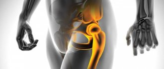
An arm orthosis is an orthopedic device for the upper limb, used to partially or completely immobilize (immobilize) the affected area or the entire arm and reduce the load on it.
It is used for the treatment and prevention of various diseases of the osteoarticular system. It is also used during the rehabilitation period after injuries or operations on the upper limb.
Why do you need a bandage and support bandages for a broken arm?
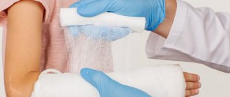
The use of various types of bandages or bandages is a necessary treatment measure for a broken arm. This is due to the fact that the patient needs to limit physical activity in the damaged area.
Previous use of fixation devices ensures immobility of the fracture and rapid healing of bone fragments.
As a result, the treatment period is shortened. In addition, these devices are used during the rehabilitation period, which will reduce the risk of complications.
Indications
The Deso bandage is necessary to immobilize the arm from the shoulder to the finger phalanges when:
- shoulder dislocation and to prevent relapses;
- fracture of the humerus;
- collarbone fracture;
- scapula fracture;
- ligament damage;
- paralysis of the upper limbs;
- arthritis, periarthritis and arthrosis;
- secondary myositis, neuritis, paresis and plexitis;
- during the recovery period after surgery on the upper limb and wearing a cast.
Can be used for simple fractures - when there is no risk of displacement and damage to soft tissues from bone fragments. Sometimes prescribed for bruises and cuts on the hands.
Types of arm bandages
In traumatology, several types are used to securely fix the hand. Each product has specific structure and functions.
Headband
A bandage in the form of a scarf for a fractured arm has the shape of an isosceles triangle. Most often they are made from natural materials, such as cotton. This helps prevent increased sweating, overheating of the skin, and the development of allergic reactions.
The scarf device is used at the prehospital stage as a method of providing first aid. It retains slight mobility of the bones of the upper shoulder girdle, which can be used as treatment after removal of the plaster. This product fixes the arm bent at the elbow joint, which is pressed to the chest.
Bandage
An arm holder for a fracture is one of the most effective ways to fix a broken limb. There are two types of devices. These are soft and hard bandages. The latter are used to immobilize a limb immediately after injury or after healing of a postoperative scar. Its design is dense with an unbendable frame that allows you to securely fix your hand.
Soft structures are used at the rehabilitation stage. They have less weight compared to a plaster cast and are small in size. The doctor can select different types of structures depending on the location of the fracture.
Retainer
This immobilization device allows you to reliably immobilize the limb and prevent displacement of soft tissues. There are several types of clamps, since products with inflatable parts or a rigid frame can be used. They are selected depending on the location of the fracture, as well as the method of treatment.
The materials from which the retainer is made are hypoallergenic and easy to clean. It is attached to the body using straps, Velcro or fasteners. The fixator on the injured arm after a fracture can be removed and also stretched when the tissues swell.
Orthosis
An orthosis is a popular device that has a rigid frame, straps, or other fixation devices. It is distinguished by its ease of use, as well as the quality of materials that prevent damage to the skin or discomfort. Cutting off the arm for an uncomplicated fracture reduces the restriction of movement, which may not limit the ability to work.
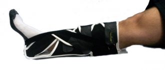
Splint
This type of splint consists of an elongated frame made of plaster, plastic or fabric. Most often they are applied for fractures of the radius in the distal part. The advantages of use in clinical practice include a reduced risk of edema, entrapment of neurovascular bundles and muscle fiber atrophy.
In addition, when applying it, you can monitor the condition of the skin.
Fracture of the hand
Fracture of the radius of the arm
The most common fracture of the hand is a fracture of the distal epimetaphysis of the radius (“in a typical location”) in combination with a fracture of the styloid process of the ulna.
This type of injury is subject to a certain seasonality: the number of victims especially increases during icy conditions. Most often it happens when falling on an outstretched arm.
Fractures of the radius according to the classification of the Association of Osteosynthesis (AO) are divided into three types - A, B and C depending on the complexity: from A (the simplest extra-articular fractures of the radius without displacement) to C (complex comminuted intra-articular fractures of the radius with pronounced displacement) . Each type of injury has its own treatment method.
Diagnosis of a fracture of the radial head is not difficult: the initial examination reveals deformation, pain, and pathological mobility. A fracture of the hand (to clarify the type and complexity of the injury) requires radiography, which gives an adequate picture of the existing situation. In some cases of displaced intra-articular radius fractures, computed tomography is sometimes performed for greater visualization.
Treatment of a broken arm
When treating closed Type A radius fractures (called impacted radius fractures), closed reduction and a plaster cast are usually used.
One of the problems that can arise with the conservative method of treating a fracture of the radius is secondary displacement of fragments. Therefore, after repositioning and applying a plaster splint, it is necessary to ensure that the bandage does not squeeze the arm, lies quite tightly, but not tightly, and after the swelling subsides (on the 5-7th day), it is necessary to perform an x-ray control to make sure that secondary displacement has not occurred.
If there is a tendency to secondary displacement, then surgical treatment is recommended. For this purpose, several osteosynthesis techniques are used:
- osteosynthesis with three knitting needles according to Kapandzhi;
- osteosynthesis with plates with angular stability, which are designed specifically for the distal radius.
Fractures of types B and C in 99% of cases tend to be displaced, that is, they are obviously unstable. Depending on their location and the degree of damage to the articular surface, two surgical treatment options are possible:
- osteosynthesis with plates with angular stability for the distal radius;
- application of distraction devices (for intra-articular, severely comminuted fractures), which are used when applying a plate causes technical difficulties associated with the presence of a large number of small fragments, osteoporosis, etc.
Osteosynthesis with wires and plates is usually combined with the mandatory application of a plaster cast for several weeks. This is necessary for less trauma to the tissues in the surgical area, for better reduction of swelling and reduction of pain.
Also in the postoperative period it is necessary to use physiotherapy and exercise therapy.
Recently, there has been a tendency that during any operation for intra-articular injuries (displaced fracture of the radius), the reposition of fragments must be controlled using an arthroscope, which makes it possible to completely eliminate the displacement of fragments and significantly reduce the risk of developing deforming arthrosis.
The most serious complication of injuries of this location is complex regional pain syndrome, which develops, as a rule, with conservative treatment, but also occurs after severely traumatic operations.
With conservative therapy, this syndrome occurs in a situation where repeated and traumatic attempts at reposition have been made, or an unmodeled plaster cast has been applied. In this case, the bandage compresses the injured limb, which leads to disruption of blood supply and, as a consequence, entails the development of complex regional pain syndrome.
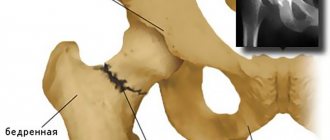
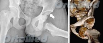
Scaphoid fracture
Fractures of the scaphoid bone of the hand are also very common. They can be either isolated or associated with a dislocation of the hand, transscaphoid-perilunar dislocations. The choice of treatment method for such a fracture of the arm depends on the time that has passed since the injury and on the degree of negative changes in the area of the scaphoid bone.
If the patient has a fresh fracture of the arm, treatment (in the absence of displacement) is indicated conservatively. A plaster cast is applied, after 6 weeks an X-ray control is performed, and if the image shows no signs of fusion, osteosynthesis of the scaphoid bone is performed. In the case of transscaphoid-perilunar dislocation, elimination of the dislocation, reposition of the scaphoid bone and osteosynthesis with a special titanium screw are performed, ensuring adequate compression of the fragments, which is necessary for fusion.
In more advanced cases, when cystic bone reconstruction is present, treatment combines various options for bone grafting.
If there are pronounced signs of deforming arthrosis in the area of the scaphoid, various treatment options for bone fractures are also possible:
- removal of the scaphoid bone and the formation of partial arthrodeses of the wrist joint;
- bone prosthetics using pericarbon prostheses (very common in foreign practice);
- complete arthrodesis of the wrist joint - in the event that no other options bring relief to the person and do not help reduce pain.
For diagnosis, in addition to visual examination, radiography and computed tomography are indicated. In some situations, it makes sense to conduct angiography of the injured limb to determine the degree of vascularization (blood supply) of the damaged bone.
Metacarpal fractures
Metacarpal fractures are an injury that most often occurs when the fist hits hard objects. Treatment for this type of injury depends on its location.
In case of a fracture of the head of the radial bone, it is advisable to perform osteosynthesis with knitting needles or osteosynthesis with a plate.
A fracture of the diaphyses of the metacarpal bones is treated using osteosynthesis with pins (stable osteosynthesis) according to a technique developed under the guidance of Professor V.F. Korshunov. This is a fairly simple and effective technique that allows you to make a reposition using two small incisions (1-1.5 cm each) and insert a pin into the bone. This technique allows you to fully restore hand function almost a week after surgery. Six months after installation, if there are signs of fusion in the fracture area, the pin is removed.
A fracture in the area of the base of the metacarpal bones is fixed mainly with the help of knitting needles. Oblique and helical fractures can be fixed with minifragment screws in combination, if necessary, with plates for external osteosynthesis.
Fractures of the phalanges of the fingers: osteosynthesis with wires and microscrews for mini-fragments
Damage to hand tendons
For fresh injuries to the tendons of the hand (both flexor and extensor tendons), a suture is performed. Unfortunately, when applying a suture to the flexor tendon, there is a certain difficulty in that the tendon is usually damaged in the area of the fibrous canal, in the “dead zone”. A seam in this area often does not lead to the desired result. Recently, special sutures have been developed that allow the patient to be quickly activated in the postoperative period to fully restore the functions of the injured limb.
In long-standing situations of flexor tendon injuries, a two-stage repair is performed. At the first stage, scar tissue is removed from the tendon canal area and a special silicone prosthesis is placed there. Six months later, after a new tendon canal has formed around it, it is replaced by a graft of another tendon (either from the foot or hand).
Nerve damage
Damage to the radial nerve most often occurs during humerus fractures. Traumatic damage to the nerve can occur even if there was no direct impact on it - only due to cicatricial processes in the area of wound healing. Treatment of damage to the radial nerve in this case is carried out using surgery aimed at releasing the nerve from compression.
In all cases of fresh peripheral nerve injury, an epineural suture is necessary. Only in this case can we count on the most complete restoration of the function of the damaged nerve.
In cases of old damage, in the absence of a nerve defect after its isolation from scar tissue, it is possible to perform a secondary epineural suture. If there is a nerve defect, it is necessary to perform nerve plasty, most often with a graft from n. suralis.
Types of Shoulder Bands
Bandages for fractures in different parts of the arm are prescribed to reduce the load on the upper shoulder girdle, as well as limit motor activity.
Deso bandage
An immobilizing bandage is applied after placing a cotton roller in the armpit. The arm should be bent at the shoulder joint and pressed to the chest. Bandages are applied in horizontal and vertical directions.
Spica bandage
This type allows you to reliably fix the affected area with a compression effect. It is used for uncomplicated fractures, as well as in the postoperative period. In order to prevent infection, it is necessary to apply a sterile napkin to the skin. Overlay involves laying rounds on the previous one with an oblique arrangement.
Kerchief
This type is the most common, since the patient can apply it independently. Due to the speed of application of the material, it is widely used in emergency situations.
Types of bandages for the wrist joint Injury to the wrist joint or hand requires the use of various types of bandages. Among the most common are:
- Circular or circular. This type is the most common and simple; a prerequisite is that the new round overlaps the previous one.
- Spiral. It is distinguished by a more complex technique, since the previous round should only be covered by half. The spiral bandage allows you to fix a larger area. Creeping. Thanks to this type of bandaging, a large surface of the affected area is covered, while it is characterized by low strength due to the fact that the rounds do not overlap each other.
- Cross-shaped. The use of dressing material in the form of 8 is indicated for fixing mobile joints. They help limit physical activity if the affected area is irregularly shaped.
The choice of type of bandage depends on the type of injury, as well as the extent of the lesion.
Available materials for constructing a sling
It is not always possible to access qualified medical assistance on time. For example, if by an unfortunate accident you were injured while hiking and you still need to get to the doctor, it is better to temporarily immobilize your arm using improvised means. You can use the following options:
- Clothes with long sleeves. Tie a shirt or sweater with the sleeves behind your head, and insert your hand into the resulting loop. This will help transfer the weight of the injured limb to the neck or back. The length of the sling can be adjusted.
- Belt. Make a loop of the required size, secure it with a buckle, put your hand through it and put the belt around your neck so that the metal elements do not put pressure on the skin.
- Tie. Using a simple knot, make a loop out of the tie and hang the injured limb around your neck. You need to adjust the loop so that your hand is at an angle of 90 degrees.
- An improvised bandage can be made from adhesive tape, wound in several layers.
Rules for constructing a bandage
Any scarf or bandage to support an arm during a fracture must be constructed in compliance with the basic rules. These include:
- Preliminary preparation of material. A bandage or any material must be used in its pure form. The only exception is emergency care.
- Application of the first fixation round, which ensures reliable fixation of subsequent sections.
- Use in a comfortable position for the patient, as well as the person providing assistance.
- Pre-treatment of the affected surface.
Trauma to the skull and face
Skull injuries are divided into calvarial fractures and basal skull fractures. Such fractures are among the most dangerous because they can lead to brain damage.
Treatment of a fracture of the base of the skull
For a fracture of the base of the skull, the choice of treatment method is based on the nature and severity of the injury. In the absence of bone displacement and damage to the meninges, conservative treatment is carried out:
For a fracture with bone fragmentation, displacement and damage to the meninges, surgical treatment is indicated.
How to make a bandage for a fracture with your own hands at home
As first aid at the prehospital stage, bandages are used if a fracture of the arm is suspected.
We make a bandage for an injury to the elbow and wrist
Support for an injured arm with a fracture in the elbow or wrist is most often done using a circular or scarf bandage. These methods are the simplest and most effective.
Making a shoulder bandage
To securely fix the shoulder at home, use the Deso bandage. To do this, a roller is placed in the armpit, and the arm pressed to the chest is bandaged around the circumference of the chest and the arm vertically.
How to wear a bandage and bandage correctly
In order to choose the right bandage after a broken arm, you need to consult a doctor. A traumatologist or surgeon selects a product based on the severity of the disease, taking into account safe material and size. In addition, to avoid complications, it is important to follow the basic rules regarding the use of the product. These include:
- Checking the integrity of the product, as well as fixing devices.
- Checking the purity and quality of materials that will adhere to the skin. This will prevent the formation of pressure sores or ulcers associated with chafing.
- First fixation in the presence of a specialist. After the bandage or fixation bandage is put on, the doctor evaluates the reliability of attachment of the parts, maintaining the arm in the desired position, as well as safety for surrounding tissues.
A children's arm bandage for a fracture is selected taking into account the patient's age. The material from which the product is made must be natural.
Features of the metacarpal bones
Our hands have as many metacarpal bones as there are fingers—usually five on each hand. These bones occupy most of the surface of the palm. On the dorsum of the hand, the metacarpal bones can be easily felt under the skin and are separated from it only by a thin layer of fatty tissue and tendons. Each metacarpal bone consists of a head (knuckle), a body (shaft) and a base.
The 1st metacarpal bone plays a special role: it has maximum mobility and moves in different directions. The remaining metacarpal bones are not as mobile. And if the 4th and 5th bones still retain a certain range of motion, then the 2nd and 3rd are firmly connected to the wrist. In this case, a more mobile bone with an incorrectly healed fracture can compensate for the angular curvature by movement, but for a stationary bone, a deviation of even a few degrees becomes fatal.
Types of typical injuries
After an injury, one or more bones may break; damage to the head, diaphysis, or base of the bone may occur. The weakest zone of the 2nd-4th metacarpal bones is the so-called neck, located under the head of the bone. It is this that breaks under load along the longitudinal axis. If the load has a rotational moment, a spiral fracture is formed; if there was direct pressure across the bone, a transverse fracture occurs.
The most common fracture is the subcapitate fracture of the 5th metatarsal bone. This injury is also called a boxer's fracture because the mechanism usually involves a forceful punch to a hard surface. The head of the bone is “folded” in the palmar direction or driven into the diaphysis. Sometimes, in addition to the 5th, other bones of the hand can break in this way.
Any injury to the 1st metacarpal bone is difficult to endure, since it is this bone that ensures the mobility of the thumb. Each type of fracture of this bone even has its own name, for example, Rolando, Bennett, Winterstein fracture. An incorrectly healed or non-healed fracture of the base of the 1st metacarpal bone completely “turns off” the ability to make precise movements and grip strength.
Posture when wearing a bandage
Damage to the bones of the upper limb is an indication for immobilization of the patient. In most cases, a fixing bandage is applied to reduce the load on the damaged bone structure, as well as reduce the load on this part. It should be taken into account that different types lead to increased load on the spine in the cervical and thoracic region.
Long-term wearing of fixing devices is accompanied by the development of pain symptoms, as well as dizziness.
In order to avoid problems associated with poor posture, you must adhere to the basic rules. Among them:
- Maintaining a straight but relaxed back with shoulders slightly back when walking. You should also keep your chin slightly raised, as lowering it increases the load on the cervical spine.
- The use of chairs with a back, as well as armrests, on which the injured arm is placed. If the patient is sitting, then his back should be in a straight position.
- Contact a specialist at the first symptoms of pain and discomfort to resolve the issue of changing fixing devices and eliminating pathological manifestations.
Using a bandage based on the severity and type of fracture allows for rapid healing and restoration of limb mobility. Errors in choosing the method of immobilization can lead to pathological fusion of bones, as well as damage to the neurovascular bundle.
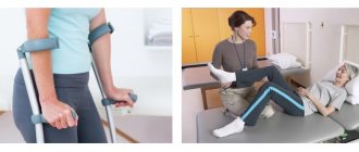
Rehabilitation treatment after a fracture
In order not only to achieve bone fusion, but also to return the normal functioning of the injured limb and restore its full motor activity, it is necessary to undergo a full course of rehabilitation treatment. Therefore, after the anatomical structure of the bone is restored, we do not just advise, we strongly recommend that each patient engage in restorative therapy.
A set of rehabilitation procedures is developed individually and depends not only on the type and complexity of the fracture, but also on the general condition of the patient, his age and physical capabilities. The main points of the therapeutic program are various physiotherapeutic procedures and physical therapy necessary to restore the functioning of the bones and muscle-ligamentous apparatus.
If discomfort and pain occur, the patient is prescribed xenon procedures aimed at increasing tone and physical activity. This allows you to go through the entire rehabilitation cycle in full force and achieve lasting results.