Bones, lungs and liver are the organs to which metastases most often spread in stage 4 cancer. Bone metastases from cancer are much more common than primary tumors.
Our expert in this field:
Sergeev Pyotr Sergeevich
Oncologist, surgeon, chemotherapist, Ph.D. Member of the international society of surgical oncologists EESG
Call the doctor Reviews about the doctor
Most often, bone metastases occur in breast, prostate and lung cancer. One of the most common locations is the spine. Remission for cancer with secondary lesions in the bones is almost impossible, but there are treatments that help slow the progression of the tumor and increase the patient's life expectancy.
Why are so few spinal surgeries performed in Russia for metastatic destruction of the vertebral bodies? Does it make sense to deal with pain and undergo surgery, since bone metastases significantly reduce the survival prognosis?
Why do bone metastases occur?
Metastasis is a complex process, its mechanisms are not fully understood. As the primary tumor grows into the surrounding tissue, some cells may break away from it and penetrate into the lumen of the blood or lymph vessels. They migrate with the blood or lymph flow, then, reaching small vessels in the bone, they exit into the tissue and give rise to new lesions. As the metastasis grows, the cancer cells release substances that stimulate the formation of new blood vessels.
The main problem with metastases is that they are usually not one or two nodes that can be removed. As a rule, there are many secondary lesions, some of them very small. Therefore, it is difficult to fight them.
Most often, cancer cells settle in bones, which have a rich blood supply. In most cases, they are found in the vertebrae, pelvic bones, femurs, ribs, and arm bones.
Risk factors
- Injury. But there is an opinion that trauma is a common occurrence in children and adolescents and is not a fully proven cause of the development of sarcoma or one of its triggers.
- Children's age and heavy physical activity. Sarcoma is most often localized in the distal femur and proximal tibia. These are areas of bone tissue growth.
- Exposure to ionizing radiation. This may be due to radiotherapy for malignant tumors of other locations or living in a radiation zone.
- The presence of benign tumors prone to malignancy. These are Paget's disease, chondromas, fibrous dystrophy and others.
What are the symptoms of bone metastases?
The main symptom is bone pain . If they begin to bother a patient suffering from cancer, suspicion should first of all fall on bone metastases. Depending on which part of the skeleton is affected, you may experience pain in the back, pelvis, arms, and legs. They often get worse at night, during physical activity. There are two causes of pain in bones affected by metastases: firstly, the tumor compresses the vessels and nerves, and secondly, an inflammatory reaction develops in response to it.
Weakened bones lead to pathological fractures , which occur in 10–30% of all cancer patients. In more than half of the cases, the femur is affected. Fractures of the ribs and vertebrae often occur - because of this, the functioning of the lungs is disrupted, and compression of the spinal cord can occur.
Get a consultation with a doctor
Message sent!
expect a call, we will contact you shortly
Due to the destruction of bone tissue, a lot of calcium enters the blood, and hypercalcemia . This leads to disruption of the nervous, digestive and kidney systems. The patient is concerned about constipation, constant thirst, increased fatigue, and an increase in the amount of urine. If the level of calcium in the blood is very high, this threatens heart rhythm disturbances and acute renal failure.
A serious complication of metastases in the spine is compression of the spinal cord . In this case, the main symptom is impaired movement and sensitivity below the point of compression, pain in the back, lower back, and loss of control over urination and defecation. If the compression is not eliminated within 1–2 days, normal functioning of the spinal cord is unlikely to be restored.
Radiation therapy
Radiation helps relieve bone pain and prevent pathological fractures. Treatment is carried out in one of two modes:
- You can carry out 1-2 procedures, during which large doses of radiation are given to the bone. This is convenient for the patient, as the number of trips to the clinic is reduced.
- Another scheme involves 5-10 sessions in smaller doses. In this case, the total dose will be slightly higher than in the first case; in such patients, pain is less likely to recur and there is a need for re-treatment.
Current questions about bone metastases
Why are patients in our country denied treatment for primary cancer sites when bone metastases appear? More often than not, they are simply sent home to die—saying, “under the supervision of an oncologist at their place of residence”?
This is the problem of our country. Doctors do not know that eliminating peripheral metastases dramatically increases the sensitivity of the tumor to chemotherapy. They do not know that standard PCT drugs practically do not penetrate bone tissue, so they have no effect on bone metastases. They don't know that bisphosphonates don't kill tumor cells in the bone. But most importantly, they do not realize that if bone metastases are present, the patient will become bedridden at any point of time due to pathological fracture.
A complex of high-quality therapy for a patient with metastases to the skeletal bones can allow the patient to live for 5 or more years. But, unfortunately, the lifespan of most patients does not exceed 1-2 years due to late diagnosis or inadequate treatment.
Are all bone metastases the same?
All the bones of our body are living - they are constantly renewed due to the dynamic balance of the processes of osteoresorption (destruction) and bone formation. Cancer cells in the area of metastasis can disrupt both processes, excessively activating either osteoblasts (young cells of new bone tissue) or osteoclasts (cells that destroy bone tissue). Therefore, there are two types of cancer metastases in the bone - osteolytic, in which destruction of bone tissue predominates, and osteoplastic, in which thickening of the bone area is observed.
In which bones do metastases most often develop?
Most often, the bones that are abundantly supplied with blood are affected - the spinal column, ribs, pelvic bones, skull, as well as the femur and humerus.
Why do bones hurt with cancer?
Initially, bone metastases do not manifest themselves. As they grow, pulling pains first appear, and then they turn into aching pains and their intensity increases. The mechanism for the development of pain syndrome is mechanical (due to compression or stretching) and chemical (as a result of the release of large amounts of prostaglandins) stimulation of pain receptors located in the periosteum. It is important to know that pain during cancer metastases intensifies in the afternoon, reaches a maximum at night, and is provoked by physical activity. Over time, the pain becomes excruciating, unbearable, and can only be relieved by the use of narcotic analgesics.
Why are bone metastases dangerous?
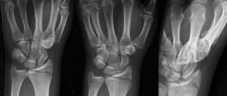
Sufficiently large bone metastases can cause visible deformation, be detected on palpation in the form of a tumor-like formation, or be visible on radiographs as an area of destruction. Pathological fractures are a serious complication of bone metastases. in 15-25% of cases, occurring in the area of tubular bones, in almost half of the cases - in the area of the vertebrae. Sometimes, as bone metastases grow, they compress nearby large vessels or nerves.
In the first case, circulatory disorders occur, in the second - neurological disorders. Severe complications of this pathology also include spinal cord compression and hypercalcemia. Local symptoms of bone metastases are combined with general manifestations of cancer: weakness, loss of appetite, weight loss, nausea, apathy, fatigue, anemia and increased body temperature.
If painkillers no longer help you with pain in the spine, RFA (radiofrequency ablation) of bones will help you!
Anonymous reviews
Anonymously. A friend was first diagnosed with breast cancer, then with lytic metastases. The doctor told her that they live with MTS for a long time, her friend goes every month to take Zometa and overall feels good.
Anonymously. The doctor also told me that the survival rate for mts in the bones is higher than if it is in the organs. I had two chemotherapy treatments, now I take bisphosphonates regularly and continue to live a full life, so there is no need to despair.
Anonymously. The grandmother lived with metastases for 10 and a half years, but there was pain and several fractures, she was active and did not sit still. She took morphine for pain.
About the RFA (radiofrequency ablation) method of bone metastases
The procedure for radiofrequency ablation of bone metastases is necessary in cases of pain caused by the destruction of the bone tissue itself and compression of nerve roots and nodes. In most patients, over a very short period of time, the pain caused by this pathology gradually becomes unbearable and unresponsive to drug therapy. In such cases, the RFA procedure is the only way to return the patient to a comfortable life.
In the presence of hard-to-reach tumors, especially in the spinal column and pelvic bones, when traditional surgery is associated with a high risk of death, we perform RFA with 3D navigation, that is, after creating a 3D model of the affected organ using special coordinates, step-by-step CT-controlled injection is performed needles, bypassing important organs and tissues. As a result, the risk of possible complications is reduced to zero, and the procedure for the patient becomes almost painless.
Video from the operating room. See how radiofrequency ablation of bone metastases works. Operating surgeon - Sergeev P.S., Ph.D.
The problem of limited thinking regarding operations for cancer metastases in the spine among most domestic oncologists is clearly illustrated by the following statistics, which are cited as justification for refusing surgical treatment of metastases in the skeletal bones:
- In lung cancer, metastasis is observed in 30-40% of cases, while patient survival after detection of metastases is about six months;
- In breast cancer, metastases are detected in 60-70% of cases, while life expectancy after detection of metastases ranges from one and a half to two years;
- In prostate cancer, the frequency of metastasis, according to various researchers, varies from 50 to 70% of cases, and the average life expectancy is about three years.
- In kidney cancer, the incidence of metastases is 20-25%, the median survival is about 1 year;
- For thyroid cancer in 60-70% of cases, the median survival is four years;
- In melanoma, the formation of metastases is 15-45%, life expectancy is on average about six months.
Hearing such numbers, even a healthy person panics. We must remember that these data are WITHOUT treatment of the underlying disease. With a successful operation and subsequent anti-relapse therapy, patients will live a long time!
RFA of tumor lesions of bones allows you to avoid large and traumatic operations and achieve an ideal analgesic effect.
Call your doctor now
Message sent!
expect a call, we will contact you shortly
Monoclonal antibody preparations
Another drug belonging to the group of osteomodifying agents (OMAs) is denosumab. It is a fully human monoclonal antibody with high affinity and specificity for human RANK ligand (RANKL). By binding to RANKL, denosumab prevents its interaction with the RANK receptor on the surface of osteoclasts and, as a result, the activation of the nuclear transcription factor NF-κB, which suppresses the process of maturation, functioning and survival of osteoclasts, the only cell type responsible for bone resorption. As a result, denosumab reduces bone resorption and destruction, acting similarly to the natural protein osteoprotegerin.
Early clinical studies of denosumab (phase I, II) showed rapid absorption of various doses of the drug, a half-life of 30–46 days, and the absence of neutralizing antibodies to the drug. The optimal regimen for administering denosumab is 120 mg subcutaneously once every 4 weeks. In phase II, the advantage of the drug compared to the active control (zoledronic acid) was revealed in delayed bone complications and a decrease in bone resorption markers, as well as a favorable tolerability profile [13–15]. An integrated analysis of 3 randomized phase III clinical trials, performed according to the same design and including 5726 patients, found that denosumab significantly reduced the risk of developing the first SSCS during the study by 17% in all types of tumors (p<0.001) [15]. Its advantage compared to control in breast cancer and prostate cancer has been shown: the risk of developing the first and subsequent SCS is reduced by 18% (p<0.001). When treated with denosumab, a significantly more significant reduction in pain was observed compared to the control (p<0.01).
When analyzing adverse events in the comparison group (zoledronic acid), acute reactions predominated: 20.2% versus 8.7%. Renal toxicity and mandibular necrosis occurred equally frequently in both groups: 11.8% versus 9.2% and 1.3% versus 1.8%, respectively. Serious adverse reactions were also reported equally frequently in both groups: 57.1% versus 56.3%, respectively [16]. According to the results of the integral analysis, progression-free time and overall survival did not differ between the groups [17]. However, with a detailed assessment of patients with lung cancer, a significant reduction in the risk of death was recorded by 20% (p = 0.01), and in the subgroup of non-small cell cancer - by 22% (p = 0.0104) in those receiving denosumab. At the same time, the difference in overall survival and a reduction in the risk of death by 32% was achieved due to patients with the squamous cell form of the disease. The median overall survival with denosumab and in the comparison group was 8.6 months. and 6.4 months. respectively (p=0.0350; RR (risk ratio) 0.68). The results obtained in the form of an increase in overall survival may be due, on the one hand, to the suppression of RANKL and changes in the bone microenvironment, on the other hand, to a direct effect on tumor cells, which in non-small cell cancer express RANK and RANKL receptors [18].
Diagnostic methods
Bone metastases help to visualize diagnostic methods such as radiography, CT, and MRI. Currently, PET scanning has become the “gold standard” for searching for secondary lesions. During this test, a radiopharmaceutical is injected into the body and accumulates in cancer cells. Then they take pictures with a special device, and all the metastases are clearly highlighted and “highlighted”.
As bones break down, levels of calcium and the enzyme alkaline phosphatase . These changes can be detected by performing a biochemical analysis.
If in a patient suffering from cancer it was possible to visualize formations in the bones on photographs, while the level of calcium in the blood is elevated, this highly likely indicates the presence of bone metastases. But in order to confirm this definitively, a biopsy must be performed; it helps to directly detect tumor cells.
You can obtain a fragment of tumor tissue using a needle. But this is often very difficult, since metastases are located in hard-to-reach places. The doctor risks damaging the tissue surrounding the bone. The needle may enter a vessel or nerve, which will lead to bleeding and pain. If the needle position is incorrect, a false positive or false negative result will be obtained.
The task is greatly simplified when the biopsy is performed under CT guidance - the current gold standard. During the procedure, the doctor makes a layer-by-layer marking of the tumor using CT, then inserts a needle into the bone. This allows you to accurately “get” into the tumor, even if it has a complex localization.
The procedure lasts approximately 30 minutes and does not require anesthesia. At the same time, the radiation load on the body is low: the body receives the same dose of X-ray radiation as when flying from Novosibirsk to Moscow. The patient can leave the clinic immediately after the doctor finishes collecting the material. After 3-5 days, the doctor will invite you for a second consultation based on the results of histological examination. Due to high accuracy, the probability of false positive and false negative results is minimized.
Radiopharmaceuticals
Radiopharmaceuticals are radioactive substances that, after intravenous administration, reach tumor tissue, accumulate in it and destroy cancer cells. This is an alternative to traditional radiation therapy. If a patient has multiple metastases, it is not advisable to irradiate every bone: this is not very effective and can cause serious side effects. It is worth giving preference to radiopharmaceuticals: they spread through the bloodstream throughout the body and reach all secondary foci.
Currently, in foreign literature there is data on the successful use of strontium-89 (Metastron), samarium-153 (Quadramet), radium-223 (Xofigo). Radiopharmaceuticals have been shown to be effective in reducing pain in affected bones for several months. If necessary, the procedure can be repeated.
Radiopharmaceuticals work best for osteoblastic metastases, when the activity of osteoblasts, the cells that form new bone tissue, is increased.
Biopsy of bone and soft tissue tumors under CT navigation
If the tumor is located in hard-to-reach places during a biopsy, complications such as damage to surrounding tissues leading to internal bleeding, pain, and a false positive or negative biopsy result may occur.
Tumors are often located in places that are difficult to reach for ultrasound, and it takes a long time for the patient to receive a correct diagnosis. With the introduction of diagnostic methods such as CT (computed tomography), diagnosis has become better.
In real time, the doctor, after layer-by-layer precise marking of the tumor using a CT machine, performs a fine-needle biopsy of the tumor in the most complex locations. The false answer when choosing this method is reduced to zero. Most often, CT navigation is used for biopsy of bone tumors: metastases and sarcomas.
Such biopsies are performed by an oncologist surgeon, Ph.D. Sergeev Petr Sergeevich is a qualified specialist in lesions of the musculoskeletal system. The photo shows a fine-needle biopsy of the pelvic bones under the control of computed tomography and a 3D reconstruction of the position of the needle in relation to the surrounding structures - the sacrum and ilium:
The method allows you to obtain a sufficient amount of the necessary material by accurately placing the needle into the center of the tumor without an incision or anesthesia. After the procedure is completed, the patient leaves the clinic.
The procedure time is usually 30 minutes. After 3–5 days, the patient is called for a second consultation based on the results of histological examination. The radiation exposure during such a procedure is comparable to a flight between Moscow and Novosibirsk.
Call and find out more about the RFA method
Message sent!
expect a call, we will contact you shortly
For what pathologies is this method used:
- metastatic bone lesions of any location,
- suspected bone sarcomas: osteosarcoma, chondrosarcoma, Ewing's sarcoma, giant cell tumor,
- soft tissue sarcomas of any location,
- chest tumors.
What diagnostics are performed before performing RFA of bone metastases?
For the most accurate localization of the lesion and three-dimensional reconstruction of the operated tissue area, we perform high-resolution computed tomography of the problem area of the spinal column or bone and make its 3D visualization.
Fig. 1 Inserting the needle as close as possible to the spinal cord, bypassing it (front view). Rice. 2 Inserting the needle as close as possible to the spinal cord, bypassing it (rear view).
Treatment of bone metastases
Bone cancer with metastases is stage 4 cancer; remission is extremely unlikely. But this does not mean at all that nothing more can be done and there is no need for treatment.
You can almost always help. There are treatment methods that help slow the progression of bone metastases, cope with symptoms, and improve the patient’s quality of life.
The choice of treatment methods depends on many factors, the main ones being: the origin of the primary tumor (in which organ the cancer initially arose), the location of metastases (which bones are affected), the presence of pathological fractures, symptoms, and the patient’s health condition.
If the diagnosis “metastasis in the spinal column”, “metastasis in the skull bone” is pronounced, it sounds like a death sentence in the head of most patients. However, you should not take it. If you contacted the Medica24 international clinic, and our orthopedic oncologist sees the point in treatment, then there is a chance. Active treatment of bone metastases will eliminate severe pain, dramatically improve the quality of life, and create the opportunity to remove the primary tumor site or perform a major palliative operation. The maximum possible cytoreduction dramatically increases the effectiveness of subsequent chemotherapy, which ultimately prolongs the patient's life.
We will call you back
Message sent!
expect a call, we will contact you shortly
Feedback on vertebroplasty of the lumbar vertebrae
The patient came to the clinic due to constant pain in the lumbar spine. She had difficulty walking and could not stand up or sit down normally. An examination showed she had a compression fracture of the fourth and fifth lumbar vertebrae. Percutaneous vertebroplasty was performed. The patient was activated and was able to walk two hours after surgery. “At first I was very afraid of this operation, but it went well... Read full review
International clinic Medica24 is the only place where they could help
Our clinic became the only place where they could help the patient. All the others refused it due to age and concomitant diseases. The reason for hospitalization was the threat of a fracture of the first lumbar vertebra due to a secondary lesion. The patient underwent CT-guided vertebroplasty and the first course of immunotherapy. Further treatment is ahead. “The only clinic where we found help and very good specialists. I express my gratitude to the doctors... Read full review
After spinal surgery, mom already tried to dance
Maria Lvovna’s mother could not walk due to severe back pain. Diagnostics revealed damage to five vertebrae, which caused excruciating pain. Andemir Olegovich Akhov came to the rescue, who performed vertebroplasty in three stages, during which bone tissue was restored. Thanks to the technique used and the professionalism of Andemir Olegovich, the recovery period was short and, as the patient’s daughter, mother, notes... Read full review
Feedback on the treatment of spinal hemangioma 12/12/2019
The patient presented with a hemangioma of the first thoracic vertebra. She complained of instability and heaviness when getting out of bed. She underwent vertebroplasty surgery. The patient was able to walk the same day. The problems she complained about disappeared! “It’s just wonderful to spend it here. Very fast, very professional. The condition recovers very quickly after this. So don't be afraid of anything. Just relax and trust your doctor. Everything will be... Read full review
Testimonial from a patient after treatment of hemangioma of the first thoracic vertebra
The causes of certain symptoms may lie far from the place where they appear. Therefore, there is a high probability of medical error and incorrect diagnosis. Some doctors, not understanding the causes of symptoms, refuse treatment. This is possible everywhere, but not in the international clinic Medica24. Here is a case where our specialists were able to help a person even when others refused. The patient came to… Read full review
Vertebroplasty for risk of pathological fracture
The patient was admitted to the clinic in serious condition due to the underlying disease. The patient’s relatives chose the clinic on the recommendation of a friend who had previously been treated here. A CT scan showed the presence of distant metastases in the bones of the spine. At the medical consultation, a decision was made to carry out staged vertebroplasty in two parts of the spine: In the cervical spine for the purpose of fixation and stabilization. In the lumbar… Read full review
Vertebroplasty at the international clinic Medica24
Patient Yuri Pavlovich came to the clinic with complaints of complications after a course of chemotherapy. A routine CT scan revealed foci of destruction of the bone tissue of the spine and ilium. This threatened the patient with a pathological fracture. Yuri Pavlovich underwent osteo- and vertebroplasty. Operating surgeon Andemir Olegovich Akhov. Read full review
Surgery
Surgical methods are used to stabilize the bone and restore its integrity in case of a pathological fracture. This helps reduce pain and improve the function of the affected part of the body (spinal column, arms, legs). Various fixing structures are used: plates, rods, screws, pins, etc. If the patient cannot undergo surgery, splints and orthopedic devices are used.
Sometimes radiofrequency ablation is used to destroy metastases. A special needle is inserted into the tumor, an electric current is applied to it, this leads to heating and destruction of tumor cells.
Fig. 1 Insertion of the electrode into the center of the metastasis after performing 3D navigation (guidance) under the control of a computed tomograph, in order to prevent burns of surrounding tissues and anatomical structures. Rice. 2 CT control (one of five) of the position of the conductor and needle of the RFA device during the procedure.
How much does it cost to treat bone metastases at the international clinic Medica24 and, for example, in Israel?
When going for treatment to Israel for the treatment of cancer metastases in the bones, you can go to one of the public clinics - Rambam Medical Center, Sourasky Medical Center, Chaim Sheba Medical Center, or to one of the private centers - Assuta Medical Center, Haddas Medical Center, Medical Shaare Zedek Center.
In Israeli government centers, the cost of medical services is strictly regulated. In private ones it is determined by the owners. The value depends on the number and size of metastatic foci, the prevalence of the metastatic process, and the presence of concomitant diseases. In most cases, the final cost after drawing up a treatment plan differs from the preliminary calculation by 50-200%, according to the experience of the patients we referred.
On average, RFA of 3 lesions in the lumbar spine with cementation and preliminary postoperative examination costs from $15,000, excluding the cost of the trip itself. RFA of 1 lesion in the thoracic region - from $8,000 US. At the international clinic Medica24, the cost of treatment will be at least 30-50% lower than in Israel, and approximately 2.5 times lower than in Germany, excluding the cost of travel.
Fig. 1 View through the viewing window of the CT operating room: anesthesia and insertion of a guide into the tumor tissue. Rice. 2 View through the viewing window of the CT operating room: before the RFA electrode starts working in the patient’s body.
If you add up the cost of your own treatment, plus transportation costs, plus the costs of the services of translators, guides and a relative accompanying you on the trip, we can say that the cost of a completed clinical case is almost 3-3.5 times less, with the same quality.
Watch an interview with an oncologist from the international clinic Medica24 about vertebroplasty of bone metastases and the treatment of pain.
Chemotherapy
Chemotherapy drugs help reduce the size of tumors, relieve pain, and improve the patient’s condition. Sometimes chemotherapy is combined with radiation therapy. Chemoradiation therapy is more effective at killing tumor cells, but is usually less well tolerated and causes more serious side effects.
Other types of drug treatments that your doctor may prescribe to treat bone metastases include:
- Hormonal therapy . It is used if tumor cells are sensitive to hormones - for breast and prostate cancer.
- Targeted therapy. Targeted drugs act more precisely than chemotherapy drugs. They block molecules that help cancer cells multiply quickly, obtain oxygen and nutrients, and hide from the immune system.
- Immunotherapy . Checkpoint inhibitors are drugs that block molecules that prevent the immune system from recognizing and destroying tumor cells.
- Radiopharmaceuticals . Contains radioactive elements. These drugs are administered intravenously, they reach the tumor tissue, accumulate in it and destroy cancer cells.
- Bisphosphonates . These drugs do not destroy tumor cells; they are used to slow down bone destruction and reduce calcium levels in the blood. Bisphosphonates inhibit the activity of osteoclasts, cells that break down bone.
The photo shows a patient with complete destruction of the phalanx of the third finger before and after 8 injections of a modern drug. The photo on the right is an image of a complete restoration of the phalanx.
Radiation therapy
Radiation therapy for bone metastases helps reduce pain and prevent pathological fractures. Typically, 1–2 sessions of high-dose radiation or 5–10 lower doses are prescribed.
Bone grafting
Bone grafting is performed when RFA is performed on large (over 3-5 cm) tumor lesions. In this case, a two-stage operation is performed. RFA makes it possible to completely eliminate metastases, after which necrotic tissue is excised. Then cementoplasty is performed using special medical cement, which restores the integrity of the bone. For larger defects, the resected area of bone is replaced with a prosthesis. Surgical bone osteosynthesis is performed less frequently.
Vertebroplasty and osteoplasty are minimally invasive treatment methods that can quickly reduce or stop the degree of pain in 85% of patients, thereby increasing motor activity and improving quality of life in 78% of patients, and reducing the threat of a pathological fracture of the affected bone segment in 100% of patients. and begin special treatment as soon as possible.
The combined performance of VP and OP eliminates the risk of pathological fractures and allows the patient to be mobilized as soon as possible after the surgical intervention.
Overall, it's important to know! Metastasis to the bones of the skeleton is not the final verdict. Yes, this is a serious complication in some types of cancer. Yes, the possibility of a radical cure for primary cancer has already been lost in most patients. But with our help at the international clinic Medica24, it will be possible to achieve significant progress - to destroy tumor foci in the bones, prevent the development of severe neurological complications, and make it possible to carry out palliative treatment. The patient gets the opportunity to live without unbearable pain and constant suffering, and his chances of prolonging his life increase.
Virtuous cycle
The process of bone tissue remodeling consists of five main stages: rest, activation, resorption, reversion, formation (Fig. 4). Each stage involves different types of bone cells [15].
Figure 4. Bone remodeling. Consists of five phases: 1) resting phase - osteocytes are motionless; 2) activation phase - differentiation of osteoclasts from preosteoclasts; 3) resorption phase - destruction of bone by osteoclasts; 4) reversion phase - differentiation of osteoblasts; 5) formation phase - osteoblasts create and mineralize bone.
[37]
The intercellular matrix consists of 85–90% collagen fibers, which provide structural support for bone mineralization. The remainder are proteoglycans, carboxylated proteins, cell adhesion proteins and growth factors.
Osteoblasts are formed from mesenchymal stem cells. During their work, they release a mixture of growth factors and proteins into the bone matrix, forming a specific microenvironment. They then undergo planned cell death or differentiate into osteocytes (Figure 5).
Figure 5. Scheme of differentiation of osteoblasts and osteocytes. Osteoblasts begin to differentiate from mesenchymal stem cells in the bone marrow.
[37]
Osteoblasts secrete bone growth factors, including transforming growth factor β (TGF-β), insulin-like growth factors (IGF), fibroblast growth factors (FGF), interleukins, platelet-derived growth factor ( PDFG ). These factors remain within the bone matrix and are released after bone degradation. This process provides feedback between bone formation and bone resorption [11].
Osteoclasts—phagocytes for bone—differentiate from the monocyte/macrophage lineage (Figure 6). Activated osteoclasts gather on the surface of the bone and attach to it through special receptors. They acidify the microenvironment at the bone-osteoclast interface and secrete enzymes involved in degradation: proteases, collagenase and alkaline phosphatase.
Figure 6. Scheme of osteoclast differentiation. Osteoclasts are giant multinucleated cells that differentiate from hematopoietic stem cells of the monocyte/macrophage lineage in the bone marrow.
[37]
The development of mature osteoclasts is a multistep process that is regulated by a complex system of cytokines and interactions within the bone stroma. Adjacent stromal cells and osteoblasts produce macrophage colony-stimulating factor (M-CSF), which acts through the colony-stimulating factor 1 (c-FMS) receptor on multinucleated progenitors, and activate RANK (Fig. 7). Its ligand, RANKL, like M-CSF, is synthesized by osteoblasts and stromal cells in response to the action of parathyroid hormone (PTH). RANKL is a cytokine from the tumor necrosis factor family. By binding to its receptor, it activates a number of important factors that regulate the expression of osteoclast genes. This creates conditions for final differentiation, fusion of precursors and the functioning of the resulting multinucleated osteoclasts. Activated osteoclasts break down bone and release TGF-β and other bone growth factors that enhance osteoblast proliferation [11].
Figure 7. Model of normal bone remodeling. Bone is constantly being remodeled. This process requires the interaction of bone-forming osteoblasts and bone-destructive osteoclasts. Details in the text.
[16]
Osteoblasts produce osteoprotegerin (OPG), a cytokine from the tumor necrosis factor family. It binds to RANKL, inhibiting the RANK-RANKL interaction, and therefore inhibits osteoclastogenesis and bone resorption. The RANK-RANKL-OPG interaction helps maintain a virtuous cycle.
Scientific comment:
Why is it necessary to treat bone metastases?
It has been noted that with metastasis only to the skeletal bones, even multifocal, but without affecting other organs and systems, the patient is much more likely to live for several years than with metastases to the lungs, and even more so the liver and brain. Bone lesions are difficult to treat, the effect is often limited to the stabilization of tumor destruction - bone destruction, however, with skillful treatment, this “fading” of cancer can last for several years. Treatment of bone metastases significantly increases the likelihood of cancer destaging—returning to the TxNxMx state for several years.
Why should I be treated in your clinic?
Result. I am results-oriented. And for the patient, the main result is relief from unbearable chronic pain - this occurs in half an hour to an hour, because all operations using RFA are performed under local anesthesia. And secondly, there is hope - after the elimination of all lesions in the bone, the body responds more actively to chemotherapy and treatment of the primary lesion or its relapses is many times more effective. I give patients hope! Read the reviews of my patients, watch them on Youtube, and you will understand everything.
Why not to the oncology center, not to EMC?
Unfortunately, they have not mastered this technique, although technically they are no worse equipped. Maybe because the administration does not want to upgrade the CT scanner to the level of a CT operating room. Perhaps doctors do not want to risk their health and “catch an X-ray dose,” because 3D navigation is impossible without the use of CT, and this is gamma radiation. It is safe for the patient, but given the number of procedures, it affects the health of the doctor.
I searched everywhere. Are you the only surgeon in Moscow who performs RFA of bone metastases under CT guidance?
Unfortunately, at the moment, yes.
Who did you study with?
I have quite a lot of experience working with leading specialists in the field of bone pathology and oncology. Let's start with the fact that my development as a specialist began at the Oncological Research Center named after. N.N. Blokhin, where the faculty of my teachers is beyond doubt. Then I repeatedly trained in Israel with Professor J. Schechter on the treatment of metastatic melanoma.
Can this procedure be done cheaper?
No. One electrode costs 2.5 thousand euros. They are not produced in Russia. 1 patient - 1 electrode. They cannot be sterilized or reused.
How long do such patients live?
Metastatic cancer is very difficult to treat:
- The 5-year survival rate (percentage of patients still alive 5 years after diagnosis) is 1–2%.
- Median survival (time after which half of the patients died, the other half remained alive) is 6-12 months (without treatment - 2-4 months).
However, such negative statistics are not a reason to give up.
Some patients (even if only a few out of a hundred) still remain alive for a very long time, many years. No one knows in advance which of those newly diagnosed with stage IV lung cancer will be among these survivors.
In addition, science does not stand still. Now doctors and scientists have high hopes for immunotherapy and targeted drugs. Treatment methods are improving, which means the chances of defeating cancer are increasing.
What is the survival prognosis for bone metastases?
The median life expectancy is known for different types of bone metastases This indicator indicates the time during which 50% of patients die:
- For breast cancer - 19–25 months.
- For prostate cancer - 12–53 months.
- For thyroid cancer - 48 months.
- For kidney cancer - 12 months.
- For bladder cancer - 6–9 months.
- For lung cancer - 6–7 months.
The international clinic Medica24 employs experienced doctors who know how to maximize the life of patients with stage 4 cancer, how to deal with bone metastases and cope with painful symptoms. We use advanced diagnostic and treatment methods. Contact us.
The material was prepared by a member of the international society of oncological surgeons EESG, candidate of medical sciences Sergeev Petr Sergeevich.
Role of osteocytes
The last stage of osteoblast differentiation is represented by osteocytes. They are localized in the bone matrix, making them ideal regulators of bone remodeling. However, this same advantage becomes a disadvantage when bone metastases develop, since they can fuel a vicious cycle.
Osteocytes are important producers of RANKL. They can also control metastatic growth by secreting factors, some of which activate the proliferation of breast cancer cells [16].