Magnetotherapy (16) Infrared and phototherapy (10) Vibroacoustic therapy (6) Laser therapy (4) Quantum therapy (2) Thermotherapy (3) Electrophoresis and Galvanization (5) Electrical stimulation (3)
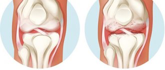
Magnetic therapy for gout helps relieve its painful symptoms and make attacks less frequent and intense. With gout, metabolism is disrupted, uric acid is poorly excreted from the body, and crystals of its salts are deposited in the joints. The result is inflammation, severe pain, and in severe cases, destruction of the joint and impaired mobility.
Magnetic therapy for gout is indicated in combination with diet and medications.
Magnetic therapy for gout gives the following results:
- relieves both sharp and aching pain;
- improves blood supply to joints;
- accelerates the utilization of uric acid salts;
- improves metabolic processes in cells;
- reduces the inflammatory response;
- promotes restoration of damaged tissues.
Regular magnetic physiotherapy procedures will help stop the progression of gout, prevent irreversible changes and preserve joints.
Magnetic therapy for gout of the legs will reduce pain when walking and help restore mobility. Magnetic therapy devices are universal and can be useful for treating a variety of diseases. Home appliances are designed specifically for use by people without medical education; they are affordable and easy to use. Gout is a chronic disease, so in case of gout it is convenient to have your own magnetic therapy device.
Devices for magnetic therapy of gout
[td]
Three stages of gout
Stage 1: High levels of uric acid in the blood
- Blood uric acid levels may be elevated, but there are no clinical manifestations.
- High levels of uric acid in the blood (hyperuricemia) may not progress, and gout symptoms may not appear at all.
- In some people, the primary manifestation may be kidney stones rather than a gout attack.
Second stage: Episodes of acute gouty arthritis alternate with periods without symptoms.
- This stage is also called interictal.
- Uric acid crystals begin to form in the synovial fluid, usually in one joint (the big toe) - and the body responds with an inflammatory response and an attack episode occurs.
- Although the thumb is most commonly affected, it is possible that other joints may also be involved. Although the big toe is the most common site for a gout attack, gout can develop in other joints.
- After an attack of gout ends, the damaged joint quickly returns to normal within 2-3 days before the next attack (as a rule, the attack must be repeated within 1-2 years).
- Over time, the intervals between attacks become shorter and the intensity of symptoms becomes more pronounced. In addition, other joints are also involved in the process.
Third stage: Chronic tophi gout
- If the symptoms of gout appear occasionally and treatment is not carried out for several years, then a chronic process occurs and other joints are involved. The intervals between attacks disappear. This stage of gout often resembles other types of arthritis (usually osteoarthritis).
- By this time, sufficient accumulation of uric acid crystals has occurred to form nodules (tophi) in various parts of the body. Tophi are dense to the touch, mobile and located under the skin. The underlying skin may be red and thinned (the color of tophi may be yellow or cream).
- As a rule, tophi are localized near the elbow or near the toes, hands, and auricle. With progression, tophi may develop near the joints or in the cartilage tissue of the auricle. Tophi located in cartilage tissue can lead to cartilage destruction.
Symptoms
Gout usually develops after years of accumulation of uric acid crystals in the joints and surrounding tissues. Symptoms include:
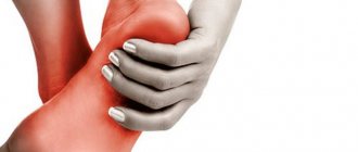
- Increased local temperature, pain, swelling, and severe tenderness in the joint, usually the big toe joint. This is a classic manifestation of gout. Sometimes a similar pattern can develop in another joint, such as the ankle or knee.
- There may be discoloration around the injured joint (bright red or purplish)
- The pain usually begins during the night and is so intense that even a light touch is very painful.
- Rapid onset of discomfort over several hours and then gradual regression over the next few days.
- Limited movement in the damaged joint.
- After a gout attack subsides, the skin around the damaged joint returns to normal and itching may persist.
- Some people may have gout without attacks, called chronic gout. Chronic gout in older age groups may not be very painful and is often confused with other types of arthritis.
- Gout may first appear as nodules (tophi) on the hands, elbows, or ears. In such cases, there may not be the classic symptoms of a gout attack.
ORION STEP
- Retail price:
16,700 rub.
Warranty from 1 year More about the terms of the guarantee
There are currently 20 people viewing this product
Compare devices Quick order Add to cart
Main characteristics
| Emitter type: | semiconductor injection AsGa |
| Laser mode: | pulse |
| Wavelength: | 880 - 910 nm |
| Laser pulse power: | constant 9 W |
| Pulse modulation frequency: | constant 1 500 Hz |
| MTBF: | 2,000 hours |
| Exposure time: | from 1 to 5 min (electronic timer with countdown) |
| Built-in photodiode: | for visualization and radiation control |
ALL CHARACTERISTICS
Characteristics
| Emitter type: | semiconductor injection AsGa |
| Laser mode: | pulse |
| Wavelength: | 880 - 910 nm |
| Laser pulse power: | constant 9 W |
| Pulse modulation frequency: | constant 1 500 Hz |
| MTBF: | 2,000 hours |
| Exposure time: | from 1 to 5 min (electronic timer with countdown) |
| Built-in photodiode: | for visualization and radiation control |
| Laser safety according to GOST R. 50723-94: | class 1 (highest) |
| Laser safety according to SANiPIN 5804-91 (radiation is safe for the eyes): | class 1 (highest) |
| Electrical safety: | class II protection BF (safety class for household appliances) |
| Device power: | from the built-in battery, charging from the network 220V, 50Hz |
| Power consumption: | no more than 5 W |
| Input voltage (battery): | 3 V |
| Device weight: | 200 grams |
| Overall dimensions of the device: | 187x60x39 mm |
| Package size: | 240x170x80 mm |
LASER TREATMENT PRECISION
- The infrared laser beam penetrates directly into the lesion
- Stops the process of joint destruction
- Relieves inflammation, pain and swelling
- Course of treatment - 10-14 days
View more details
OPTIMAL SOLUTION
- Reduces recovery time by 2-3 times
- Has no age restrictions
- Increases periods of remission
- Does not cause addiction
- Does not cause allergic reactions or side effects
View more details
CLINICALLY PROVEN EFFECT
- Therapeutic effect - 87-93%
- Central Military Clinical Hospital No. 2 named after. P.V. Mandryka
- GBUZ "Moscow Regional Research Clinical Institute (MONIKI) named after. M.F. Vladimirsky"
View full list of institutions
MAXIMUM READINGS
200 diseases are indicated for treatment, including:
- 200 diseases are indicated for treatment:
- Cardiovascular
- Neurological
- Urological
- ENT and bronchopulmonary
- Diseases of the veins and arteries
Full list
MADE IN RUSSIA
- Approved in Europe - Gold medal at the exhibition in Brussels “BRUSSELS EUREKA 97”
- Recommended by the Ministry of Health of the Russian Federation since 1993.
- The ORION device has been approved by the Ministry of Health of the Russian Federation since 1993
- Designed to the strict standards of the Soviet military industry. Warranty and post-warranty service.
Free doctor consultation
MINIMUM CONTRAINDICATIONS
- Laser safety class I
- Malignant neoplasms
- Blood diseases
- Active tuberculosis
- Fever of unknown etiology
View more details
Diagnostics
The most reliable confirmation of gout is the analysis of synovial fluid obtained by arthrocentesis. But if there is inflammation in the joint area (redness, pain), it is not advisable to perform a puncture. In such cases, the diagnosis is made based on a blood test for uric acid.
The diagnosis of gout is made based on medical history and blood and urine tests for uric acid.
X-ray is informative only in the late stages of gout and is necessary for differential diagnosis with other types of arthritis.
Difficulties in managing patients with gout
For citation. Bashkova I.B., Madyanov I.V. Difficulties in managing patients with gout // RMZh. 2015. No. 25. pp. 1508–1514.
Gout is a systemic tophi disease characterized by the deposition of monosodium urate crystals in various tissues and the resulting inflammation in individuals with hyperuricemia (HU) caused by environmental and/or genetic factors [1]. In the last two decades, there has been a steady increase in the incidence of gout, probably associated both with the characterological characteristics of patients (increased living standards, changes in diet, excessive alcoholism) [2] and with the spread of metabolic diseases in the population (obesity, metabolic syndrome, diabetes mellitus (DM), arterial hypertension (AH)), often accompanying gout [3–5]. Despite advances in diagnosis, understanding of pathogenesis and development of effective drugs for the treatment of gout, managing this category of patients can present certain difficulties for internists. The diagnosis of gout is made in the first year of the disease only in 1/4 of cases, but on average 7–8 years pass before the correct diagnosis is made [1]. The reason for late diagnosis may be the natural course of the disease - the alternation of rare, and short-term attacks of monoarthritis, with longer “bright intervals” of the disease due to the inherent self-limiting nature of gouty arthritis. Another explanation for the late diagnosis of gout can be the fact that the “first contact” doctor for this disease is often a surgeon or traumatologist, who, seeing a patient with acute, extremely painful arthritis, sends him for an X-ray examination of the affected joint, with the help of which diagnosis can be made. the debut of gout is often not possible, since large cysts in the subchondral zone of the bone (the so-called “puncture” symptom or X-ray negative intraosseous tophi) appear much later - on average 7–8 years from the moment of the first attack of gout [6, 7]. In the absence of bone-destructive changes on radiographs of the distal foot, the condition is regarded as a “soft tissue contusion,” which is usually accompanied by recommendations for taking nonsteroidal anti-inflammatory drugs (NSAIDs), which, due to their effectiveness in this situation, convince the doctor and patient of the validity of the incorrect diagnosis. In some cases, it is possible to predict the scenario for the further development of the disease: the patient will try to stop each subsequent attack of monoarthritis on his own, taking the NSAID that he “liked the first time,” until either an adverse reaction to the drug appears, or the NSAIDs cease to have the expected effect. Among the typical mistakes that doctors make when treating patients with acute gouty arthritis are: prescribing physiotherapeutic procedures, analgesics, ointments/gels applied locally in the area of the affected joint, prescribing allopurinol at the time of an attack, delay in prescribing effective analgesic and anti-inflammatory therapy [8 ]. Untimely and ineffective treatment of gout contributes not only to the aggravation of comorbid pathology, which is typical for this category of patients, in particular coronary heart disease (CHD) [9], but also to the development of fatal complications. We present our own observation when a combination of gout, GU, disorders of carbohydrate and lipid metabolism, hypertension, as well as the patient’s inattention to his health caused the death of a young man.
Patient S., 42 years old,
was hospitalized in the rheumatology department of the Republican Clinical Hospital (Cheboksary) on April 29, 2014 with complaints of constant intense pain in the left knee, ankle, metatarsophalangeal joints of the first toes of both feet, a number of proximal interphalangeal joints of the hands, worsening at night. From the medical history: the first attack of acute arthritis of the metatarsophalangeal joint of the 1st toe of the right foot developed 10 years ago, subsequently the arthritis recurred, the articular process gradually became polyarthritic in nature, and there was a natural reduction in the intervals between attacks to 2–3 months. The patient did not consult a doctor; he occasionally took nimesulide on his own. In 2009, he suffered an acute anteroseptal-apical myocardial infarction with a Q wave; in the same year, hyperglycemia (fasting blood sugar level 13.6 mmol/l) and GU (serum uric acid level 572 μmol/l) were first detected. Prescribed antiplatelet (acetylsalicylic acid 100 mg/day, clopidogrel 75 mg/day), oral hypoglycemic (metformin 1000 mg/day), lipid-lowering (atorvastatin 20 mg/day), uricodepressor (allopurinol 100 mg/day) drugs, β-blockers ( The patient did not take bisoprolol 5 mg/day after discharge from the hospital. Since 2010, there has been a rapid growth of tophi localized on the ears, in the area of the joints of the hands and feet, and an exacerbation of gouty arthritis with an increase of up to 5–6 rubles/year. In April 2014, after an alcoholic excess (alcohol abuse for 7 days), a severe exacerbation of chronic gouty arthritis developed: pain, swelling, local hyperemia and hyperthermia of the left knee joint, ankle, metatarsophalangeal joints of the first toes, a number of metacarpophalangeal and proximal interphalangeal joints of both hands. Intramuscular administration of diclofenac 75 mg 2 times / day and oral administration of nimesulide 400 mg / day - without effect. In the left heel area there is a wound measuring 1.5x1.0 cm (ulcerated tophi) with a white curdled discharge. The patient independently consulted a surgeon; on an outpatient basis, an increase in blood sugar level to 27.8 mmol/l and uric acid level to 686 μmol/l was detected, and therefore the patient was sent to a rheumatology hospital on April 29, 2014. Upon admission, the patient’s condition severe, forced position due to pain in the left knee joint, deformation of the knee (more than the left), ankle joints, multiple non-ulcerated tophi localized on the auricles, in the area of the joints of the hands (Fig. 1) and feet (Fig. 2), ulcerated tophi in the left calcaneal area. There is no peripheral edema. Body mass index – 25.6 kg/m2. On percussion there is a clear pulmonary sound throughout all pulmonary fields. In the lungs there is vesicular breathing, there are no wheezes at the time of examination. The boundaries of relative dullness of the heart: right - along the right edge of the sternum, upper - in the third intercostal space, left - 1.0 cm outward from the midclavicular line. Blood pressure (BP) – 160/100 mmHg. Art. Heart sounds are rhythmic, systolic murmur on the left edge of the sternum. The abdomen is soft, sensitive to palpation in the right hypochondrium. Liver - along the edge of the costal arch. The effleurage symptom is negative on both sides.
In the general blood test: red blood cells – 4.93×10¹²/l, hemoglobin – 149 g/l, platelets – 61×109/l, leukocytes – 4.2×10⁹/l, leukoformula shift to the left (young neutrophils – 2%, band neutrophils – 35%), absolute lymphopenia (0.38×10⁹/l), ESR according to the Panchenkov method – 36 mm/h. General urine analysis: proteinuria (0.36 g/l), microscopy of urinary sediment revealed: leukocyturia (10–12 per field of view), microhematuria (6–8 red blood cells per field of view), cylinduria (hyaline and granular), urates a lot. In the biochemical blood test: total bilirubin - 21 µmol/l, ALT - 89 U/l (with the upper limit of normal up to 40 U/l), AST - 135 U/l (with the upper limit of normal up to 40 U/l), urea – 14.4 mmol/l, creatinine – 215 µmol/l, total protein – 62 g/l, total cholesterol – 3.64 mmol/l, glucose – 15.0 mmol/l, uric acid – 761 µmol/l, high-sensitivity C-reactive protein (CRP) – 426 mg/l (with the upper limit of normal up to 5 mg/l). On a radiograph of the joints of the feet in a direct projection (dated April 28, 2014): large cysts in the subchondral zone of the metatarsal and proximal phalanges of the 1st toe and small erosions on the articular surfaces, significant compaction of the periarticular soft tissues in the area of the metatarsophalangeal joint of the 1st toe right foot (Fig. 3).
A diagnosis was made of “Primary chronic tophi gout, predominantly of a severe metabolic type. Chronic gouty polyarthritis, activity level 3, radiological stage II. Functional joint insufficiency of the 3rd degree. GU (761 µmol/l). Multiple large non-ulcerated tophi localized on the ears, in the area of the joints of the hands and feet, ulcerated tophi in the left heel area. Chronic gouty nephropathy, azotemic stage, CKD stage 3B. Type 2 diabetes, diabetic nephropathy, diabetic macroangiopathy, target level of glycosylated hemoglobin less than 7%. Risk category for developing diabetic foot syndrome 2. IHD: post-infarction cardiosclerosis (acute myocardial infarction with Q wave from 2009). Stage III hypertension. Degree of hypertension 2. Risk 4 (very high). Chronic heart failure (CHF) stage IIA (FC3).” Taking into account the decrease in excretory function of the kidneys due to the patient's renal failure (glomerular filtration rate according to EPI - 35 ml/min), type 2 diabetes, the use of NSAIDs and glucocorticoids (GC) to relieve exacerbation of chronic gouty polyarthritis was contraindicated. For analgesic purposes, intramuscular injections of tramadol 200 mg/day were prescribed, and therapy with short-acting insulin (10 U every 4 hours) was started. On April 30, 2014, at 8:30 p.m., the patient felt severe general weakness, increased thirst, dry mouth, and cold sweat. Blood pressure – 85/40 mm Hg. Art. An urgent blood test for sugar was performed - a slight hyperglycemia (9.3 mmol/l) was detected, the patient was administered a 5% glucose solution, prednisolone 90 mg, after which the patient's condition improved somewhat, but after 1 hour there was again a sharp increase in general weakness, profuse sweating When trying to get up, the patient fell, lost consciousness, cyanosis of the face and shoulder girdle appeared, blood pressure was 40/15 mm Hg. Art. The electrocardiogram recorded signs of overload of the right parts of the heart (deviation of the electrical axis of the heart to the right, P-pulmonale, deep S wave in lead I, negative T wave in lead III, right bundle branch block). To exclude acute myocardial infarction, the serum level of troponin I was examined, which was 0.148 μg/L (normally less than 0.5 μg/L). At the same time, the concentration of D-dimer in the blood serum turned out to be increased several times and amounted to 2021 μg/l (with the norm being up to 500 μg/l). Resuscitation measures were started, but they were unsuccessful - with increasing symptoms of acute cardiopulmonary failure, biological death was declared after 80 minutes. The cause of death was pulmonary embolism.
Gout is often accompanied by comorbid diseases [10–12], which can affect its course and prognosis. Hypertension and coronary artery disease are complicated by the development of chronic heart failure (CHF), which, undoubtedly, requires the doctor to prescribe diuretic drugs. In this situation, it is important to remember that all diuretics increase uric acid levels. In a population-based study of 4249 UK residents over the age of 30 years, a clinically verified diagnosis of gout was established in 164 patients, while in 25 (15%) patients, gout was induced by taking diuretics, 16 (64%) of them still continued take diuretics [13]. GU that occurs during treatment with diuretics may be due to an increase in reverse reabsorption and a decrease in urate secretion in the renal tubules, which, against the background of decreased renal circulation, leads to an increase in the production of angiotensin, which further inhibits the secretion of uric acid [14]. In addition, furosemide leads to an increase in lactic acid levels, which in turn inhibits urate excretion [15]. There is an immutable rule - diuretics prescribed for health reasons (CHF) to patients with gout must be compensated by taking allopurinol with dose titration under the supervision of a rheumatologist [7]. It is necessary to take into account the increasing incidence of abuse of diuretics by young women for cosmetic reasons (to improve their figure, etc.) [14, 16]. We present our own observation - a case of autoiatrogenicity - the development of secondary gout in a young woman, induced by long-term use of furosemide.
Patient A., 34 years old
, was admitted to the rheumatology department of the Republican Clinical Hospital (Cheboksary) on October 5, 2014 with complaints of constant intense pain, swelling and redness in the area of the metatarsophalangeal joint of the 1st toe of the left foot, which persisted for a week. From the medical history: in 2000 (at the age of 20 years), the patient, for cosmetic purposes, in a short period of time, adhering to a strict diet with limited animal protein, reduced her body weight by 20 kg. Subsequently, in order to maintain the “ideal” body weight, she independently began to take furosemide, ½ tablet 1 time/2–4 weeks. Without taking a diuretic, the patient began to subjectively feel shortness of breath, heaviness in the heart area, linking these phenomena with fluid retention, she increased the dose of the drug to 10-12 tablets per dose. In 2005 (5 years of constant use of furosemide), she first noted the appearance of pain, swelling and local hyperemia in the area of the right ankle joint, which stopped spontaneously after 3 days, but despite this, she continued to take furosemide at the same dose. In 2010, pain and swelling in the area of the right ankle joint arose again and subsequently recurred every 2–3 months, and arthritis of the metatarsophalangeal joint of the 1st toe of the left foot was added. To relieve joint pain, the patient increasingly began to resort to intramuscular injections of ketorolac. Due to the lack of effect from the use of NSAIDs, in 2014 she turned to a rheumatologist at the RCH. When examining the patient, rheumatoid arthritis, urogenital form of reactive arthritis, spondyloarthritis, and systemic lupus erythematosus were excluded. In the biochemical blood test, attention was drawn to an increase in serum levels of urea (25.5 mmol/l), creatinine (128 μmol/l), total cholesterol (9.4 mmol/l), and uric acid (495 μmol/l). To clarify the diagnosis, the patient was hospitalized in the rheumatology department. From physical examination: body mass index – 18.2 kg/m2; for organs and systems – without features; pain on palpation and deformation of the metatarsophalangeal joint of the 1st toe of the left foot with local hyperemia and hyperthermia (Fig. 4). An additional laboratory study revealed an increase in ESR to 54 mm/h, hypoisosthenuria, nocturia, hyper-alpha-2-globulinemia (14%), Fredrickson type IIb hyperlipoproteinemia, GU (654 µmol/l), a decrease in daily uric acid excretion to 0.8 mmol/day (with a norm of 1.5–4.5 mmol/day). According to esophagogastroduodenoscopy, mucus and clear bile were detected in the gastric cavity; the gastric mucosa was diffusely hyperemic and edematous. An X-ray of the foot joints in a direct projection (dated October 12, 2014) showed a slight narrowing of the joint spaces of the metatarsophalangeal joints of the 1st toes, and single cyst-like lucencies in the head of the 1st metatarsal bone on the left (Fig. 5).
A diagnosis was made of “Secondary gout induced by long-term use of furosemide, renal (hypoexcretory) type. Chronic gouty arthritis of the right ankle joint, metatarsophalangeal joint of the 1st toe of the left foot, activity of the 3rd degree, X-ray stage I. GU (654 µmol/l). Chronic interstitial nephritis, latent course. CKD stage 2. Hyperlipoproteinemia type IIb. Chemical gastropathy caused by bile reflux.” Furosemide was immediately stopped, and the drinking regimen was expanded to 1.5–2 l/day (previously I drank no more than 300–500 ml). The uric acid level dropped to 631 µmol/L within 14 days. To relieve arthritis, the patient was prescribed amtolmetin guacil (AMG) at a dose of 1200 mg/day in 2 divided doses. The drug was well tolerated. After complete relief of inflammatory phenomena in the joint (after 7 days) against the background of continued use of AMH in the hospital, therapy with allopurinol was initiated at an initial dose of 100 mg/day with recommendations for titrating the dose of the latter until normouricemia is achieved (the target level of uric acid in the blood serum is less than 360 µmol/ k) on an outpatient basis.
Errors made in the management of patients with chronic gouty arthritis are also quite common. Among them: 1) prescription of allopurinol during the acute period of the disease; 2) in some cases, the dose of allopurinol is not titrated to the most effective one, allowing to achieve the target value of uric acid level; 3) discontinuation of allopurinol when normouricemia is achieved; 4) unreasonable prescription of glucocorticoids (GC) to relieve exacerbation of chronic gouty arthritis, which in turn can lead to aggravation of metabolic disorders associated with gout (hypertension, type 2 diabetes, dyslipidemia), the development of secondary adrenal insufficiency when treatment is discontinued, and the rapid growth of intradermal tophi with long-term use, a decrease in the effectiveness of GCs after 1 year of therapy (the strength of their action of GCs becomes comparable to NSAIDs) [8, 17, 18]; 5) resection of subcutaneous tophi without achieving normouricemia, which creates a threat of infection of long-term non-healing skin defects [19]. Similar therapeutic errors made by primary care physicians in the management of patients with gouty arthritis are pointed out by M.V. Sklyanov and A.N. Kalyagin. When they conducted an anonymous survey of 50 local therapists in Irkutsk, it turned out that 56% of doctors admit the possibility of prescribing allopurinol in the acute period of the disease, every fourth (26%) indicated the necessary withdrawal of allopurinol after achieving normouricemia, 10% of doctors in their clinical practice have never prescribed patients with gout received allopurinol, while 44% of respondents indicated the advisability of using allopurinol in people with asymptomatic HU [20]. We present our own observation when the uncontrolled use of GC in the absence of achieving normouricemia served as the reason for the surgical removal of subcutaneous deposits of uric acid salts, which was inevitably accompanied by an exacerbation of chronic gouty arthritis and led to long-term non-healing of the wound surface.
Patient A., 60 years old,
was admitted to the rheumatology department of the Russian Clinical Hospital (Cheboksary) on October 19, 2015 with complaints of constant intense pain, swelling and redness in the area of the right ankle joint, a number of metatarsophalangeal joints of the right foot, a wound that did not heal within 1 year in the area of the 2nd toe of the right foot after amputation performed on September 8, 2014. From the medical history: at the age of 45 years, acute arthritis of the metatarsophalangeal joint of the 1st toe of the right foot developed for the first time, which resolved on its own after 7 days. The first visit to a general practitioner was several years after a series of recurrent arthritis of the same metatarsophalangeal joint. The examination revealed GU (serum uric acid level at that time was 456 µmol/l). A diagnosis of gout, chronic gouty arthritis was made; after the gouty attack was relieved by NSAIDs (diclofenac 150 mg/day for 10 days), therapy with allopurinol at a dose of 100 mg/day was started. The dose of the latter drug has not been revised for 12 years. Against this background, the patient notes the addition of arthritis of the joint of the same name in the left foot, ankle, elbow, wrist joints, and metacarpophalangeal joint of the 2nd finger of the right hand. Exacerbation of the disease - more than 5 rubles / year; in the area of the joints of the hands and feet, subcutaneous formations (tophi) have appeared in the area of the joints of the hands and feet over the last 7-8 years, with an increase in their size and number. Attacks of arthritis, on the recommendation of a general practitioner, were treated with intramuscular injections of ketorolac, less commonly, diclofenac or taking nimesulide. I constantly used ointments/gels on the area of the affected joints at the time of joint attacks, and took dietary supplements widely advertised in the media in order to reduce the level of uric acid in the blood. Retrospective review of outpatient medical records revealed persistent HU with a minimum uric acid value of 571 μmol/L. In September 2014, he consulted a surgeon due to the appearance of intense pain, swelling, redness in the area of the 2nd toe of the right foot with swelling and hyperemia spreading to the back of the foot, as well as an increase in body temperature. Blood tests revealed neutrophilic leukocytosis without a shift to the left (11.0×10⁹/l), increased ESR (46 mm/h) and CRP level (85.8 mg/l). An X-ray of the joints of the right foot in a direct projection (dated 09/08/2014): multiple cysts of the epiphyses of the metatarsal bones and phalanges of the fingers and erosions on the articular surfaces, compaction of the periarticular soft tissues in the area of the metatarsophalangeal joints of the right foot (Fig. 6). The condition was regarded as “acute purulent arthritis”, as a result of which on 08.0.2014 amputation of the 2nd toe of the right foot was performed (Fig. 7), followed by the prescription of antibacterial therapy. At the time of surgery, a large amount of cheesy white discharge was obtained in the absence of purulent exudate. The wound healed by secondary intention; to this day, a skin defect remains at the base of the 2nd toe of the right foot with periodic discharge of tophi masses from the wound. In the last 2 years, the patient noted frequent relapses of arthritis of the right wrist, knee and ankle joints; the use of NSAIDs (diclofenac, ketorolac, nimesulide, meloxicam) became ineffective, as a result of which intramuscular injections of prolonged betamethasone were used to relieve chronic gouty arthritis in the following months. Rapid growth of tophi was observed (Fig. 8). In March 2015, due to an increase in body temperature to 39°C, the appearance of pain in the lumbar region, and an increase in general weakness, he was hospitalized in the nephrology department of City Clinical Hospital No. 1 (Togliatti). Laboratory testing revealed leukocytosis (13.0×10⁹/l) with a shift of the leukoformula to the left (band neutrophils - 8%), an increase in ESR to 55 mm/h; in urine analysis - proteinuria (1.19 g/l), leukocyturia and bacteriuria during microscopy of urinary sediment. There was a decrease in the glomerular filtration rate according to EPI to 61 ml/min against the background of hypercreatininemia (125 µmol/l) and renal concentration function (diurnal fluctuations in the relative density of urine from 1004 to 1009, nocturia - nighttime versus daytime diuresis - 1.49 l versus 0. 8 l). An ultrasound examination of the kidneys revealed a decrease in the thickness of the parenchyma with an increase in its echodensity with multiple foci of fibrosis up to 4–5 mm, tuberosity of the kidney contour, and an expansion of the renal collecting system, which indicated that the patient had chronic pyelonephritis, which was probably secondary. A diagnosis of “Chronic gouty nephropathy with kidney damage similar to chronic interstitial nephritis” was made. Secondary chronic pyelonephritis, exacerbation. CKD stage 2." Against the background of antibacterial (ceftriaxone 2 g/day) and antihypertensive (amlodipine 10 mg/day and losartan 100 mg/day) therapy, improvements in general blood and urine tests, normalization of blood pressure, and a decrease in serum creatinine levels to 102 μmol/l were observed. (GFR according to EPI - 79 ml/min). Upon discharge, the patient was recommended to consult a rheumatologist to select a dose of allopurinol, but the patient continued to take the drug at a dose of 100 mg/day, followed by a series of exacerbations of chronic gouty arthritis, for the relief of which GCs were repeatedly used. From the life history it is known that the patient suffers from gastric ulcer with rare recurrence of ulcers. Denies alcohol abuse. During a physical examination at the time of hospitalization in the rheumatology department: body mass index – 23.2 kg/m2; for organs and systems – without features; pain on palpation and deformation of the right wrist and ankle joints with local hyperemia and hyperthermia, swelling of the dorsum of the right foot, mainly at the base of the fingers, multiple tophi localized on the hands and feet. An additional laboratory study revealed an increase in ESR (according to the Panchenkov method) to 21 mm/h and CRP to 18.9 mg/l, hypoisosthenuria, GU (551 µmol/l) with a decrease in daily excretion of uric acid to 1.0 mmol/day (at a norm of 1.5–4.5 mmol/day). On the radiograph of the joints of the hands in a direct projection: cyst-like clearings in the epiphyses of the bones, small erosions on the articular surfaces, narrowing of the joint spaces of a number of metacarpophalangeal and interphalangeal joints, significant compaction of the periarticular soft tissues in the projection of the metatarsophalangeal joints of the 3 fingers of both hands (Fig. 9 ).
A diagnosis was made of “Primary chronic tophi gout, predominantly of a severe metabolic type. Chronic gouty polyarthritis, activity level 2, radiological stage II. Functional joint insufficiency of the 2nd degree. GU (551 µmol/l). Multiple large non-ulcerated tophi localized in the joints of the hands and feet. Chronic gouty nephropathy of the type of chronic interstitial nephritis, azotemic stage, stage 2 CKD. Condition after disarticulation of the 2nd toe of the right foot. Stage II hypertension. The achieved degree of hypertension is 1. Risk 3 (high).” To relieve arthritis, the patient was prescribed AMG 600 mg twice a day (with recommendations to take the drug strictly on an empty stomach). Against the background of continued AMH intake, the dose of allopurinol was increased to 200 mg/day after 1 week. The serum uric acid level was re-determined to be 370 µmol/L, which required further titration of the allopurinol dose until normouricemia was achieved (target uric acid level less than 360 µmol/L) while continuing preventive anti-inflammatory therapy for AMH.
At the time of exacerbation of chronic gouty arthritis, the doctor is faced with the task of stopping the arthritis as quickly, effectively and safely as possible. In this case, NSAIDs become the first-line drugs. According to M.S. Eliseev and V.G. Barskova, it can be either a non-selective or a selective inhibitor of cyclooxygenase-2, provided that its anti-inflammatory and analgesic effect is realized as quickly as possible with a minimum incidence of adverse reactions [18]. In recent years, a fundamentally new non-selective NSAID has appeared in the arsenal of domestic doctors - amtolmetin guacil (Naisylat®, Dr. Reddy's Lab), which, along with a pronounced analgesic and anti-inflammatory effect, has a gastroprotective effect. The latter became possible thanks to the modification of the tolmetin molecule by introducing the amino acid vanillin, which made it possible to achieve the protective effect of AMH in relation to the mucous membrane of the gastrointestinal tract (GIT) while maintaining the class-specific positive effects of NSAIDs. The gastroprotective properties of the drug are associated with stimulation of capsaicin receptors (the so-called vanilloid receptors, the name of which comes from the presence of a vanillin group in the AMH molecule) located in the walls of the gastrointestinal tract. Excitation of capsaicin receptors leads to the release of a peptide encoded by the calcitonin gene, which in turn leads to a local increase in the production of nitric oxide (NO) in the gastric mucosa, and not in other organs and tissues [21, 22]. As is known, NO is one of the important components of the endogenous system for protecting the mucous membrane of the upper gastrointestinal tract from damage; it maintains the integrity of the structure by increasing mucus formation and bicarbonate secretion, increasing blood flow and repair of epithelial cells, reducing neutrophil chemotaxis and lipid peroxidation. The effectiveness and safety of AMH have been demonstrated in a number of clinical studies. Thus, A. Tavella and G. Ursini showed greater effectiveness of 4-week therapy with AMH compared to diclofenac in patients with musculoskeletal pain in reducing the severity of pain at rest, during movement, as well as dysfunction [23]. A meta-analysis of 18 clinical studies by R. Marcolongo et al. to evaluate the safety of AMH in patients with osteoarthritis and rheumatoid arthritis compared with other non-selective NSAIDs, such as diclofenac, tolmetin, indomethacin, naproxen, piroxicam and ibuprofen, showed that the incidence of side effects and cases of premature discontinuation of therapy was lower in the AMH group . The odds ratio (OR) for adverse reactions of AMH compared with those of other NSAIDs assessed together was 0.2 (95% CI 0.1–0.3). During endoscopic assessment, the frequency and severity of damage to the mucous membrane of the upper gastrointestinal tract in the group of patients taking AMH were significantly lower than in the control groups - the OR for moderate and severe erosive and ulcerative changes was 0.1 (95% CI 0.1–04 ) [24]. Another study on the safety and effectiveness of AMH in patients with rheumatoid arthritis was conducted by a group of Croatian scientists. The 24-week randomized, placebo-controlled trial included 180 patients with rheumatoid arthritis, of which 85 patients received AMH 1200 mg/day and 95 received celecoxib 200 mg/day. Both NSAIDs demonstrated the same clinical efficacy (according to ACR-20) when assessed at 4, 12 and 24 weeks. therapy. When analyzing endoscopic data, there was no statistically significant difference in the groups using AMH or celecoxib; any gastrointestinal changes according to esophagogastroduodenoscopy were noted in 24.8% and 22.3% of patients, respectively. While the clinical manifestations of the symptom of dyspepsia (pain and discomfort in the epigastric region) after 4 weeks. therapies were less common when taking AMH [25]. The recommended dose of AMG is 600 mg 2 times / day (maximum daily dose - 1800 mg), the maintenance dose is 600 mg 1 time / day; to maintain the gastroprotective effect of the drug, AMG should be taken on an empty stomach. Studies have shown that AMH was well tolerated by patients even with long-term use (for 6 months). Among the registered rheumatological indications for the use of AMG are not only rheumatoid arthritis, osteoarthritis, ankylosing spondylitis, damage to periarticular soft tissues (bursitis, tendovaginitis), but also articular syndrome with exacerbation of gout. And although there are currently no studies on the use of AMH in patients with gout, our positive experience of using a new effective non-selective NSAID with its own gastroprotective effect in relieving exacerbations of chronic gouty arthritis gives us the right to hope for the successful use of the drug in the fight against acute and chronic pain.
Treatment of gout
The goal of gout treatment is to reduce pain and prevent recurrent attacks and complications (damage to joints and urolithiasis). The following treatment methods are used:
Diet. If you are overweight, you need to reduce your caloric intake. In addition, significantly reduce the presence of meat products and seafood in the diet. Reduce your consumption of alcohol and especially beer.
Drug treatment. There are drugs that increase the secretion of uric acid (uricosuric drugs - for example benemid), reducing the production of uric acid (uricodepressive drugs - for example allopurinol), drugs that relieve an acute attack - colchicine, NSAIDs, corticosteroids.