Osteochondral exostoses are considered congenital pathologies. But they begin to grow actively under the influence of provoking factors. This happens especially often in adolescence. Most exostoses do not cause pain or other discomfort to the patient. But exostosis of the calcaneus is a little different from them. This pathology can appear at any age. The peculiarities of the location of the growth lead to severe pain, which often makes normal human movement impossible.
Features of the pathology
Exostosis is a pathological proliferation of osteochondral tissue caused by increased deposition of calcium salts, rapid skeletal growth or other provoking factors.
Such a growth, or, scientifically, osteochondroma, consists of cartilage cells and grows on the surface of the bone. It can have different shapes and grows to a size of 1.5-2 cm. If it does not compress the surrounding tissues and does not cause pain, it is not touched. But in the area of the heel bone, exostosis usually makes walking very difficult. And the only treatment for this pathology is surgical removal of the growth.
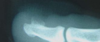
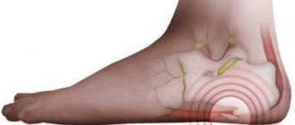
The growth of bone tissue in the heel area can be localized on its plantar part or behind. In this case, even a small formation interferes with walking and causes severe pain, as it irritates the surrounding tissues. If it is mushroom-shaped, it can compress the nerve plexuses, which will lead to numbness of the foot and loss of skin sensitivity; a semicircular growth always causes severe pain, and a linear growth in the form of a spike damages soft tissues and leads to the development of inflammation.
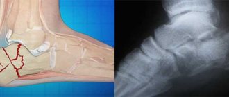
At the initial stage, pathology is very difficult to detect. As long as it does not cause pain or changes in soft tissues, patients do not even see a doctor. The growth itself first consists of cartilage tissue, so it is not visible on an x-ray. Gradually, dense bone tissue forms inside the soft shell of hyaline cartilage. Exostosis increases due to the proliferation of cartilage tissue. This distinguishes it from osteophytes, which are sharp bone growths that most often form in the joint area. They also form on the heel, but always after prolonged inflammation or injury.
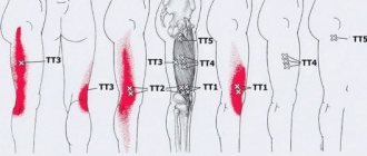
An osteochondral growth may form on the plantar surface of the heel bone
Complications of exostosis
With large exostoses, they may put pressure on neighboring bones, and bone defects and deformation of the bones of the extremities are sometimes observed. In very rare cases, fractures of the exostosis stalk are observed.
The most serious complication is the transformation of exostosis into a malignant tumor. Most often, malignant transition occurs with exostoses of the hip, scapula, pelvis, and vertebrae; Histologically, such osteogenic sarcoma can have the structure of chondrosarcoma, chondromyxosarcoma and spindle cell sarcoma, i.e., a malignant tumor of a very different morphological structure. If there is a sudden acceleration in the growth of exostoses in adults, they are surgically removed.
Varieties
Often, osteochondral exostosis on the plantar part of the heel is called a “heel spur.” This name has taken root among patients, although a “spur” is more of an acute osteophyte growth. And exostosis is an osteoma made of bone and cartilage tissue. In addition to the plantar surface, such a growth can form on the upper part of the heel tubercle. This pathology is also called posterior calcaneal exostosis, or Haglund’s deformity.
According to their structure, such formations can be of several varieties:
- hard osteoma is a layer of bone tissue on the surface of the bone;
- spongy osteoma consists mainly of soft cartilaginous tissue, it can be spherical or mushroom-shaped;
- medullary osteoma contains bone marrow and does not form on the heel.
Diagnostics
Diagnosing calcaneal exostosis at the stage of formation is difficult due to the absence of obvious disorders and symptoms that may indicate the proliferation of cartilage tissue.
Detection of an anomaly is possible through a visual examination, palpating the heel area, as well as an x-ray examination, which reveals the size, shape, stage of development and features of the course of the growth.
In severe cases, CT and MRI are used. It happens that patients undergo this examination for another reason, and as a result they are diagnosed with tumors.
Causes
In most cases, osteochondral exostosis develops in those patients who have a hereditary predisposition or any congenital pathologies of bone and cartilage tissue. But in the first years of life, growths do not form. They begin to grow under the influence of provoking factors. This could be due to injury or increased stress on the foot. Therefore, growths often form in athletes, ballerinas or people who work on their legs. It has been noted that women are more predisposed to the appearance of exostosis of the calcaneus.
Most often, exostosis occurs for the following reasons:
- after injury to the heel bone, which leads to severe inflammation or abnormal cell growth;
- constant wearing of narrow, uncomfortable shoes, frequent walking in high heels or completely flat soles;
- heavy weight and other increased loads on the foot;
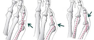
- flat feet or hallux valgus;
- circulatory disorders leading to deterioration of tissue nutrition;
- impact on cartilage tissue of infections - syphilis, gonorrhea, influenza, osteomyelitis, periostitis;
- endocrine diseases and metabolic disorders.
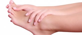
A growth on the heel bone irritates surrounding tissues when walking, causing severe pain.
Symptoms
The osteochondral formation on the heel grows gradually. Usually, until it reaches a size of 1 cm, it does not cause any discomfort. If the growth is large, it can already be felt and even noticed. Such a compaction can form on the back of the heel bone or on its plantar part. But in any case, the growth greatly interferes with walking.
More article:How to treat heel pain
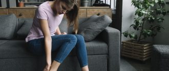
Most often, patients go to the doctor because of pain. They are strongest in the morning or after prolonged immobility. Then they calm down a little. And with increased physical activity, they intensify again in the evening. If the size of the growth on the plantar surface is more than a centimeter, it causes severe pain when walking. Therefore, patients are often forced to use a cane.
In addition to pain, due to constant irritation by the growth of soft tissues, swelling develops, and inflammation of the ligaments and tendons often occurs. For example, plantar fasciitis is a natural consequence of exostosis on the plantar part of the heel. The Achilles tendon is also often inflamed. The skin over the growth becomes rough, and calluses appear. Hyperemia is often noticeable, and this area is painful on palpation.
The skin over the site of growth becomes rough, and soft tissue inflammation often develops.
Constant pain can lead to deformation of the fingers, dysfunction of the joints, and the development of flat feet. The consequences of the pathology are also numbness of the skin of the foot, a tendency to fractures and dislocations of the joint.
Treatment
Some patients, especially older ones, do not consult a doctor with this pathology, preferring to relieve pain on their own. This approach threatens with many complications, the most serious of which is the degeneration of tumor cells and its transformation into a cancerous tumor.
After all, exostosis very rarely disappears on its own; only in adolescence is it possible to reduce the growth. But usually the pathology gradually progresses, the formation grows, increasingly irritating the surrounding tissues.
Therefore, it is very important to consult a doctor promptly if heel pain occurs. After all, treatment of exostosis is possible only surgically; no medications or folk methods will reduce the growth. They have only a symptomatic effect, alleviating the patient's condition. At the same time, constant monitoring of the growth of the formation is carried out to prevent complications.
In addition, it is very important to eliminate the causes that led to the increased proliferation of osteochondral tissue. Without this, even after surgical removal of the growth, after some time it may form again.
In many cases, only surgical treatment of exostosis on the heel bone is possible.
Operation
The only way to get rid of osteochondral growth is through surgery. But the operation is not performed on all patients with this pathology. Indications for surgical treatment are severe pain, development of inflammation, and rapid growth of formation. Surgery is also necessary if the growth interferes with walking or prevents you from wearing regular shoes.
The operation is performed under local anesthesia. Often the patient has many growths. In this case, only the largest ones and those that compress the surrounding tissue are removed. After anesthesia, a small incision is made and the growth is removed. After this, the surface of the bone is smoothed and a cosmetic suture is applied. The operation is considered uncomplicated, so the patient’s full return to normal life occurs within 1-2 weeks.
Conservative treatment
If the growth is not yet very large and does not cause much discomfort, symptomatic treatment is possible. Its task is to eliminate pain, swelling and inflammation. First of all, it is necessary to avoid trauma to soft tissues. For this purpose, choose comfortable, preferably orthopedic shoes. You can place a special insole or felt pads under the heel. This will help reduce pain when walking. In addition, it is recommended not to stand for long periods of time.
To relieve pain, you can use non-steroidal anti-inflammatory drugs in the form of tablets or ointments. Diclofenac, Ibuprofen, Ketoprofen, Voltaren gel or Dimexide solution are especially good for pain relief. Sometimes it is necessary to carry out a blockade by injecting hormonal agents into the heel area: Hydrocortisone, Diprostpan or Kenalog.
Physiotherapy procedures are effective for relieving inflammation of soft tissues and ligaments. The most commonly used are:
- warm foot baths, best with healing mineral water;
- foot massage;
- physiotherapy;
- electrophoresis with potassium iodide or Novocaine;
- shock wave therapy;
- ultrasound;
- magnetic therapy;
- laser heating;
- UHF;
- cryotherapy.
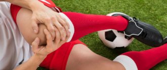
Shock wave therapy has been successfully used to relieve pain and inflammation.
Traditional methods
At the initial stage, if the pain is not severe, and the growth does not compress the nerves and does not interfere with blood circulation, it is possible to use traditional methods. They will help relieve pain and reduce inflammation. Most often, various compresses, ointments, and foot baths are used for this. The compresses should be warming, so the leg is wrapped in polyethylene. In addition, for better penetration of medicinal substances, it must first be steamed. After using foot baths, it is useful to apply an iodine net to the sore spot and wear warm socks. It is better if the procedure is carried out at night.
- A fat compress will help relieve pain and soften rough skin. You can use bear, badger or pork fat. This compress is applied at night.
- Grate raw potatoes and apply to the sore spot. Wrap and keep for 4-5 hours.
- The following composition for compresses is effective: 100 ml of aloe juice, the same amount of alcohol, a bottle of valerian, half a teaspoon of red pepper and 2 tablets of Aspirin and Analgin. The composition is mixed well and infused in a dark place for 2 weeks.
- It’s good to make a compress of medical bile at night.
- Foot baths with salt effectively relieve fatigue, swelling and pain. Make a strong brine from 5 liters of water and 1 kg of salt. You can add a few drops of iodine or soda.
- Clay baths help remove salts and relieve inflammation.
- Massage with coarse salt is useful. To do this, a kilogram of salt needs to be heated and sprinkled on a flat surface. You need to walk on warm salt with bare feet.
Oral products can also be used. They are needed to normalize metabolic processes, nourish bone tissue, improve blood circulation and strengthen the immune system. For this purpose, it is best to use a tincture of cedar grains along with shells in vodka or a tincture of lilac flowers.
To prevent the growth of bone tissue on the heel, it is necessary to avoid increased stress, wear comfortable shoes, and treat pathologies of the musculoskeletal system in a timely manner. During adolescence, you need to be regularly examined by a doctor in order to detect this disease in time. Then it can be cured without complications.
Traditional methods
To get rid of exostosis and its original factor, traditional treatment methods are used at home. Initially, before using folk remedies, you need to consult a doctor, and then choose the appropriate recipe.
Initially, they deal with the disease that led to the growth, since fighting the symptoms will not help in eliminating the pathology. Folk recipes use lotions on the heel tubercle.
Popular ones include mixtures of badger and bear fat, mumiyo, and golden mustache infusion.
- Take a spoonful of infusion and fat, crushed mummy pill. All ingredients are mixed, applied to the growth, and a napkin is placed on top. The application is covered with foil and secured with adhesive tape and a bandage.
- Taking elderberry and hawthorn tinctures. Take 3 large spoons of plants, pour in 750 ml of hot water and infuse. Take 2-3 times a day, 1/3 cup.
Also often used for treatment:
- Raw potatoes
- Lilac flowers
- Aloe
- Fat
- Pine nuts
- Medical bile
It is important to consider that folk remedies do not cure the disease completely, but are only an auxiliary component in the treatment.