A calcaneal fracture is a severe but infrequently diagnosed pathology in the practice of traumatologists. Violation of the integrity of the heel has a characteristic clinical picture and clear radiographic signs, which allow one to immediately identify the location of the injury and determine treatment tactics.
Untimely immobilization and non-compliance with the doctor’s recommendations can lead to deformation of the foot after a fracture, the development of complications and a decrease in the supporting function of the leg.
Main Causes of Injury
The heel bone is the largest and strongest structure of the foot, so its destruction requires the application of great force and often occurs in combination with other injuries. If it is determined that the patient has broken his heel and the history suggests a complex injury, the doctor may order x-rays of other bones of the legs, spine, etc.
The cause of a fracture can be the following:
- falling from a great height or jumping with landing on the entire surface of the feet,
- compression of the heel by other bones and the traumatic body during transport accidents and industrial accidents,
- direct blow to the heel tubercle area,
- prolonged high loads on the leg against the background of an existing heel defect (professional jogging or sports involving jumping, military service, etc.).
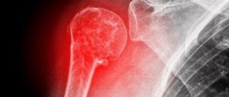
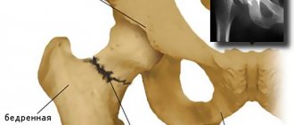
The most common cause of heel fracture is a fall or jump from a height. When landing on your foot, the weight of the body is first placed on the ankle and lower leg, and then transferred to the strong talus bone. Under great pressure, this structure wedges into the heel and splits it into several fragments. With a high intensity of impact after a fracture, displacement of the fragments occurs.
The risk of breaking a heel increases in the presence of genetic defects that affect osteosynthesis and age-related changes in skeletal strength.
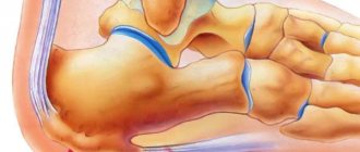
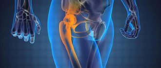
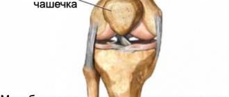
Anatomy of the heel
The heel bone, due to its size, is the largest of all human legs. When in motion, it acts as a sort of springboard for a rebound.
The calcaneus muscle is located under the plate of the foot, as well as under the anterior sagittal and fibrofemoral ligaments. In the posterior part of the bone there is a large tubercle, from which the lateral and medial processes extend. The transverse process connects to the longitudinal plantar ligaments. The medial process connects to tendons that allow the fingers to move.
Classification of heel fractures
Depending on the nature of the injury, heel fractures can be closed or open. Damage is also classified according to the location of the fault, the presence of damage to nearby joints, the number and displacement of fragments. In 80% of cases, a crack in the heel spreads to the subtalar joint, which is located above.
Closed
The formation of a wound and the release of bone fragments when the heel is damaged occurs infrequently. This is due not only to the nature of the injury, but also to the high strength of the structure itself.
A closed fracture is a less dangerous injury because not complicated by wound infection and severe bleeding.
Open
A strong blow or compression of the bone can cause damage to soft tissues and blood vessels, but rarely leads to disruption of the integrity of the skin. An open injury can occur when the leg is damaged by sharp protrusions of a traumatic object.
With offset
Trauma accompanied by displacement of debris leads to damage to nerve fibers, blood vessels and soft tissues of the foot. This provokes leg deformation, increases the risk of complications (post-traumatic edema, impaired motor function, etc.) and increases the duration of treatment.
The most severe injury is a displaced comminuted compression fracture.
Based on the nature and severity of the injury, the doctor determines whether surgery is needed or whether placing bone fragments in the correct position (reposition) and immobilizing the limb is sufficient.
No offset
Violation of the integrity of the heel without subsequent displacement of the resulting fragments is classified as a mild fracture. It is the least traumatic for the soft tissues of the foot, heals well and does not provoke complications in the form of improper bone fusion.
Closed reduction and minimally invasive osteosynthesis of a calcaneal fracture
Given the peculiarities of the blood supply to soft tissues and the high risk of infectious and ischemic complications, minimally invasive methods of surgical treatment of calcaneal fractures are gaining popularity all over the world. The basic principles of minimally invasive osteosynthesis are: restoration of the height and length of the heel bone, elimination of valgus/varus displacement of the heel tubercle, elimination of displacement of the posterior facet, restoration of the width of the calcaneal tubercle, without the use of large skin incisions and exposure of the bone. Elimination of displacement is carried out taking into account the principle of ligamentotaxis - due to traction in one, two or three directions, followed by fixation of fragments with screws or knitting needles.
Ligamentotaxis – due to traction in one, two or three directions, reposition of calcaneal bone fragments is achieved.
Elimination of displacement of calcaneal bone fragments due to ligamentotaxis
Depression of the area of the posterior facet of the calcaneus is eliminated with the help of an impactor from the approach to the subtalar sinus or from access along the plantar surface.
Elimination of displacement of the posterior facet of the calcaneus using an impactor
Percutaneous fixation of fracture fragments using screws.
Also, thick Kirschner wires or Steinman or Shants pins can be used for reposition; after installation in a bone fragment, such a “joystick” allows you to easily manipulate the fragment under the control of the image intensifier.
Intra-articular fracture of the calcaneus with avulsion of the tuberosity
Installation of a thick Kirschner wire into the calcaneal tuberosity
Reposition of fragments under x-ray control
Temporary fixation of fragments with a knitting needle
Sequential fixation of a calcaneal fracture with three cannulated screws
Restoring the Gissan angle
What are the signs that indicate a broken heel?
Symptoms of a heel fracture are:
- swelling that covers the entire surface of the foot and extends upward from the heel to the ankle (most pronounced with combined injury to the ankle and other bones of the foot),
- hematoma at the site of injury,
- heel pain radiating to the lower leg (irradiation of pain, as well as massive subcutaneous hemorrhage, is typical for displaced injuries and comminuted fractures),
- deformation (flattening of the arch) of the foot, blurriness of the contour of its rear part due to the displacement of debris and the formation of edema,
- increased pain when palpating the heel and passive movements of the fingers, difficulty in palpation with a fresh injury due to severe pain,
- loss of supporting function of the injured leg, inability to move and turn the foot.
The manifestations of heel injuries of varying severity can be similar, so it is important during the initial diagnosis to distinguish a fracture from a bruise, so as not to aggravate the patient’s condition by dislodging fragments. Making a final diagnosis and determining the degree of deformation is possible only with hardware examination or surgical intervention.
The differences between bruise and bone destruction are observed in the following aspects:
- Prevalence of edema. In case of a bruise, moderate swelling of the injured area or the entire foot is observed (with a combined injury). Fractures are characterized by swelling spreading over the entire surface of the foot and leg to the level of the ankle. In the heel area, the swelling is most pronounced and is complicated by a hematoma.
- The nature of the pain syndrome. When a minor injury occurs that is not accompanied by bone destruction, the patient experiences moderate pain, the intensity of which decreases over time. A fracture is manifested by a sharp pain syndrome, which intensifies due to the pressure of swelling on the damaged area and the displacement of debris during movement.
- Possibility of movement and support on the foot. If you bruise your heel, the damaged area will hurt when you try to walk, but the supporting function of the limb is preserved. Violation of the integrity of the bone leads to the inability to support the affected foot.
You should not try to palpate or step on your leg on your own before making a differential diagnosis, because this can provoke displacement of the debris, increased swelling and damage to soft tissues.
Restoring the previous capabilities of the limb
Rehabilitation after a heel fracture is, first of all, careful treatment of the damaged area, especially in the first days after the plaster is removed. During this period, pain can still be quite strong, so the increase in load is carried out gradually, and do not forget about the timely implementation of procedures in the required quality.
Incorrectly chosen recovery measures can lead to serious complications:
- Incorrectly fused bone;
- The appearance of flat feet of a post-traumatic nature;
- The feet are often deformed;
- Development of joint diseases of a destructive, dystrophic nature;
- Formation of growths on bones;
- The foot may lose its ability to function fully.
Incorrectly fused bone
Flat feet of post-traumatic nature
Feet become deformed
Formation of growths on bones
If a displaced heel fracture occurs as a result of an injury, rehabilitation can be carried out according to a special schedule. In this case, loads can be allowed no earlier than at least three months have passed after the injury.
Diagnostics
Diagnosis of a heel fracture includes:
- determining the nature of the injury (collecting anamnesis data),
- examination and palpation of the heel and ankle area,
- radiography in 3 projections (differential diagnosis of a bruise and determination of the degree of displacement of debris),
- MRI, CT (if necessary).
With a combined injury, a heel fracture may go unnoticed against the background of damage to the spinal column and ankle, which are more pronounced. All patients admitted with these injuries after falling onto their feet from a height of more than 1.5 m should have their feet x-rayed to rule out cracks in the heel bones.
If there is a bilateral heel injury and the patient is unconscious, it is necessary to analyze the condition of the pelvis, spinal column and knees.
Prevention
To prevent this type of injury, general safety rules must be followed:
- Before intense exercise, you should do a thorough warm-up;
- Do not jump from great heights;
- For all types of jumps, land with your feet together and your knees slightly bent;
- Only suitable shoes should be worn when running or exercising. Shoes with thin soles are not suitable for sports;
Do not run or jump on hard surfaces. Asphalt is also not suitable for running.
How to provide first aid
If you suspect a violation of the integrity of the heel, you must:
- Move the patient away from an unsafe area if there is a possibility of further injury and there is no evidence of spinal injury.
- Call the ambulance dispatcher, calling the team, and carefully listen to the instructions for providing assistance until the doctors arrive.
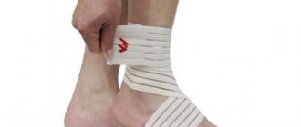
- Keep the injured leg completely immobile by placing the foot higher than the body to reduce pain and swelling. To do this, you need to use rolled up clothing, a pillow or other supports that are placed under the calf muscle to eliminate pressure on the heel.
- Unlace your shoes completely so that removing them does not dislodge bone fragments. Cut the sock off your foot. It is necessary to expose the sore leg as soon as possible, because increasing swelling makes any manipulation more painful.
- If there is a wound on the foot, it is necessary to stop the bleeding and treat the damaged area with an antiseptic solution (for example, Chlorhexidine or Miramistin). If possible, a loose sterile dressing should be applied after treatment. Trying to remove a traumatic object or bone fragments from a wound is prohibited.
- If there is heavy bleeding from the wound, apply a tourniquet to the knee area. The duration of a tight tourniquet on the leg should not exceed 2 hours.
- If the injury is closed, apply an ice compress to the injury site.
- Cover the patient with a light blanket and provide him with access to drink.
It is not recommended to give painkillers to the victim before the ambulance team arrives, because the occurrence of pain when moving the foot reduces the risk of displacement of bone fragments. If necessary, the patient can be given sedatives.
Treatment methods
The fracture treatment method is selected depending on the nature of the injury, the presence of complications and the displacement of the broken fragments.
Painkillers and limb immobilization are required.
Conservative therapy
If there is no displacement of the fragments or a slight displacement (deviation of the heel angle by no more than 5-7°), a circular gypsum splint is applied and modeling of the longitudinal arch is carried out. The cast covers the surface of the leg from the toes to the knee joint or mid-thigh. If necessary, before applying a splint, large bone fragments are compared (closed reduction) under local anesthesia.
To support the arch and eliminate axial load on the heel, metal instep supports can be used, which are located between the sole and the plaster splint. The use of arch supports reduces the risk of post-traumatic deformity.
Drug therapy for damage may include the following groups of drugs:
- analgesics (Nise, Ketanov, Analgin, etc.),
- complexes of vitamins and minerals (calcium and D3, B vitamins),
- preparations with chondroitin and glucosamine (Osteogen),
- immunomodulators,
- antibiotics,
- sedatives.
Before removing the immobilizing bandage, the patient is given an x-ray. If there is no damage or deformation of the bone, the doctor removes the plaster and draws up a plan for developing leg mobility.
Surgery
For complex and compression injuries, which are accompanied by displacement of debris and changes in the angle of the heel, surgical repositioning techniques are used. These include:
- Skeletal traction. If closed reposition of the fragments is ineffective, a needle made of biocompatible metal is passed through the heel fragments, which stabilizes the position of the bone. Small weights are attached to the protruding end of the structure, under the weight of which the fragments are displaced and installed in the correct position.
- Operations with external osteosynthesis (Ilizarov apparatus). In case of complicated open fractures, as well as severe displacement of fragments, surgical intervention is performed. During the operation, the doctor dissects the soft tissues, opens the talocalcaneal, calcaneocuboid and talonavicular joints, compares parts of the bones and inserts metal knitting needles into them. To secure these knitting needles, a design of hemispheres is used, which gradually stretches and corrects the shape of the bone. If necessary, the gaps between the fragments are filled with autograft.
- Three-joint resection of the leg. For old injuries and deformities resulting from improper treatment of a fracture, an operation is performed in which the crooked bones are trimmed, the arch is re-formed, the valgus deformity is eliminated and the width of the heel is normalized. After surgery, the heel is strengthened with metal or biodegradable structures, the wound is sutured, and the limb is immobilized with plaster.
During treatment, it is necessary to adhere to a special diet, which includes a large amount of dairy products, dishes with gelatin, lean sources of protein, calcium, silicon, omega acids and vitamins A, D3 and C. The daily dosage of calcium in the diet of patients with a heel fracture should be up to 2000 mg.
Types of surgery
Medical practice has developed two types of restoration of the calcaneus after a traumatic fracture. Depending on the type of damage to the heel bone, the following types of surgical treatment are used:
- Percutaneous screw fixation method. Used in the presence of large bone fragments. In this case, reduction can be performed without large incisions. Small incisions are used to insert special mounting screws inside.
- A method of open reduction of internal fixation, when the normal shape of the heel bone is restored using open access. Fixation is performed using knitting needles, screws, and metal plates for a heel fracture .
The decision to use one or another type of treatment for an injury is made by the attending physician, according to the results of the examination of the patient.
How long does it take for a fracture to heal and when can you step on your heel?
The length of time spent in a cast depends on the individual characteristics of the patient, the nature and severity of the injury. The immobilizing bandage is removed only after a control x-ray.
The average duration of immobilization is:
- for uncomplicated mild injury – 4-8 weeks,
- for a displaced fracture and treatment using skeletal traction – 11-12 weeks of immobilization after 4-5 weeks of fusion with the wire,
- in case of severe injury with the formation of fragments - 9-10 weeks after 6-8 weeks of wearing the Ilizarov apparatus.
After the cast is removed, the surgeon may recommend that the patient wear a brace.
In order for the bone to heal properly, it is necessary to use crutches: this will completely eliminate pressure on the heel. Even after the injury site stops hurting, you should not rely on the immobilized limb until a repeat x-ray is taken, which confirms complete healing of the bone tissue.
It is recommended to load the front part of the injured foot no earlier than after 4-5 weeks (for minor injuries) and 3-5 months (for complex compression fractures). The timing of complete healing of the heel and the moment when you can safely step on the entire foot are determined individually.
Physiotherapy – what is the effectiveness?
Rehabilitation after a heel fracture, both uncomplicated and displaced, will be better if massage and physiotherapeutic measures are added to the restorative procedures.
Immediately after the plaster is removed, you can begin treatment procedures. At first, it is not very convenient for the patient to move - due to prolonged immobilization of the limb, which often results in a condition akin to muscle atrophy, blood circulation slows down. By taking comprehensive massage sessions and physiotherapeutic procedures, you can normalize blood circulation and restore the ability to move freely.
Physiotherapy procedures are selected individually - the final option remains at the discretion of the surgeon. The type of injury and the general physical condition of the patient influence the decision. The procedures could be:
- Various types of heating, for example, UHF;
- Electrophoresis, for which a calcium solution is used;
- Magnetic therapy;
- Phonophoresis;
- Laser treatment.
Recovery period after a fracture
After complete fusion of the bone tissue, it is necessary to begin to develop the leg in order to prevent muscle atrophy and restore the mobility of the foot. To activate blood circulation in the injured limb, prevent lymph stagnation and stimulate muscle tissue, physical therapy, physiotherapy and massage are used.
Foot development is not recommended to be done at home. The duration of bone healing and rehabilitation ranges from 6 to 12 months. During this period, you need to wear special orthopedic shoes with arch supports.
Physiotherapy
The exercise therapy program is compiled individually depending on the patient’s condition and the complexity of the injury he received. The load on the injured foot increases gradually: in the first classes, only the knee joint, toes and forefoot are used.
In the absence of swelling and pain during exercise therapy, the patient is allowed to first exercise on an exercise bike, and then slowly walk. The first walks last no more than 5-10 minutes. If pain or discomfort occurs in the heel area, the load is limited.
Physiotherapy
Physiotherapy can reduce the period of tissue restoration and prevent trophic disorders in the injured limb. To improve blood circulation and lymph flow in the foot, the following procedures are prescribed:
- electrophoresis,
- UHF therapy,
- phonophoresis,
- laser therapy,
- magnetic therapy, etc.
Before physical therapy, you should do a light warm-up to warm up your injured leg.
Massage treatments
Foot massage stimulates blood circulation and helps restore the functionality of muscle tissue. The procedure should only be carried out by a specialist, because illiterate impact on damaged joints and the heel prevents the correct formation of callus and reduces the effectiveness of therapy.
What exercises can you do in a cast and immediately after it is removed?
If a patient is recovering from a heel fracture, they are interested in how they can help their foot recover. When the plaster has already been removed, what is allowed to be done next - should you step on the limb or save it, how to redevelop the leg? The decision to prescribe exercise must be made by the doctor.
During the first week after the fracture, there is no need to perform any movements with the injured limb. If you try to do this, you will not only provoke the appearance of painful sensations, but you can also displace bone fragments, touching them with the Achilles tendon. After two weeks have passed, gradually begin to move your fingers. This will help maintain better motor activity of the metatarsal joints.
When a month has passed, you can carefully step on the injured foot. In this case, the leg must be plastered or fixed with a splint.
It's time to start walking on crutches to gradually restore motor activity. After some time, the plaster can be removed - after this, they bend and straighten the knee, using not only the injured leg, but also the healthy leg. Thanks to such exercises, it is possible to restore elasticity and tone to the muscles of the lower extremities, preventing the displacement of fragments.
Try rolling a small ball on the floor with your injured foot. At first, gymnastics inevitably causes pain. Don’t be afraid of this and stop exercising completely – the pain gradually goes away if you don’t overload yourself with exercise. But by performing these simple steps you can achieve excellent results, and very quickly. Another effective and simple option is to pull the sock towards yourself and then pull it away from you, performing rotations with the heel and toe of the foot alternately.
Negative consequences after a fracture
If pathology is detected late, improperly treated, or if doctor’s recommendations are not followed during bone healing, the following complications may occur:
- pain when putting weight on the foot,
- osteoporosis,
- flat feet,
- deforming osteoarthritis of the foot joints adjacent to the heel,
- valgus curvature of the foot,
- impairment of the supporting function of the foot due to the formation of bony protrusions,
- wound infection with an open type of injury,
- lymph flow disorders,
- pinched nerves, neuralgia,
- muscle atrophy.