General information
A radial bone fracture is a violation of the integrity of the radial bone caused by various types of traumatic effects.
ICD-10 radial fracture code: S52.1; S52.3; S52.5. A fracture can occur at different levels of the radius: in the head/neck area, in the lower/middle third, or a fracture of the styloid process. Fracture of the radius occurs in people of both sexes and of any age, however, fractures of the diaphysis/damage to the upper part of the radius are found more often in children and young/middle-aged people, and fractures in a typical location (fracture of the distal metaepiphysis) are found more often in elderly people. In 75% of cases, a closed radial fracture occurs. In practice, the most common occurrence is the so-called fracture of the radius in a typical location. By “typical location” we mean fractures of the radius in the area slightly above the wrist (distal part of the radius, with the damage localized closer to the hand). It accounts for about 72% of all radial fractures. A fracture in a typical location is most common in women aged 70-75 years, which is largely due to the phenomenon of osteoporosis (decreased bone mass, structural disorders - disruption of trabecular microarchitecture/appearance of microfractures, increased porosity of the cortical bone), which contributes to a decrease in bone strength and structural impairment / quality of bone tissue.
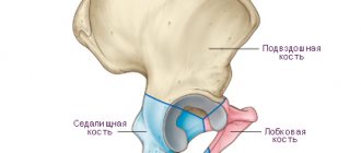
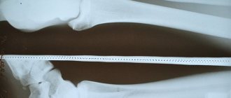
The radius of the arm is a long, fixed paired tubular bone within the bones of the forearm, having a triangular shape with anterior/posterior and lateral surfaces and anterior/posterior/interosseous edges. It is located on the side of the first finger, next to the ulna bone with which they are interconnected and dependent on each other. These bones connect below with the bones of the wrist, forming the wrist joint, and at the top these bones participate in the formation of the elbow joint. The radius is responsible for the functional mobility of the forearm at the elbow joint. However, at the same time, fractures of the radius are much more common than the ulna. The figure below shows the radius and ulna bones and their dislocation relative to each other.
Radial fractures can be either isolated or combined with other injuries. In practice, combined fractures of the shoulder (radius/ulna) are more common. At the same time, complete fractures of the radius/ulna are usually accompanied by displacement of bone fragments along the length, width, at an angle or around the longitudinal axis. With an atypical mechanism of injury (fall from a great height, road/work injuries), combined injuries to other bones of the extremities are possible (fractures of the spine, ribs, pelvic bones, damage to internal organs, kidney damage, chest damage, bladder damage and blunt abdominal trauma).
Stages of bone regeneration
In medical practice, the following regeneration stages have been identified:
- Catabolism of tissue structures and cellular infiltration. After damage, the tissue dies, cells disintegrate into elements, and hematomas appear.
- Stage of cell differentiation. This stage is characterized by primary bone fusion. With good blood supply, fusion occurs according to the type of primary osteogenesis. The process takes 10-15 days.
- Stage of primary osteon formation. Callus begins to form on the damaged area. Primary fusion takes place. The tissue breaks through with capillaries, its protein base hardens. A chaotic network of bone trabeculae grows, and they, connecting, form the primary osteon.
- Stage of callus spongiosis. This stage is characterized by the appearance of plastic bone cover, the cortical substance appears, and the damaged structure is restored. Depending on the severity of the damage, this stage can last several months or up to 3 years.
A prerequisite for a normally healing fracture is that the recovery stages proceed without disturbances or complications.
Pathogenesis
The pathogenesis of a pathological fracture is based on quantitative/structural changes in bone tissue, which significantly reduce bone strength. These include a decrease in bone mass and, as a consequence, a decrease in the mechanical strength of the bone and structural changes - disturbances in the microarchitecture of the trabeculae, an increase in the porosity of the cortical bone and the accumulation of trabecular microfractures, which directly affects the strength of the bone, regardless of its mass. Thus, the pathogenesis of pathological fractures involves a significant decrease in bone mass per unit volume and a violation of the strength/structural characteristics of bone tissue.
First aid for a fracture
The rate of healing of broken bones is greatly influenced by the provision of first aid for fractures. If it is an open fracture, it is very important that the wound does not get infected in order to avoid inflammation and suppuration in this area. Therefore, the damaged area needs to be disinfected; for this, the circumference of the wound should be treated with an antiseptic and covered with a sterile napkin until the medical team arrives.
To transport the victim to a medical facility, it is necessary to organize immobilization of the limb. To complete the task, they use available means - plywood, flat boards, tree branches, which are secured to the injured limb with a cloth or bandage. If a person has a spinal injury, then a solid stretcher or improvised means, such as flat boards, on which the patient must be carefully laid, are used for transportation.
The timing of consolidation of fractures directly depends on the provision of first aid and emergency transportation of the victim to the hospital.
Classification
The classification of radial fractures is based on factors such as the nature and nature of the fracture, fracture lines, displacement of fragments, the presence of fragments, the presence of a fracture of the ulna, etc., according to which the following types of radial fracture are distinguished.
According to the nature of the fracture, there are:
- Traumatic fractures with and without displacement that occur when the radius is exposed to a mechanical factor in the form of a fall, twisting, blow, or excessive physical activity.
- Pathological fractures resulting from a decrease in mineral density/impairment of the strength/structural characteristics of the bone (osteoporosis).
Depending on the fracture line, transverse, oblique, helical (spiral fracture), longitudinal, impacted, T-shaped, and comminuted fractures are distinguished.
Depending on the presence/absence of a violation of the integrity of the skin, the following are distinguished:
- Open fracture with damage (violation) of the skin. In this case, fragments of the radial bone come out.
- Closed fracture without skin damage.
Depending on the relationship of the radial bone fragments, the following are distinguished:
- Fracture of the radius without displacement of bone fragments (crack-type damage); Fractures of this type in most cases do not require additional interventions (except for fixation).
- Displaced fracture of the radius (complicated fracture with a high risk of re-displacement).
Anatomical classification, according to which:
- extra-articular fractures - fracture of the body (diaphysis) of the bone;
- intra-articular fractures - fracture of the head of the radial bone (neck);
- fracture of the styloid process.
Treatment (conservative, surgical)
There are two treatment options:
Conservative. It is used for simple fractures, without fragments or displacements. The patient is given a plaster cast, and the arm heals under it on its own. Sometimes conservative treatment is used to restore fragments and displacement. The procedure may fail and the limb may become deformed over time. The entire treatment takes from 4 to 6 weeks plus a rehabilitation period.
Operational. It is carried out under anesthesia, cutting the arm and restoring the anatomical structure of the bone. The fragments are fixed with special titanium plates and screws. Transosseous osteosynthesis according to Ilizarov is also performed, and a special fixation apparatus is used to restore the arm. At the Laditsen Medical Center, the operation is performed using Dr. Veklich’s improved design, without the use of traumatic needles. After the operation, you do not need to wear a cast, just an elbow bandage. Recovery occurs faster, and the patient returns to his usual lifestyle.
Causes of radial head fracture
The most common cause of a radius fracture is a household/work/sports injury. Less commonly, fractures are caused by osteoporosis. The level of fracture of the radius is determined by the nature/location of the traumatic impact:
- Fractures of the lower third of the radius - the main cause is a fall of a person with emphasis on the palm/dorsum of the hand, less often the injury is provoked by a strong blow at the level of the dorsal surface of the wrist.
- Fractures of the middle third of the radius—the main cause is a blow to the radial side of the forearm.
- Fractures of the upper third—the main cause is a person falling on an outstretched, slightly abducted arm.
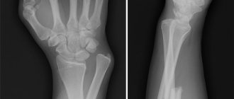
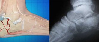
Fractures in a typical location without displacement/with displacement of fragments usually occur in cases of a person falling while leaning on an outstretched arm. Taking into account the nature of the displacement, it is customary to distinguish several types of injuries - Colles fractures , in which the distal fragment is displaced to the rear, and Smith fractures , in which the distal fragment is displaced to the palm (Fig. below).
The main cause of a fracture of the radius in the diaphysis area is a blow to the radial side of the forearm.
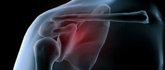
A fracture of the radial bone in the head region usually occurs as a result of a person falling onto an outstretched, slightly abducted arm. In 60% of cases, there is combined damage to other anatomical structures with dislocation of the bones of the forearm.
Combined fracture of the radius with dislocation in the wrist joint of the ulna? (Galeazzi damage) are formed when a person falls on an extended pronated hand, accompanied by concomitant compression of veins, arteries and nerves by edematous soft tissue. Factors that significantly increase the likelihood of fracture include:
- Osteoporosis / osteomalacia (decreased bone density/impaired ossification of the bone matrix).
- Weakness of the muscles/ligaments of the forearm and hand.
- Insufficient physical training.
- Increased body weight.
- Diseases of the elbow/wrist joint.
- Previous history of upper extremity injuries.
- Taking medications that affect bone metabolism (cytostatics, glucocorticoids, antidepressants, anticonvulsants, etc.).
The reasons that increase the likelihood of falls as a main factor can be divided into external and internal:
- External reasons are due to everyday and situational factors: uneven/slippery surface, poor lighting, etc.
- Internal causes are caused directly by the physical condition of a person: poor vision, asthenia , hearing loss, orthostatic disorders, dementia , decreased joint mobility, low physical activity, and the use of medications that affect the neurological status.
Fracture healing rate in adults
The process of bone fusion is complex and takes a long time. With a closed fracture in one place of the limb, the healing rate is high and ranges from 9 to 14 days. Multiple injuries heal on average in about 1 month. An open fracture is considered the most dangerous and takes the longest to recover; the healing period in such cases exceeds 2 months. When the bones are displaced relative to each other, the duration of the regeneration process increases even more.
Healing of fractures of the upper extremities occurs slowly, but they pose less danger to humans than injuries to the lower extremities. They heal in the following periods:
- phalanges of fingers - 22 days;
- wrist bones - 29 days;
- radius - 29-36 days;
- ulna - 61-76 days;
- forearm bones - 70-85 days;
- humerus - 42-59 days.
Healing time for fractures of the lower extremities:
- calcaneus - 35-42 days;
- metatarsal bone - 21-42 days;
- ankle - 45-60 days;
- patella - 30 days;
- femur - 60-120 days;
- pelvic bones - 30 days.
The reasons for the low healing rate may be improper treatment, excessive stress on the broken limb, or insufficient calcium levels in the body.
Symptoms
The main symptoms of a radius fracture include:
- Soreness. Patients complain of acute pain, which intensifies when attempting to rotate the forearm (perform rotational movements). The pain syndrome is especially intense with an open fracture, especially with displacement of fragments.
- Crepitation of bone fragments. It appears under the fingers when trying to move the bones in the form of a characteristic crunching sensation, however, it is not recommended to check for the presence of crepitus on your own as this can lead to even greater displacement of bone fragments.
- Local tissue swelling. It is caused by a cascade of reactions that contribute to the development of the inflammatory process, in which there is an expansion of blood vessels and partial sweating of fluid into the tissue, which contributes to the appearance of swelling. If a hematoma appears at the fracture site, over time the swelling site acquires a purplish-bluish tint.
- Pathological mobility in the hand. It is an absolute sign of a fracture, but only a medical professional can check it.
- Shortening the arm. The symptom occurs in cases of fracture of the radius/ulna with displacement of the fragments along the length.
However, each location of the radial bone fracture is characterized by specific symptoms. Thus, in the case of a fracture of the head of the radial bone, sharp pain appears in the elbow joint, which intensifies with palpation or an attempt to bend/rotate the arm. On examination, there is joint deformation, limitation of movements, hemarthrosis and swelling . A particularly sharp limitation is observed when performing rotational movements with the forearm. In cases where a fracture of the head of the radius is combined with a dislocation of the forearm, a more severe deformity is present, movements in the joint are completely absent, and there is often a disturbance in the blood supply/sensitivity in the distal parts (numbness of the fingers).
In isolated fractures of the diaphysis of the radius, the symptoms are often erased: swelling occurs in the area of the fracture, complaints of pain, which intensify with rotational movements and palpation of the fracture site. Pathological mobility/crepitus is usually absent, which is due to the retention of radial bone fragments by the interosseous membrane/whole ulna.
A radial fracture in a typical location is accompanied by severe pain, hemorrhage and swelling. Possible pathological mobility and crepitus. In cases of displacement of fragments, there is a visible deformation in the projection slightly above the wrist joint. Palpation/movement is sharply painful.
If Galeazzi is damaged, there is pain in the middle/lower third of the forearm, and when pulling on the fingers, the pain syndrome increases, the appearance of pronounced swelling, and the formation of subcutaneous hematomas is possible. Any movement in the wrist joint is extremely limited. Distinctive features of this type of fracture are frequent nerve damage, as well as the development of compartment syndrome, manifested by compression of nerve fibers, veins and arteries by edematous soft tissue. Often accompanied by loss of sensitivity/movement in the hand area. Increasing tension of soft tissues, excruciating increasing pain indicate the presence of compartment syndrome.
Rehabilitation and recovery
Complete recovery after a fracture of the radius consists of restoring bone structure and limb function (mobility and sensitivity). Even with absolutely adequate treatment, prolonged immobility in the joints and muscles of the upper limb leads to the fact that it is difficult for the patient to make movements in the joints that were previously accessible to him. The process of recovery from injury takes a long time and requires patience and desire from the patient to work.
Rehabilitation specialists at the Yusupov Hospital begin to develop joints and muscles in case of a fracture of the radius as early as possible. The timing of the start of rehabilitation measures depends on the type of fracture and method of treatment. If the fracture is treated conservatively, then after 3-5 days, after the swelling subsides, they begin to work on the fingers.
First, passive movements are performed. Take the finger on the broken limb with your healthy hand and carefully begin to bend it in all joints. In this way, stretch all fingers except the thumb for 5-7 minutes 3 times a day. After a week of such training, they move on to active movements. The patient can begin to bend his fingers independently, without the help of a second hand. It is very important to distribute the load correctly. If pain or swelling appears again during the exercise, the exercise should be stopped.
Simultaneously with the beginning of passive movements in the fingers, active movements in the elbow and shoulder joints begin. The patient raises and lowers his arm, bends it at the elbow. Do these exercises for 3-5 minutes at least 2 times a day, gradually increasing the load. After 3-4 weeks, if active movements of the fingers do not cause pain, they begin to increase the load on these joints. You need to take a lump of plasticine and knead it in your fist. This exercise should be done as often as possible, throughout the week. After removing the cast, proceed to exercises with a wrist expander. You should exercise at least three times a day for 5-7 minutes.
A physical therapy instructor teaches a patient fine motor skills exercises. By the end of the fourth week, the patient may begin to draw or write with the affected hand. You can sort out rice or buckwheat grains one grain at a time. This will preserve not only the strength and mobility of the joints, but also the coordination of movements of the fingers. You can type texts on a computer keyboard as a coordination exercise. If the patient does exercises while he has a plaster splint installed, then after its removal, the rehabilitation period will be significantly reduced.
Exercises should involve all joints of the injured limb. It is important to regularly warm up your fingers. To relieve tension in the affected limb, some activities after removing the plaster cast should be done in water. The duration of the course of physical therapy is determined individually by rehabilitation specialists at the Yusupov Hospital. The average duration of a course of therapeutic exercises is 1.5 months.
Rehabilitation therapists teach patients exercises that can be performed at home. Water gymnastics involves performing simple physical therapy exercises in water. Classes can be carried out in the bathroom. Cosmetic sea or table salt should be added to warm water. It will simplify the process of doing the exercises.
The complex consists of the following exercises:
- flexion and extension movements of the palms in the water;
- circular movements of the hand;
- rotations in the elbow joint;
- clenching and unclenching the palm into a fist.
The duration of the procedure is from 10 to 15 minutes. It is recommended to do 2-3 approaches daily.
Patients can also perform the exercise therapy complex recommended by the instructor at home. You should take the starting position “sitting at the table”. You need to place your elbows on the table, and place a thin, flat mat under your hands. The hands must be bent and unbent, and made rotational movements. Place your hands on the table edgewise, and then tilt your hand so that the little finger first touches the surface of the table, and then all fingers successively. Rest your elbows on the table, clasp your palms and tilt them alternately towards your left and right wrist.
Tests and diagnostics
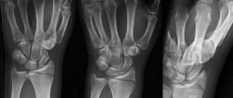
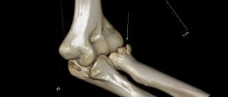
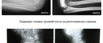
The diagnosis of “radial bone fracture” is made on the basis of anamnesis (presence of exposure to a traumatic factor), physical examination (pain/swelling in the forearm, forced position of the arm, deformity in the forearm, shortening of the upper limb, pathological mobility, crepitus of fragments, etc. ) and instrumental examination data - an x-ray in several projections, allowing to assess the nature and severity of the fracture (localization, with or without displacement, the presence of intra-articular injuries and concomitant fractures of the ulna). If soft tissue damage (muscles, blood vessels, nerve fibers) is suspected, CT/MRI may be prescribed.
Factors influencing the rate of bone fusion
The healing of a broken bone depends on a number of factors that either speed it up or hinder it. The regeneration process itself is individual for each patient.
First aid is critical to the speed of healing. With an open fracture, it is important to prevent infection from entering the wound, because inflammation and suppuration will slow down the regeneration process.
Healing occurs faster when small bones are broken.
In open fractures, callus forms much longer if a wound infection develops, which is accompanied by bone sequestration and post-traumatic osteomyelitis. That is why, if the fracture is treated incorrectly, the formation of callus slows down or does not occur at all. In such situations, fractures that do not heal for a long time, characterized by slow consolidation, as well as false joints appear:
- If patients suffer from hypovitaminosis and vitamin deficiency (osteomalacia in pregnant women, rickets, scurvy).
- If there are disturbances in the activity of the parathyroid glands (decreased calcium concentration in the blood) and adrenal hyperfunction.
- The presence of concomitant diseases occurring in the chronic stage, as well as inflammatory processes. Any pathological processes in the body significantly delay the recovery period after a fracture.
- The presence of excess body weight negatively affects the healing process of bone tissue.
- Metabolic disorder.
- Failure to comply with the terms of wearing a plaster cast. Many cases of bone tissue taking too long to heal are due to the fact that a person does not want to walk in a cast for a long time and removes it before the time prescribed by the doctor. The fused area of the bone is under pressure and displacement occurs.
How quickly bones heal depends on factors such as the need to install an implant. This occurs in cases where there are too many bone fragments, they are very small, and it is not possible to reassemble them.
Diet
Diet for fractures
- Efficacy: therapeutic effect after a month
- Timeframe: 2 months
- Cost of food: 1600-1800 rubles per week
Throughout bone fusion, dietary nutrition is indicated, the main task of which is to accelerate the fusion process. The diet for fractures should contain animal protein in an amount of about 100 g/day, which includes amino acids involved in the formation of bone tissue ( arginine , lysine , glutamine , proline , glycine , cystine ). The main products containing animal protein are dietary red meats, chicken, fish, chicken eggs, cottage cheese and dairy products.
Calcium, magnesium, phosphorus and zinc play a vital role in the formation of bone tissue, so the diet should include foods containing these elements. Sources of calcium are dairy products (cottage cheese/cheese), milk, fermented milk products, hazelnuts, lettuce, sesame seeds, spinach. It should be taken into account that for effective absorption of calcium, the diet must contain foods rich in vitamin D (fish oil/fatty sea fish).
The main sources of organic phosphorus compounds are meat, fish, milk, beef liver, beans, yolk, sturgeon caviar, walnuts, buckwheat/oatmeal, and dairy products. The required amount of magnesium in the body will be ensured by the inclusion in the diet of such products as wholemeal products, legumes, wheat, oatmeal and buckwheat (kernels), hazelnuts, milk powder, bananas, coffee beans, almonds.
And zinc is rich in bran, yeast, legumes, seafood, cereal grains, beef, dairy products, mushrooms, cocoa, sesame seeds, pumpkin seeds, peanuts, sunflower seeds, potatoes, onions.
B vitamins in the diet ; D ; WITH ; A , E , which are catalysts for the reactions necessary for the healing of fractures. Thus, vitamin D from fish oil, chicken yolk, fatty sea fish (sprats); vitamin C is found in rose hips, sea buckthorn, fruits/berries; group vitamins in offal (kidneys, pork/beef liver), cereals, walnuts, sweet peppers, hazelnuts, milk, yeast, garlic; vitamin E - in cold-pressed vegetable oils. A fracture of the radius heals on average in 27-35 days, and throughout this entire period, as well as for another 1-2 months, you must follow a diet.
Healing mechanism
Healing of fractures begins immediately after injury. Fusion can be of two types:
- Primary fusion. If the bones are reliably connected, there is no need to build up a callus on the broken area; the fracture heals easily and with good blood circulation.
- Secondary fusion. In this case, it is necessary to build up the callus due to the active movement of bone fragments.
The mechanism of fracture healing is very complex, and therefore is divided into certain stages:
- The first stage involves the formation of a clot formed from the blood surrounding the damaged area. After some time, they transform into new tissue for bone building. This clot forms within a few days after the injury.
- At the second stage, this clot is filled with osteoblasts and osteoclasts. They greatly support healing and restoration. By filling the clot around the fracture, they smooth out and align the bone fragments, after which a granular bridge is created. It is he who will hold the edges of the bone to prevent displacement.
- The third stage is characterized by the appearance of callus. After a few weeks (2-3) from injury, the granular bridge turns into bone tissue. During this period of time, it is still very fragile and differs from ordinary bone tissue. This area is called callus. To prevent damage, it is important that the fracture is securely immobilized.
- During the fourth stage, complete healing of the fracture occurs. After a certain time after the incident, depending on its severity and area (3-10 weeks), blood circulation in this place is completely normalized, which helps strengthen the bone. The tissue takes a little longer to recover (6-12 months).
At the end of all stages, the fused bone regains its strength and is able to withstand various loads.
Consequences and complications
The most common complication of a radius fracture is its malunion. At the same time, the severity of the consequences can vary widely depending on the location of the fracture/degree of bone deformation and can be represented by:
- Sudeck-Thurner syndrome (chronic pain syndrome/reflex sympathetic dystrophy). The pathology is progressive in nature and is accompanied by chronic pain syndrome, limb deformation with impaired function, trophic disorders, development of stiffness of adjacent joints/osteoporosis and often ending in disability.
- Instability of the wrist joint/impaired rotational function of the wrist joint.
- Shortening the forearm.
- Decreased grip strength in the hand.
- Slowing growth of the radius/development of radial clubhand.
- Deforming arthrosis of the wrist joint.
- Volkmann's contracture , developing against the background of long-term compartment syndrome.
Complications are observed mainly in the absence of timely/adequate treatment for complex radial fractures and non-compliance with recommendations for the duration of bone fixation and lifestyle correction for the healing period.
Healing rate of childhood fractures
In a child, fracture treatment occurs 30% faster than in adults. This is due to the high content of ossein and protein in the children's skeleton. The periosteum is thicker and has excellent blood supply. The skeleton of children is constantly increasing, and the presence of growth plates accelerates bone fusion even more. In children from six to twelve years of age, with damaged bone tissue, correction of fragments is observed without surgical intervention, and therefore, in most situations, specialists make do only with the application of plaster.
The most common fractures in children:
- Full. In such cases, the bone is separated into several parts.
- Compression fractures occur due to strong compression along the axis of the tubular bone. Healing occurs in 15-25 days.
- Green branch type fracture. The limb bends, causing cracks and fragments to form. Occurs when excessive pressure is applied with insufficient force to cause complete destruction.
- Plastic bending. Appears in the knee and elbow joints. Partial destruction of bone tissue without scars and cracks is observed.
List of sources
- Ashkenazi A.I. Fractures of the radius in a typical location // Carpal joint surgery. - M, 1990.- P. 124-138.
- Vorontsov P.M. Treatment of fractures of the distal metaepiphysis of the forearm bones / P.M. Vorontsov // Man and his health: Materials / Russian National Congress. - St. Petersburg, 1997. - P. 80.
- Angarskaya, E.G. Features of fractures of the radius in a typical location / E.G. Angarskaya, B.E. Munkozhargalov, Yu.N. Blagoveshchensky // Siberian Medical Journal. - 2008. - No. 3. - P. 33-35.
- Ardashev, I.P. Surgical treatment of improperly consolidated fractures of the distal metaphysis of the radius / I.P. Ardashev, V.N. Drobotov, A.V. Ivanov et al. // Modern high technology. - 2009. - No. 12. - P. 19-21.
- Bakhovudinov, A.Kh. Current state of the problem of complex pain syndrome with a fracture of the radius in a typical location / A.Kh. Bakhovudinov, V.A. Lanshakov, A.A. Panov et al. // Siberian Medical Journal. - 2009. - No. 3. - P. 104-110.
Etiology of radial fractures in a typical location.
In most cases, this is a fall supported by a straight arm; in the case of young patients, high-energy trauma, falls during sports, from bicycles or other rolling devices, and road traffic accidents are more common. In patients over 50 years of age, especially women, fractures of the distal radius are more often of a low-energy nature and occur due to osteoporosis. Low-energy fractures of the distal metaepiphysis of the radius are an indication for densitometry and subsequent consultation with an endocrinologist and significantly increase the risk of subsequent osteoporotic fractures.
Nutrition during the rehabilitation period
The issue of nutrition during the rehabilitation period is no less important than sets of exercises. The diet should be enriched with foods high in calcium, collagen, and magnesium. These are important components for bones and joints, which are important not only during the recovery period.
It is worth recalling that alcohol flushes calcium and other important components from the body. This can provoke prolonged healing of the fracture and a long rehabilitation process. It is better to avoid alcoholic drinks altogether. The same applies to salt, sugar, smoked, pickled, spicy and fried foods. There is a desire to quickly restore health to your hand, which means you need to eliminate everything harmful.
There are products that can saturate the body not only with components important for bones, but also with other substances that are no less important for the rehabilitation period. These are vitamins A, D, E. Therefore, dietary nutrition should contain:
- dairy products;
- hard cheeses;
- sea fish, seafood;
- lean meats;
- fruits, berries;
- nuts;
- dried fruits, figs;
- seeds (pumpkin, sesame);
- eggs;
- liver.
Food is taken in small portions in 4-5 meals. It’s not just alcohol that interferes with the absorption of important components. Oxalic acid, which is found in parsley and spinach, helps flush out important components. This must be taken into account when preparing your daily diet. If it is difficult to create a menu on your own, a nutritionist or attending physician will help with this.
Why does swelling occur after rehabilitation?
Swelling of the limbs after a fracture is normal. It often appears for several months after the cast is removed. In such a situation, it does not pose a great danger and occurs after heavy loads on the hand, when weather conditions change. This situation can also occur if a person is immersed in too hot or cold water. Muscle spasms cause swelling.
Also, the cause of this picture can be improperly fused bone. I don’t want to scare people who have suffered such an injury, but in such situations the problem can only be solved through a forced fracture and re-treatment.