Subluxation of the radial head in children often occurs between the ages of 1 and 3 to 6 years. In the pre-school period, subluxation is observed among girls 2 times more often than among boys. The right hand is much less likely to be injured. The left limb is much more likely to be damaged. School-age children practically do not encounter this injury.
Subluxation as a common injury has been described by many pediatric surgeons. However, not even all modern doctors are well aware of this injury.
Anatomical features
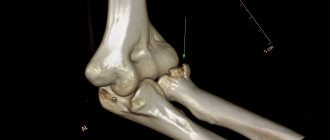
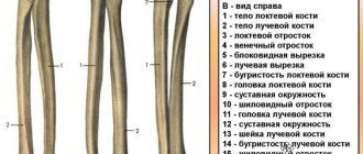
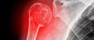
The ulna and radius bones are located in the forearm. With the palm facing up, the radius is on the outside. The body of the radius ends at an upper and lower end. The head of the radius is at the upper (proximal) end. The glenoid fossa (flat depression) serves as a dynamic connection to the head of the condyle of the humerus.
Due to the reduced volume of mineral salts, children's bones are more elastic. The connective periosteal tissue in children is structured in a special way. It has sufficient thickness and is vigorously supplied with blood. A kind of case is formed that protects the bone from unwanted damage.
At both ends of children's tubular bones there are epiphyses. These terminal sections are connected by germinal cartilage to the metaphyses.
Subluxation of the radius in the elbow joint in children is characterized by divergence of the superficial articulations. But contact remains between the displaced surfaces, the places of contact are preserved. These points disappear with complete dislocation. At the same time, the articulated surfaces shift.
Mechanisms of injury
Subluxation of the radial head has synonymous names. To designate this type of injury in medical practice, the concepts of “painful pronation of small children” or “dislocation from traction” are used.
The popular consciousness determined the essence of the injury in the name “Nanny's elbow.” Adults (nannies) often become the unwitting culprits of subluxation of the radius in children. The injury is caused to the baby according to the principle of “they wanted what was best.” Adults, saving the baby from falling, sharply pull him up by the hand. An adult, as a rule, leads the child by the left limb with his right hand.
The mechanism of injury is reduced to traction along the axis of one of the upper limbs. Children are injured:
- In play situations (pulled strongly by the hand; rotated the baby, holding both hands);
- In case of unexpected falls on a straight or bent upper limb (the baby’s arm tucks under itself);
- When walking with adults by the hand (the elders try to hold the child, pulling strongly on the upper limb);
- When taking off or putting on clothes with narrow and tight sleeves;
- Bruises to the arm;
- If a child's hand gets caught in rotating devices.
Typologically, the causes of subluxation come down to the situation when the baby’s outstretched arm is pulled by the lower part of the forearm or hand. As a result of the rough movement, the head of the radius slips out. It falls out of the annular ligament.
Subluxation of the radius is directly related to anatomical features. Underdeveloped muscles, a thin joint capsule, and underdevelopment of the distal condyle in the outer part lead to injury. The pathogenesis of the damage is overcome as the baby grows physiologically.
Emergency medicine
Subluxation of the radial head occurs exclusively in children under 3-5 years of age and is also called “dislocation from traction” or “painful pronation of small children.” Although the injury has long been described by pediatric surgeons and is common, it is still not well known to doctors.
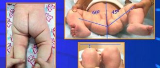
The damage is observed mainly in children aged 1 to 3 years. Subsequently, the frequency of this damage drops sharply, and in children over 6 years of age it is an exception. In girls, subluxation occurs 2 times more often than in boys. The left hand is affected more often than the right (60 and 40%, respectively).
The reason that causes subluxation of the head of the radial bone is usually a movement in which the child’s arm, which is in an extended position, is subjected to a sharp stretch by the hand or the lower end of the forearm along the longitudinal axis of the limb, often upward, sometimes forward. From the anamnesis, it is possible to establish that the child stumbled or slipped, and the mother, who was leading him, holding his left hand, pulled it to keep the child from falling (Fig. 53). Sometimes in a small child this stretching of the arm occurs while playing or putting on and taking off a narrow sleeve. In some cases, the mother indicates that the hand crunched.
Rice. 53. The mechanism of occurrence of subluxation of the head of the radial bone in children under the age of 3-4 years. a - on a walk; b - when dressing.
Whatever the reason that caused the damage, according to the mother, the child screams in pain, after which he immediately stops moving his arm and has since held it in a forced position, stretched along the body, slightly bent at the elbow joint.
When you try to force the child to move his arm, he protests and complains of pain in the elbow, and sometimes in the wrist area.
When collecting anamnesis, you should always try to understand the mechanism of injury and remember that subluxation occurs when there is a sharp stretch along the axis of the limb. If it is possible to establish the fact of such a sprain, the doctor immediately receives very valuable diagnostic instructions.
The clinical picture is always typical. The arm hangs along the body, like a paralyzed one, in a position of slight flexion at the elbow joint and pronation. An attempt to move the elbow joint causes the child to cry, as the movements are painful. However, you can carefully slowly flex and extend the elbow without changing the position of the forearm.
When palpating, it is sometimes possible to determine that pressing on the head of the radial bone is painful, but no visible changes are noted in this area. No pathological changes are visible on the x-ray.
There are various anatomical explanations for this lesion. L. Ombredan believes that the head of the radius remains half pinched in the annular ligament and cannot free itself and take a normal position. A. Ya. Masterman, studying the anatomy of this department on children's corpses, came to the conclusion that the subluxation of the radial bone cannot be explained by pinching the head in the annular ligament alone. He believes that this damage is due to age-related characteristics of the ligamentous and musculoskeletal system, which in children under 3 years of age is less developed; they have incomplete bone development, in particular the later development of the capitate eminence of the humerus, muscle weakness and thinness of the articular capsule. In addition, the joint capsule between the humerus and the head of the radius in children is wider and has a synovial fold - a duplicate that protrudes into the joint cavity. While studying the articulation of the head of the radius with the humerus, this author discovered several variations in the shape and size of the duplication.
So, when the joint is stretched, due to these features, the head of the radial bone slides from its normal place, and the duplicate, due to suction by the stretched joint, is retracted and pinched between the articular ends of the bones. Thus, A. Ya. Masterman concludes that the pathogenesis of subluxation of the radial bone is not determined by the infringement of the head of the radial bone in the annular ligament, but by the presence of the indicated age-related anatomical features, which change as the child develops, which explains the sharp decrease in this type of damage after 3 years.
When differential diagnosis of subluxation of the radius, one must remember the fracture of the clavicle and the neck of the humerus.
When the collarbone is fractured, the patient also sometimes keeps his arm lowered along the body and spares it, which gives reason to mistake one fracture for another. A clavicle fracture can easily be ruled out by examining it and feeling it.
When the neck of the humerus is fractured, the area of the shoulder joint swells, the contours of the joint are smoothed, palpation causes sharp pain, while movements in the elbow joint are completely free. Nerve damage can be easily ruled out by examining the patient and making sure that he can move his hand and fingers. The immobility of the arm is explained by the fear of pain that the child experiences when moving the elbow joint. Thus, the diagnosis is not difficult if the doctor remembers the possibility of subluxation of the radial head.
Treatment . Reduction in most cases is very easy. The forearm is carefully moved to a position of flexion at a right angle in the elbow joint, which is painful for the child, the patient’s hand is grabbed with the same hand, while fixing the wrist, and with the other hand they clasp the elbow and, lightly pressing the thumb on the head of the radius for control, make the movement complete supination. In this case, the child experiences some pain, and the setter’s finger feels a clicking or slight crunch. The patient immediately calms down and literally after 1-2 minutes freely, independently makes movements in the elbow joint and begins to use the arm as if it were healthy. In some cases, reduction is not immediately successful and the described technique must be repeated 2-3 times. Failure usually occurs from improper fixation and insufficient flexion of the arm or from incomplete supination.
After reduction, the arm is suspended on a scarf for 1-2 days. Parents are advised to be careful and not to lead their child by the affected arm. It is advisable to recommend the use of “reins” when walking with toddlers.
Subluxations on two arms in turn and recurrences of this injury are observed. Such cases confirm that the cause of subluxation should be sought in the congenital weakness of the ligamentous-muscular apparatus of the arm. In case of relapses, it is recommended to fix the arm bent at a right angle at the elbow joint with a bandage (plaster splint) for up to 7-10 days to give rest to the joint and promote contraction of the ligaments and joint capsule.
Isakov Yu. F. Pediatric surgery, 1983.
Clinical picture of injury
When an injury occurs, adults feel a crunch in the child's upper limb. The child literally screams in pain. Then the baby’s arm hangs along the body. The limb assumes a straightened position. The baby cannot move his injured arm painlessly.
When an injury occurs, the child manages to gently bend his arm to an angle of 90°. Rotational movements become unpleasantly painful. When the baby is asked to move his arm, he complains about painful sensations in the elbow area or wrist.
Diagnosis of injury
Recognizing a subluxation or dislocation of the radius in a child is not easy. Palpation (feeling) is made difficult by connective subcutaneous tissue .
A common mechanism of injury does not involve the use of x-rays. Ossification nuclei are absent in the epiphyses. This pathology cannot be diagnosed by radiographs.
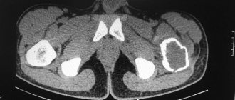
In children of preschool age, most of the bone epiphysis is cartilaginous material. X-rays penetrate the tissue. The ossification nucleus casts a dot-shaped shadow.
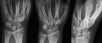
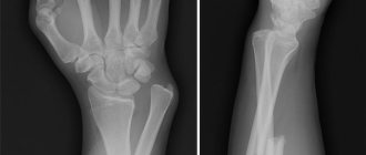
To diagnose subluxation of the radial bone, a radiograph taken in 2 projections may be required. For comparison, a photograph of the uninjured hand is used.
Palpation helps to identify painful sensations in the area of the radial head. No visible changes are observed.
When making a differential diagnosis of subluxation, one should not exclude a violation of the integrity of the neck of the humerus or clavicle. The anamnestic method helps to clarify the causes and types of damage.
Loss of integrity at the neck of the humerus causes swelling in the shoulder joint. A clavicle fracture is excluded by palpation. If the patient moves his hand and fingers, then the nerves are not damaged.
When studying the anamnesis, it is necessary to determine the fact of sprain. This becomes very valuable evidence for diagnosis. You need to know and remember the mechanism of subluxation, dislocation of the head of the radial bone.
Radial head: diagnosis
During diagnosis, parents of an injured child should tell the traumatologist about the event that led to the injury. Before making a final diagnosis, the doctor must examine the patient for the following pathologies:
- Congenital or acquired nerve damage;
- osteomyelitis;
- congenital dislocations;
- Fracture of the clavicle or neck of the humerus;
- Rheumatoid arthritis (juvenile or septic); osteoarthritis;
- fracture of the wrist or ulna.
Sometimes a trauma surgeon may recommend taking an x-ray of the child. Additional diagnostics are usually necessary if a fracture is suspected or if correct reposition of the radial head has not been achieved.
If the doctor has doubts about the diagnosis, additional diagnostics are simply necessary, so parents should not refuse to have their child x-rayed. If a radial dislocation is confirmed, no changes in the joint are visible in the photo.
If your child has recurring injuries, the doctor may recommend an MRI.
Radial head: first aid
If your baby has an upper limb injury, parents should first reassure him. Until medical assistance is provided, the only thing that can be done is to alleviate the condition of the little patient and try to relieve his pain.
To do this, apply a cotton cloth soaked in cold water or an ice pack to the stretch. If the pain is unbearable, your child may be given a dose of ibuprofen or paracetamol depending on their age.
However, the best first aid method is to go to a trauma center immediately. Adults should be aware that they may not be able to treat sprains at home. Moreover, self-medication can lead to the most serious consequences.
Therefore, in order not to further injure the injured arm, a child’s radial dislocation should be treated by a traumatologist. After all, only a doctor who knows the child’s physique can quickly and painlessly correct the bone.
First aid
After injury, the child must be reassured. The baby is distracted from what happened and his attention is switched to other objects.
A towel is applied to the injury site. It is moistened in cold water. An alternative is an ice compress. Useful means or splints to immobilize the joint can help alleviate the pain.
Painkillers and antipyretic medications, including ibuprofen, reduce the effects of pain. The dosage is determined by the baby’s age and body weight.
After the preparatory procedures, the baby is transported to a medical facility. There the victim is provided with qualified assistance. This is the only way to avoid serious and undesirable consequences.
Treatment algorithm
Reduction of a subluxation can be performed without previous anesthesia. This is done promptly throughout the day.
Before reduction, the victim's shoulder is secured. The forearm is gently bent at the elbow joint at a right angle. At the same time, supination is performed and finger pressure is applied to the head of the radial bone. Actions are performed smoothly. The moment of correction is indicated by a click.
The child feels pain. The baby screams and calms down almost immediately. Active movements are resumed after a short time.
Pronation subluxation of the radial head in children is best reduced in a trauma center. It is not advisable to use painkillers. Additional immobilization is not performed. 5–15 minutes after the subluxation has been reduced, the baby can move the injured limb freely.
Undiagnosed subluxation is accompanied by increasing pain. Swelling may develop around the elbow joint. In such cases, reduction is carried out with great effort.
For late reductions or recurrent subluxations, short-term immobilization is recommended. A plaster splint is used to create immobility. This gives the joint a state of rest and promotes contraction of the joint capsule and ligaments. Immobility of the affected area should not last more than a week.
For repeated reductions of the subluxation, a plaster splint is applied. The patient needs to walk with it for 2–3 weeks. Sometimes it is necessary to use pins to fix the head of the radius. In rare cases, it is not possible to reduce a subluxation using the closed method. We have to resort to surgical intervention.
Radial head: when dislocation recurs
If the sprain returns after repair of the radial head, the trauma surgeon may apply a bandage to the injured arm, which the patient must wear for two weeks. This way, the weakened joint will recover at rest.
For preventive purposes, active or passive exercises for joints should be performed to prevent re-damage. However, in order not to harm the baby, exercises should be performed under the supervision of medical personnel. Therapeutic exercises will help strengthen the muscles of the growing body of a preschooler.
Rehabilitation and recovery
After the subluxation has been reduced, the child is protected from excessive physical exertion. The hand is developed gradually. This is helped by activities aimed at strengthening muscles and ligaments. This prevents recurrent subluxations.
The doctor has the right to prescribe physiotherapeutic measures. Treatment:
- Laser therapy;
- Magnetotherapy;
- Monophoresis;
- Ultrasound;
- Massage.
At the rehabilitation stage, the child’s diet is carefully monitored. The baby should eat cereals, vegetables, fruits and other foods with increased amounts of minerals and vitamins. Be sure to include calcium-rich dairy products and meat in your diet.
Additionally, they use nutritional supplements and vitamin complexes. They must contain significant magnesium and calcium parts.
Relapse Prevention
To prevent new subluxations, they resort to physical therapy. A set of exercises is agreed upon with a rehabilitation doctor. Their main task is to strengthen the muscle frame for reliable fixation of the joints. It is recommended to conduct systematic massage sessions.
Parents should remember about the injured hand. Do not drag by the injured limb or lift by the wrist. When walking, you should not hold your baby’s arm that has a subluxation.
Parents need to reconsider their own treatment of their child. Participate in avoiding actions that could potentially lead to injury.
If a subluxation is suspected, contact a traumatologist as soon as possible to avoid serious consequences. It is better to trust the reduction of a subluxation to an orthopedic traumatologist.
Prevention of recurrent dislocations
If the baby is just learning to walk and cannot stand on his feet, mom and dad should help him overcome his fear. For example, do not hold your child's hand, but use harnesses to support him.
If radial dislocations occur systematically, adults should carefully analyze the child’s actions that lead to traumatic situations.
There is also a possibility that a sprain may occur as a result of parents' mistakes, so they should analyze their behavior.
You should not lead a preschooler holding him by the injured arm, sharply pull a limb, or lift the preschooler while holding him by the wrist. The congenital weakness of deformed annular ligaments can lead to frequent recurrence of injuries.