Diagnosis of a radius fracture
The first diagnosis occurs on the spot - upon injury. Usually, it's hard not to notice. A characteristic crunch and terrible pain arise immediately, the person himself realizes that he has broken his arm.
But for an accurate diagnosis, you need to consult a doctor. After all, wrist injuries are very different, and their treatment is also different.
Symptoms of a radial fracture:
- crunching sound caused by a fall or injury;
- immediately after a fracture of the radius, the hand does not bend, it is impossible to clench a fist and grasp an object;
- swelling after 30-120 minutes;
- if the joints are affected, then hemorrhage occurs and a hematoma is visible.
In some situations, the hand may only hurt during physical activity and show no other symptoms. This is dangerous because the bones may not heal properly. Then patients turn to Ladisten with a complaint: after a fracture of the radius, the arm is crooked.
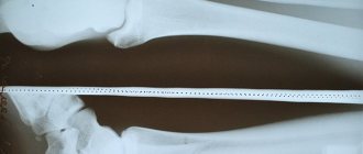
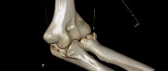
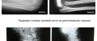
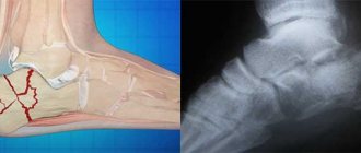
X-ray confirms the diagnosis. It also determines the severity of the injury, the presence of displacements, fragments, and exact location. MRI and CT scans show whether joints and muscles are affected.
Restoration of wrist bones
In the complex of measures for treating a wrist fracture, the rehabilitation period is very important. In the first months after removing the cast, it is not recommended to lift heavy objects with the affected arm or make sudden movements. During the recovery period, it is advisable to undergo therapeutic massage and exercise therapy. To relieve pain, you can use special pain-relieving ointments, gels and compresses. At home, you should regularly do light exercises to develop your hand and restore its mobility. For example, you can knead plasticine, squeeze a special expander or tennis ball with your sore hand. Another effective exercise is tilting the closed, straight palms towards the wrists.
After complete restoration of wrist mobility and the disappearance of pain, you should avoid falling on a straight arm and do not lift heavy objects with one hand.
Types of radius fractures
Doctors distinguish two main types:
- Kollesa . Prevails in all cases. When a person falls, he leans on his palm reflexively. If a fracture occurs, the bone fragments are displaced to the back.
- Smith . Occurs to a lesser extent. The exact opposite of the Colles injury is a fall on a hand that is concave inward. Therefore, the fragment moves towards the palm.
Both types are equally dangerous and risk causing complications if not treated promptly and correctly. Even if you visually do not see a fracture, your arm hurts only slightly, still consult a doctor after falling on your wrist.
Prevention
To avoid serious injury, you must adhere to safety rules when performing various manipulations. To prevent falls, wear comfortable shoes. Use protective equipment when playing sports. Proper nutrition and timely treatment of chronic diseases will prevent bone fragility, which contributes to the development of injury. If an accident occurs, a visit to the doctor should not be delayed. A fracture of the hand can lead to immobility, so the assistance provided must be effective and immediate.
Classification of fractures
How much your arm hurts after a fracture of the radius largely depends on the type of fracture. They can be complicated or simple. Pain of a different nature occurs, joints and soft tissues may be affected, or you may not feel the problem at all.
There are 5 main types of radial fracture:
- Open. The skin is touched and damaged, bleeding occurs. There is a high risk of infection and urgent surgery is required to restore the limb. Additionally, they check whether the patient has a tetanus vaccination, if not, they will be vaccinated;
- Splintered. A complex type that takes a long time to heal and is difficult to recover. The name means that the bone is crushed into several fragments - 3 or more. Without surgical intervention, it is impossible to “assemble the puzzle”;
- With offset. The situation is complicated by the displacement of fragments inside the broken limb. They may fuse in the wrong position and prolong the treatment period. The limb becomes deformed and may remain that way forever without treatment. If a displaced fracture of the radius occurs, rehabilitation also becomes more complicated. The patient has to get used to the new position and shape of the bones;
- Intra-articular. The injury affects the wrist joint, which also needs to be restored;
- Extra-articular. It is considered the simplest, does not affect the joint, and is easily corrected if there are no fragments or displacements.
Most carpal radius fractures require surgery.
Characteristic symptoms
Violation of the integrity of one or several bones of the hand leads to the development of a vivid clinical picture, the diversity of which depends on the location of the fracture. All manifestations can be divided into general and specific signs.
Classic symptoms:
- piercing pain – there are numerous pain receptors on the palms and in the fingers;
- swelling that is not limited to the injured hand, but extends to the wrist joint and forearm;
- hematoma - subcutaneous hemorrhage can be localized or occupy a large area;
- bluish skin over a broken bone;
- hand movements are impossible;
- unnatural deformation of part of the hand or finger, as well as protruding bone fragments visible under the skin;
- open fractures are characterized by the presence of a wound in which damaged muscles and bones are visible;
- The skin on the injured area is hot to the touch.
A fracture of the hand is easy to suspect if there is a history of a fall or crushing of the upper limb.
Considering the localization, the following specific signs are observed:
- Wrist area – the victim’s fingers “do not work”; it is impossible to clench the hand into a fist. When trying to move, a piercing pain impulse appears with irradiation into the phalanges of the 3rd and 4th fingers, as well as into the scaphoid bone.
- If the bones at the base of the fingers are destroyed, attempting to move 2 fingers causes acute pain. In this case, the first finger takes a forced position: pressed to the hand in a bent form.
- Violation of the integrity of the fingers is accompanied by deformation of the phalanges with deviation from the vertical axis.
When bone fragments are displaced, a hard formation is noticeable under the skin.
On palpation, it is painful and easily moves with a crunching sound, which indicates crepitus due to friction of parts of the broken bone against each other.
Causes of fractures
The main reason is a fall on an outstretched arm. This is an innate reflex that a person uses when he loses his balance. When falling, people stretch out their arms to support and protect their internal organs, face, and head. But the protective mechanism does not always work; the radius bone cannot withstand the force of the blow or the weight of the body and breaks.
Trauma can happen to anyone at any age.
Risk group : athletes, people over 50 years of age, patients with diseases of bones and joints.
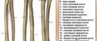
Structure and functions of the metacarpal bones of the hand
There are a total of 5 metacarpal bones on each hand. They have a tubular structure.
The bases of the metacarpal bones are cuboidal in shape, connecting through ligaments to the distal row of carpal bones.
The body of the metacarpal bone is slightly curved towards the palmar surface and resembles a rocker. Interosseous muscles are attached to the lateral surfaces of the body, which provide fine motor movements of the hand. In case of a transverse fracture of the metacarpal bone, the displacement is difficult to eliminate and maintain precisely because of these muscles, because they constantly pull the fragments onto themselves. The extensor tendons of the fingers are attached to the dorsal surface. The body of the metacarpal bone passes into the neck and then into the head, on which there is an articular surface for connection with the proximal phalanx of the finger. The weakest point of the metacarpal bone can be called the neck; it is at this level that most fractures occur, due to the anatomy and mechanism of injury.
The first metacarpal is unique in that it is shorter and wider than the others and has more extreme angles with the carpus, opposing the axes of the other bones, which characterizes the primary function of the first digit. The second and third metacarpal bones are most rigidly attached to the bones of the wrist with their base. In contrast, the fourth and fifth are more loosely attached and allow the hand to perform more movements and securely grip objects and tools of various shapes.
Treatment (conservative, surgical)
There are two treatment options:
Conservative. It is used for simple fractures, without fragments or displacements. The patient is given a plaster cast, and the arm heals under it on its own. Sometimes conservative treatment is used to restore fragments and displacement. The procedure may fail and the limb may become deformed over time. The entire treatment takes from 4 to 6 weeks plus a rehabilitation period.
Operational. It is carried out under anesthesia, cutting the arm and restoring the anatomical structure of the bone. The fragments are fixed with special titanium plates and screws. Transosseous osteosynthesis according to Ilizarov is also performed, and a special fixation apparatus is used to restore the arm. At the Laditsen Medical Center, the operation is performed using Dr. Veklich’s improved design, without the use of traumatic needles. After the operation, you do not need to wear a cast, just an elbow bandage. Recovery occurs faster, and the patient returns to his usual lifestyle.
First aid in case of injury
Immediately after a traumatic situation occurs, emergency medical assistance should be called. After this, it is necessary to promptly and correctly try to help the victim, since first aid for fractures of the hands ensures a quick diagnosis and rapid healing of the resulting injury.
If the fracture is open, it is important to immediately try to stop the bleeding that occurs. To do this, you will need a tourniquet, which can be an ordinary trouser belt, and a sterile or clean bandage made of gauze or bandage. You need to step back 15 centimeters from the wound and bandage your hand. Also, in the case of open fractures, you should remember to disinfect the wound.
After stopping the bleeding or immediately after the injury, if the fracture is closed, you should apply the broken arm to something cold to prevent the swelling from growing. If the arm has taken an unusual shape, hangs atypically or sticks out to the side, it is important to promptly understand that a displaced fracture has occurred and apply a handy splint to the affected bone. The hand on the hand must be completely immobilized (immobilized), for which it must be placed between two small boards and wrapped tightly.
If severe pain occurs, the victim should be given a painkiller to drink. If it is impossible to call an ambulance, the patient with the injury in question should be transported to the hospital independently.
When providing first aid and until medical control is established over the victim, make sure that he is conscious.
Loss of consciousness can occur due to pain, shock, blood loss and other factors. It is dangerous because during a long period of unconsciousness, the victim’s internal processes may be disrupted, as well as the tongue may become stuck, which is why he begins to choke when returning to consciousness. If all recommendations for first aid for a broken hand are followed, treatment will proceed quickly and without complications.
Consequences and complications
Timely seeking qualified medical help rarely leads to complications. Serious consequences arise from self-treatment or non-compliance with doctor’s recommendations after removing the plaster cast. Sudden physical stress on the injured limb can lead to relapse (re-fracture).
Complications of the injury itself
Complications more often occur with improper immobilization of the limb or an open fracture. The sharp edges of a broken bone injure muscles and nerve fibers - impulses do not reach the brain, which leads to impaired control of the hand and loss of sensitivity.
Possible complications:
- Tendon rupture – fingers completely or partially lose motor ability;
- Infection of the wound surface - inflammation develops;
- Damage to the main vessel - interstitial bleeding and impaired blood supply to the hand develop.
A displaced wrist fracture can cause pathological swelling in the injured hand - the consequences of this complication lead to reflex immobility of the fingers and the development of osteoporosis. Turner's edema is very rare.
Complications that may appear in the distant future as a result of long-standing injury
Surgical treatment of fractures is considered the most effective and reduces the risk of complications in the future. But a complication in the form of improper bone fusion cannot be ruled out, after which repositioning will be required. There is also a risk of tissue infection when installing fixing devices - a purulent cavity is formed in the bone (osteomyelitis).
Possible complications:
- Formation of a false joint - the cast is weakly fixed, a callus has formed with a gap between the bone, this leads to the formation of an inflammatory process;
- Abnormal size of the callus – the motor function of the wrist joint is impaired;
- Volkmann's ischemic contracture occurs with strong fixation with a plaster splint. Impairment of arterial blood flow leads to deformation of the wrist and fingers (claw deformity).
With a closed fracture, an intra-articular blood clot often forms, and the hand loses its physiological mobility over time.
Therapeutic measures
Treatment for a fracture of the wrist joint bone is aimed at restoring its anatomical position and ensuring complete rest for the injured limb. Further treatment tactics and the need for inpatient observation depend on the severity of the injury. Typical fractures that do not have complications are monitored on an outpatient basis.
Conservative treatment
The conservative method is used for uncomplicated fractures; the traumatologist administers local anesthesia and sets the bone (if it is displaced, but without signs of pathology). Fixation of the anatomically correct position of the bone is ensured using a plaster splint (from the fingers to the elbow joint). The thumb on the injured hand must also be fixed. To ensure complete rest, the injured limb is placed in a scarf or orthopedic bandage.
Surgical treatment
Bone deformation, or a fracture with multiple splintered fragments, is an indication for instrumental intervention. Comparison with a bone fragment (reposition) is carried out using auxiliary devices.
Types of surgical reduction:
- Closed - needles or plates are passed through the skin, necessary for fixing the fragments (local anesthesia is used);
- Minimally invasive – bone fragments are fixed with screws in the bone and a plate located subcutaneously (local anesthesia is used);
- Open - the skin layers are cut to remove small fragments manually. The bones are given an anatomically correct position using staples and pins (medicated sleep is used);
- Compression-distraction fixation – a device is used for external fixation of bone fragments (general anesthesia).
After removal of the fixing devices, follow-up diagnostics (X-ray or CT) are performed to check the adequacy of bone restoration and evaluate the reduction performed.
Types of injury
A fracture of the hand with displacement in traumatology has several varieties depending on which bone is damaged:
- A fall on a straight arm in most cases results in a fracture of the scaphoid bone.
- Violation of the integrity of the lunate bone occurs due to the fact that the bent hand receives the blow.
- The metacarpal bone is the least likely to be damaged; usually the thumb is affected, the fracture of which is also accompanied by displacement.
- The smallest percentage of hand injuries involves fractures of the phalanges of the fingers.
Injury to any element of the hand can be either open (with a violation of the integrity of the soft tissues) or closed. Any injury may be accompanied by displacement of bone fragments, which can overlap each other or diverge under the influence of muscle force.
Differences between a fracture and a dislocated arm
Any injury is accompanied by a reaction from the receptors of the peripheral nervous system - pain occurs after the blow. It is possible to differentiate a dislocation from a wrist fracture based on characteristic signs. The traumatologist conducts a physical examination (external) of the injured limb; if a violation of the integrity of the bone is suspected, additional diagnostics are prescribed.
Signs of dislocation:
- Acute pain when moving in the joint area;
- The mobility of the hand is limited;
- The damaged arm is visually different from the healthy one - shortening of the limb;
- The position of the limb is unnatural - the patient spares the injured arm, choosing the most painless position;
- Swelling in the joint area.
Normally, the joints are adjacent to each other; dislocation leads to complete divergence of the articular ends. Traumatic dislocation occurs as a result of a sharp contraction of muscles; its main difference is the localization of the damage - remote from the joint area.
Attention! Only a doctor should correct a dislocation; independent manipulations will lead to complications in the form of rupture of the joint capsule or tendons.