Coxarthrosis is a gradual destruction of the hip joint: disruption of tissue nutrition, loss of strength of the cartilaginous layer, its division into fibers, abrasion. As a result, the head of the bone is left unprotected. From constant injury to the acetabulum, bone growths (osteophytes) appear as an attempt by the body to compensate for the lack of cartilage tissue. The structure of the bone tissue is disrupted, necrosis of the femoral head and acetabulum occurs (the contours of the head and the edges of the acetabulum become denser). On this basis, cysts form, in this case being a direct consequence of coxarthrosis of the hip joint. As a result, motor function is weakened up to the point of disability, when the only option is endoprosthetics.
Cyst on MRI.
The mechanism of appearance of a bone cyst
A bone cyst is a cavity formed in the bone as a result of metabolic disturbances against the background of increased activity of lysosomal enzymes that break down proteins. A cavity is formed with high-pressure fluid inside, which contributes to the progression of bone tissue destruction. With coxarthrosis, both single and multiple bone cysts can occur.
Cysts that appear against the background of coxarthrosis cannot be filled with bone tissue using conservative methods. Their presence increases bone fragility and complicates endoprosthetic surgery. Doctors usually resort to this type of intervention when joint mobility is almost completely lost and constant pain is present. Although many experts are inclined to believe that it is better to replace a joint at those stages when the destructive process has not yet gone far. In the end, everything depends on the patient’s condition, since some even with stage 2 coxarthrosis have pain that interferes with normal movement.
Causes
Experts identify several factors that influence the occurrence of hip cysts:
- inflammation caused by diseases (osteomyelitis, arthritis);
- degenerative-deforming process;
- previous injuries;
- heredity;
- disorders ;
- fragility of bone tissue;
- lack of nutrients ;
The risk group includes teenagers and older people.
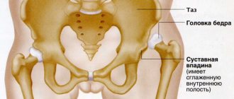
Installation of the acetabular cup
A series of X-rays are taken, in which it is important to correctly identify and evaluate the signs of arthrosis, since, for example, different methods of fixing endoprostheses are used for dysplastic and hyperplastic coxarthrosis. Sclerosis and cysts directly influence the choice of arthroplasty technique, in particular the method of cup attachment. It is impossible to integrate the coating of a cementless cup into sclerotic bone that has lost its elasticity.
For a more reliable fixation of the cup, the cavity is cleaned down to the level of the spongy substance, since this tissue is able to effectively participate in the process of osseointegration. Cement fixation is also more stable when placed on a spongy substance. It is important to remove as much sclerotic tissue as possible.
After treating the surface of the acetabulum with special cutters, holes for attaching the cup are drilled to the level of the spongy substance, and all cystic formations are cleaned out. The cavities remaining after curettage are filled with a substance obtained either from the head of the femoral bone or from the bone canal. The cavity is washed, tamponed, and a cup is installed. When planting on cement, the required thickness of the mantle layer under the bowl is taken into account.
If a cup of cementless fixation is installed in the presence of cysts, then after all the stages of cleansing the surface of the cavity from osteophytes and scraping out the cystic cavities, the remaining defects are filled with spongy autologous bone - a substance extracted from the head, as well as remaining during the treatment of the medullary canal. The substance is compacted at the bottom of the depression, strengthening it. If the patient’s own autologous bone is not enough, then a special synthetic hypoallergenic material is used. In order to increase reliability, the bowl is additionally secured with screws.
Minimally invasive endoprosthetics in the Czech Republic: doctors, rehabilitation, terms and prices.
Find out more
Treatment of cystic formation
Before going on the operating table, the doctor tries to localize the disease using a conservative method. The traditional method of therapy is used if the presence of a cavity does not cause severe discomfort to the patient. Actions are aimed at preventing capsule rupture and damage to adjacent healthy tissues. The treatment algorithm involves performing the following manipulations:
- Application of a tourniquet, tight bandage, elastic bandage after puncture. Immobilization of joints helps reduce the production of synovial fluid and relieve swelling.
- Taking corticosteroids and anti-inflammatory drugs.
- Applying warming ointments. This will help increase microcirculation in the joint.
- Injection of an ozone-oxygen mixture helps saturate cells with beneficial microelements, and the introduction of hyaluronic acid has a lubricating effect on joints and helps reduce friction.
The disadvantage of conservative therapy is that the risk of recurrence of the pathology is high. Therefore, drug therapy is temporary.
Manual physiotherapy
Exposure to a cyst in the hip joint with natural and artificial factors helps strengthen muscle muscles and restore tone. The procedure or complex is prescribed depending on the nature of the pathology, location, degree of development, and includes the following manipulations:
- Electrophoresis is the electrolysis of medicinal substances into the hip joint area using an electrophoretic preparation. For a visible effect, it is enough to limit yourself to 5 procedures.
- Magnetotherapy – increasing the permeability of membranes under the influence of magnetic fields. Exposure to impulses helps relieve pain, relieve swelling, and stimulate regeneration processes in bone and cartilage tissue.
- Laser therapy – the irritants are monochromatic light. This approach has an immunostimulating effect and normalizes cell metabolism. It is recommended to perform 10-12 sessions.
Physiotherapeutic effects on joints, treatment with physical means are popular because they have no specific contraindications, are painless and safe.
The importance of exercise therapy for cystic formation of the hip joint
Therapeutic gymnastics will help restore joint mobility. Modern medicine knows many methods for developing muscle groups. But the Pilyuiko set of exercises and the Bubnovsky and Dikul method are recognized as the most effective. Before starting the exercises, the patient should be notified of contraindications. To avoid harm to the body, you should adhere to the following recommendations:
- Physical activity is allowed only during the period of remission, after pain has been relieved.
- You should start and end gymnastics with a light massage to reduce the risk of joint injury.
- If you feel discomfort or pain, you should stop exercising.
- It is recommended to perform the exercises smoothly with a small amplitude, gradually increasing the load.
Head installation
If there are cysts, growths and other defects in the bone tissue of the femoral head, it is removed, and a femoral canal is formed in the bone using special devices under the stem of the endoprosthetic component. Next, a bone plug is installed at the bottom of the canal, the material for which is the removed head. A femoral endoprosthesis is installed; the use of cement is determined by indications. The head is immersed in the formed acetabulum. The capsule, muscles, and soft tissues are sutured.
Differential diagnosis
Baker's cyst can be mistaken for several other knee pathologies. The patient's medical history, as well as clinical examination and imaging, allow for the correct diagnosis of the disease.
Conditions that are accompanied by the appearance of a space-occupying formation in the popliteal fossa/tibia
| Muscle strain or tear | Palpable formation/tenderness, swelling/hyperemia, pain when contracting and/or stretching the muscle. |
| Muscle contusion or hematoma | Local muscle damage accompanied by bleeding or swelling, painful muscle contraction/stretching. If the hematoma is old, an organized compaction develops. |
| Muscle spasm or cramp | Painful, palpable cord/thickening, possible limited range of motion, pain when stretching muscles. |
| Fascial tear with muscle hernia | Palpable soft formation, acute muscle pain with increased activity, local swelling after physical activity. |
| Myositis ossificans | Palpable painful tightness in the muscles, micro-tears in the muscle fibers that cause pain and swelling when the muscles contract or stretch, impaired movement due to limited muscle function. |
| Deep vein thrombosis | Constant pain, pain with passive dorsiflexion of the foot (Homan sign), local hyperemia, local tenderness on palpation of the calf and possible swelling, increased body temperature. |
| Benign tumor | Local pain and tenderness, possible palpation sensation of softness or stiffness of tissues, there may be a disturbance in movement (depending on the location). |
| Malignant tumor | Generalized malaise, possible sudden weight loss, localized pain and/or swelling of varying sizes and consistency. |
| Hemangioma | Present for a long time, slow changes in size over time, palpation - swelling, may be painful, may/may not limit movement. |
| Baker's cyst | Palpation reveals swelling with possible pain in the area of the popliteal fossa and the posterior inner part of the calf. |
| Rupture or enlargement of Baker's cyst (pseudothrombophlebitis) | Can simulate deep vein thrombosis, swelling in the lower leg, acute pain that intensifies with compression. |
A popliteal cyst may also be confused with a lipoma, which will present less pressure resistance compared to a Baker's cyst or aneurysm differentiated by ultrasound.
Differential diagnosis of compactions in the calf muscle
| Possible diagnosis | Supporting Evidence | Negative evidence |
| Old muscle strain or injury with scar tissue | Palpation - compaction | No history of muscle tear or injury, no pain when running |
| Fascial tear with muscle hernia | Pain when walking over a distance of more than 0.5 km, tenderness on palpation | No connection with injury, latent onset of the disease, no pain when running or performing basic exercises |
| Local muscle spasm | Pain when walking more than 0.5 km, noticeable soreness | No pain when passively stretching the calf, no pain when resisting muscle contraction, no limitation of motion in the foot or knee |
| Deep vein thrombosis | Palpable tenderness, pain when sitting cross-legged, family history of increased coagulation factor XII, oral contraceptive use | No history of deep vein thrombosis and recent immobilization, no edema, no elevated body temperature, negative Homan test, presence of pulsation at the site of injury |
| Benign tumor | Palpable induration, palpable pain, latent onset of the disease | Intermittent pain |
| Malignant tumor | Palpable induration, palpable pain, latent onset of the disease | No weight loss, no night pain or feeling unwell, intermittent pain, good general health |
| Hemangioma | Hidden onset, unknown cause, hormonal changes (oral contraceptives) present for a long time, slow change in size over time, palpable swelling, may be painful, not always accompanied by restriction of movement |
Accurate differentiation of a patient's symptoms can be achieved using ultrasound.
If a popliteal cyst becomes infected, it often results in a painful lump forming behind the knee. In such cases, it may be difficult to make a diagnosis, and an infected cyst may be mistaken for a neoplasm. The cyst may rupture (open up), causing severe pain in the calf, decreased ankle motion, and causing symptoms similar to deep vein thrombosis (seen on ultrasound or venogram).
You may be interested in the article: Prepatellar bursitis: causes, symptoms, treatment.
It is important to diagnose a ruptured Baker's cyst as early as possible to determine the best treatment and avoid complications such as compartment syndrome, and to differentiate it from the following conditions:
- thrombophlebitis;
- popliteal aneurysm;
- inflammatory arthritis;
- calf muscle strain;
- soft tissue swelling or muscle rupture.
It should be considered that swelling along the anterior surface of the proximal tibia in a patient with a previous history of ipsilateral total knee arthroplasty may be a consequence of a Baker's cyst.
Rehabilitation
The rehabilitation period is no less important than the proper execution of joint replacement surgery. Not all clinics offer a full program, limiting it to a few days, and patients are often not conscious enough to continue the recovery complex on their own. Therefore, after the operation, it is recommended to go to a specialized sanatorium, or it is better to have a joint replacement performed in a clinic, which guarantees not only high-quality endoprosthetics, but also provides a full rehabilitation course.
All this can be guaranteed by specialized medical institutions in the Czech Republic, where the high professionalism of doctors, the European level of service provision and the availability of natural resources for the restoration of the musculoskeletal system are combined with low prices for Europe. For many years, he has been organizing treatment for patients from the Russian Federation with orthopedic problems in the best clinics in the Czech Republic. The client receives full information support, assistance in paperwork and transportation. All financial issues are discussed in advance, the results are fixed in the contract and no longer change throughout the entire process of treatment and rehabilitation.
Symptoms
The first sign of cyst development is a feeling of discomfort in the area of the disease. Over time, the cavity increases in size and the following symptoms appear:
- Local swelling and swelling of the problem area.
- Feeling pain. Unpleasant sensations increase when standing for a long time. Exacerbation occurs during physical activity. Relief occurs during rest. Soreness indicates damage to the joint capsule and the tendons connecting to it.
- Changes in the shape of the joint in cases where the pathology is caused by arthritis.
- Restrictions in physical activity, stiffness. There is no possibility of hip abduction.
- Numbness and decreased sensitivity of the skin.
It is worth noting that pain may increase at night and in the morning.