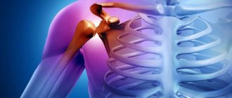
Arthrosis of the shoulder joint code according to the ICD. General information about the disease
Have you been trying to heal your JOINTS for many years?
Head of the Institute for the Treatment of Joints: “You will be amazed at how easy it is to cure your joints by taking the product every day for 147 rubles...
Read more "
Arthrosis is the abbreviated name for a disease such as osteoarthritis or deforming arthritis of the articular surfaces. The pathology is characterized by a chronic course and leads to the gradual destruction of articular cartilage with transition to bone tissue. As the disease progresses, it irreversibly deforms the joint, which is why timely diagnosis and treatment comes to the fore.
OUR READERS RECOMMEND!
Our readers successfully use Sustalaif to treat joints. Seeing how popular this product is, we decided to bring it to your attention. Read more here...
Arthrosis of the ankle, elbow, shoulder and other localizations is characterized by the same clinical picture. The disease is characterized by the following symptoms:
- soreness (at first only during physical exertion, hypothermia or prolonged exposure to an uncomfortable position, and then becomes constant);
- stiffness (decreased range of motion in the joint apparatus, bothers you in the morning);
- crunching (almost the first sign of a pathological process is periodic clicks or crunches in the affected area, which people rarely pay attention to);
- pathological mobility (the more the pathology progresses, the less limited the articular surface is by physiological norms);
- deformation (the latest sign of pathology, avoiding which is the priority task of the attending physician).
Since the arthrosis code in ICD 10 is represented by a larger number of symbols than other pathologies, you should carefully approach the issue of therapeutic measures, choosing the right protocol.
During therapy, systemic medications are used to stop tissue destruction, as well as physiotherapeutic procedures. In advanced cases, radical surgery is performed, the indications for which are determined individually.
Save the link, or share useful information on social media. networks
Source mkbkody.ru
When making a diagnosis, maintaining a medical history, issuing sick leaves, epicrisis, not only the name, but also the disease code according to the international classification (ICD) is used. The use of codes makes the process of accumulating, summarizing, and analyzing information about various diseases and maintaining medical statistics more convenient. Thanks to the system of alphanumeric codes developed by WHO, the diagnosis made by a Moscow doctor will be unambiguously interpreted in any country in the world where the ICD is used.
In ICD 10, arthrosis of the shoulder joint also has its own code. Patients with osteoarthritis of large joints often prefer to go to foreign clinics for endoprosthetics. The medical history using generally accepted codes is understandable to foreign specialists.
Osteoporosis. Prevention of osteoporosis
Osteoporosis is a disease the development of which we can prevent on our own and prevention is necessary throughout life.
What is included in the prevention of osteoporosis:
- Increase your intake of calcium-rich foods (calcium tablets if necessary).
- Vitamin D intake (sun exposure, foods rich in vitamin D, vitamin D in solution).
- Adequate physical activity (walking, Nordic walking, gymnastics).
- Stop smoking, moderate alcohol consumption (up to 2 glasses per day).
- Limit coffee (up to 2 cups per day).
- Maintain normal body weight.
- Avoid falls.
In order to reduce the risk of falls, it is necessary (this is especially important for older people): While outside:
- try to avoid icy sections of the road, walk on paths sprinkled with sand
- wear stable shoes with low heels and anti-slip soles
- if the road is wet, it is better to walk on the grass
- If you have difficulty walking on your own, use a cane. Canes and walkers must be stable, with wide legs.
At home:
- All carpets or rugs must have a non-slip base or be secured to the floor
- It’s better to wear non-slip slippers at home
- maintain order, unnecessary things on the floor, bent corners of the carpet/linoleum can cause a fall, also loose wires, cords that can get caught on (they must be removed)
- place a rubber mat in the kitchen near the stove and sink
- The lighting in the room should be good. A rubberized shower mat is also necessary.
Rubber suction pads in the bathroom will help prevent falls. Shower floors must be dry. all drugs that cause drowsiness, dizziness, weakness can cause a fall.
Discuss with your doctor replacing them with safer alternatives. See an ophthalmologist regularly; if you have vision problems, use glasses/lenses.
Physical activity in patients with osteoporosis
For osteoporosis, three types of exercises are used: aerobic, strength and balance training, as well as their various combinations:
- aerobics to strengthen leg muscles - climbing stairs, dancing, walking;
- strength training to strengthen your back, as well as hanging on a horizontal bar;
- swimming and water gymnastics, which have a beneficial effect on all muscle groups;
Ideally, weekly physical activity should certainly include alternate exercises from all of the above-mentioned 4 groups. The most important thing with any physical activity is regularity. Exercising for 5 minutes every day will benefit you more than half an hour once a week.
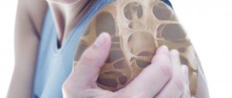
Osteoarthritis of the knee joint
Arthrosis of the knee joint (gonarthrosis) is a chronic degenerative-dystrophic disease of the knee joint, which is characterized by the destruction of articular cartilage, deformation of the knee joint with subsequent limitation of movements in it.
Destruction of cartilage due to arthrosis of the knee joint
Arthrosis of the knee joint ranks first in prevalence in the overall structure. The disease mainly affects people over forty years of age - in this age group, arthrosis of the knee joint is more common in women; among younger patients, males predominate. In approximately 6-7% of cases, arthrosis of the knee joint leads to disability.
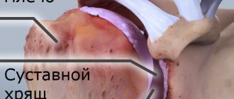
The knee joint is formed by the articular surfaces of the femur and tibia. The front surface of the knee joint is protected by the patella (kneecap). The articular surfaces of the femur and tibia, as well as the posterior surface of the patella, are covered with densely elastic hyaline cartilage, the thickness of which is 5-6 mm. During shock loads, cartilage performs a shock-absorbing function, and during movements it reduces friction. In clinically healthy people, the processes of synthesis and destruction of cartilage tissue are in balance; when the balance is disturbed in the direction of increasing destruction, arthrosis develops. With arthrosis of the knee joint, blood circulation in the small intraosseous blood vessels that feed the hyaline cartilage is disrupted, as a result of which the surface of the cartilage becomes drier and, over time, loses its smoothness. Cracks form on the surface of the hyaline cartilage, which causes regular microtrauma to the cartilage during movement. As the pathological process progresses, the cartilage tissue becomes thinner, and the articular platform flattens, adapting to the load. Along the edges of the articular surfaces, osteophytes appear, which are bone outgrowths formed due to compensatory growth of bone tissue. The pathological process involves the synovial membrane and joint capsule. The joint fluid becomes more viscous and performs its functions worse, which leads to increased destructive processes in the joint. With further progression of the disease, the cartilage thins even more, and in some areas disappears completely. This leads to a sharp increase in friction between the articular surfaces and deformation of the bones that form the joint. The knee joint is deformed; the deformity can be valgus, i.e. X-shaped, or varus, i.e. O-shaped. Movement in the affected joint is limited until it disappears completely (irreversible and complete destruction of the joint is formed).
ICD code 10 arthrosis of the shoulder joint. Symptoms
The main symptoms of the development of DOA include:
Joint pain is the main reason for a visit to a specialist. Initially, it appears irregularly, mainly during movement (running, walking), hypothermia, or prolonged uncomfortable body position. Then the pain becomes non-disappearing and its intensity increases.
At an early stage, gonarthrosis is characterized by a feeling of “stiffness” that appears after a long period of rest (sleep, rest). The knee joint becomes less mobile, its sensitivity decreases and pain of varying intensity is felt. All these manifestations decrease or completely disappear with movement.
Another characteristic symptom is creaking, clicking and other extraneous sounds that occur during prolonged walking or a sudden change in body position. In the future, these sounds become a constant accompaniment when moving.
Often arthrosis of the knee joint leads to its pathologically hypertrophied mobility. According to ICD 10 code: M25.2, this is defined as a “loose joint.” This manifests itself in linear or horizontal mobility that is unusual for it. A decrease in the sensitivity of the end parts of the limbs was noted.
The main functions of the knee joint are movement (motor function) and maintaining body position (support function). Arthrosis leads to functional impairment. This can be expressed both in limited amplitude of its movement and in excessive mobility, “looseness” of the joint. The latter is a consequence of damage to the capsular-ligamentous apparatus or hypertrophied muscle development.
With the development of the disease, the motor function of the diarthrosis joint degrades, and passive contractures begin to appear, characterized by limited passive movements in the joint (ICD code 10: M25.6 Stiffness in the joint).
Musculoskeletal dysfunction
The degenerative-dystrophic changes that occur over time develop into dysfunction (motor and support) of the entire lower limb. This manifests itself in lameness and stiffness of movement, unstable functioning of the musculoskeletal system. Irreversible processes of limb deformation begin, which ultimately leads to loss of ability to work and disability.
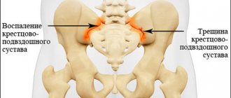
Arthrosis of the hip joint. General information
Coxarthrosis is a progressive degenerative-dystrophic pathology of the human musculoskeletal system, which affects the articular articulation of the head of the femur with the pelvic acetabulum, or, more simply, the hip joint. This disease is characterized by a chronic course with consistent destruction of the cartilaginous structure and bone elements of the joint and, if not treated in a timely manner, ultimately leads to disruption of its functionality, and sometimes to complete immobilization.
According to medical statistics, coxartosis accounts for more than 40% of all musculoskeletal problems diagnosed in humans, which, together with the severe consequences of this disease, in terms of the high chance of the patient becoming disabled, brings it to a socially significant level.
The most susceptible to arthrosis of the hip joint are people over 40 years of age, who have previously experienced intense physical stress on the lower limbs or the entire body as a whole. As a result, many orthopedic doctors consider these two etiological factors (age, hard work/sports) to be paramount in the development of this pathology.
Gender predisposition to the occurrence of coxarthrosis is not traced, since the frequency of its detection in men and women is approximately the same. Children and adolescents suffer from it much less often than adults and mainly under certain unfavorable circumstances (congenital anomalies, injuries, infections, etc.). For older people, the best way to avoid this problem is to prevent it in advance, since treating arthrosis of the hip joint is quite difficult.
Structure of the hip joint
In the human body, the hip joint plays the role of the most powerful and multifunctional motor mechanism, which is directly responsible for upright posture and therefore experiences the maximum load. The structural features of this complex joint allow it to perform the entire range of movements necessary for human life in three planes, including abduction/adduction, flexion/extension, and outward/inward rotation.
The hip joint itself consists of the following structural elements:
- acetabulum - a hemispherical concavity on the outer side of the pelvic bones, which is a hip articular fossa lined with hyaline cartilage tissue;
- femoral head - the upper spherical part of the femur, covered with similar cartilage and its surface entering the acetabulum;
- articular capsule - dense fibrous-synovial tissue of a cylindrical shape, on one side along the circumference adjacent to the acetabulum, and on the other to the neck of the femur;
- articular cavity - a hermetically closed slit-like space surrounding the articular osteochondral surfaces and covered from the inside with a synovial membrane;
- synovial fluid is a viscous substance produced into the joint cavity by the synovial membrane that nourishes the intra-articular elements and ensures their smooth sliding;
- ligamentous apparatus - internal (femoral heads) and external (pubofemoral, iliofemoral, ischiofemoral) ligaments, providing shock absorption and strength of the joint;
- periarticular tissues - muscles, vessels, tendons and nerves directly enveloping the joint from the outside, which are responsible for its nutrition and movement.
Degrees of osteoarthritis
In modern medicine, there are 3 degrees of development of osteoarthritis.
During the first stage, the manifestation of symptoms is minimal. Painful sensations occur only with prolonged load on the joint and cease after rest. An X-ray examination reveals subchondral sclerosis of the articular surfaces.
With osteoarthritis of the second stage, the pain intensifies and can occur even with minor loads, for example, while walking up the stairs. An X-ray image shows growths on the joint and narrowing of the joint space.
The most severe form of osteoarthritis - the third stage - is accompanied by constant pain and limited joint mobility. Examinations show complete destruction of cartilage, absence of joint space, a large number of bone growths and deformation.
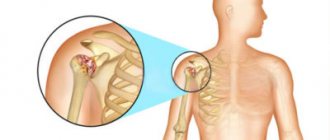
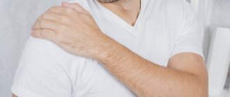
Bursitis of the shoulder joint. Subdeltoid bursitis of the shoulder joint: causes of pathology
There are a considerable number of synovial bursae in the shoulder area. Each bursa received its own name.
Among them there are subacromial, double subdeltoid, subcoracoid, subcutaneous and several others.
One of the bags is called subdeltoid. It is located under the deltoid muscle on the back of the shoulder.
When it becomes inflamed, subdeltoid bursitis of the shoulder joint is detected: the causes of the development of pathology, the symptoms and methods of treatment of which will be discussed further.
The main reasons for the development of the disease
The joints located in the shoulder area are strong. Many people believe that they are capable of carrying quite large loads.
However, more often than not the assumptions are not justified. Without calculating the strength when planning loads, adults, children and adolescents get subdeltoid bursitis.
The causes of the disease, accompanied by inflammation of the joint capsule, are associated with injuries.
They are often obtained during training, training on simulators, in the gym, with certain equipment. Repeated exercise can lead to the development of pathology.
The second reason for the development of subdeltoid bursitis of the shoulder joint is damage to the bursa by infection. Pathogenic organisms most often enter the bursa through an open wound or a cut in the skin.
Development of subdeltoid bursitis of the shoulder joint
Bruises also lead to the development of the disease. It may appear after an accidental fall or collision with hard objects.
Some athletes are susceptible to developing inflammation of the shoulder bursae. Weightlifters, shot throwers, discus throwers, and wrestlers often suffer from the pathology.
To this group you can add loaders who have to lift and move heavy objects every day.
Rare atypical causes of this type of bursitis include gout or autoimmune diseases.
Symptoms of pathology
Aching pain in the shoulder area is the main sign indicating that the patient has an inflamed bursa.
Subdeltoid bursitis is accompanied by incessant pain, sometimes of a pulsating nature.
They may decrease, but they do not disappear. They become especially noticeable when performing any hand action.
Pain with subdeltoid bursitis of the shoulder joint
With an effort of will, one manages to move the limb back and perform a rotational movement. The pain spreads to neighboring areas, radiates to the shoulder blade, neck, worsening the general condition.
Other signs of bursitis caused by inflammation of the subdeltoid bursa include:
- swelling in the shoulder joint;
- limited movement;
- weakness;
- chills.
In the acute form of bursitis, patients may experience uncharacteristic signs of pathology: diarrhea, high fever, vomiting.
Diagnosis of the disease
Shoulder pain does not always indicate inflammation of the synovial bursa, therefore, if subdeltoid bursitis of the shoulder joint is suspected, treatment is prescribed after diagnosis.
It is performed by a traumatologist or surgeon. The doctor performs an examination and asks the patient about his health.
A general blood test is prescribed. The puncture is performed if purulent bursitis is suspected.
Diagnosis of subdeltoid bursitis of the shoulder joint
The synovial fluid is examined, in which traces of infection, blood clots, pus, and mucus can be found.
An ultrasound is prescribed to study the condition of the joint. X-rays are added to rule out bone injuries.
Treatment of subdeltoid bursitis of the shoulder joint
Treatment of subdeltoid bursitis involves immobilization of the limb. A fixing bandage or tight bandage is applied.
During the course, it is prohibited to make any sudden movements with your hand or lift objects.
The course of treatment for bursitis is usually at least 7 days. The products must be used in accordance with the instructions.
The ointment must be applied before bedtime after hygiene procedures.
The therapeutic course includes non-steroidal anti-inflammatory drugs.
They relieve attacks of pain and stop the inflammatory process. Ointments or tablets may be prescribed.
The best NSAIDs for bursitis:
- Nise gel;
- Meloxicam;
- Diclofenac;
- Voltaren gel;
- Celebrex;
Having identified pus in the synovial bursa, the doctor will definitely prescribe antibiotics. They are offered in the form of tablets, injections or ointments.
Celebrex for the drug treatment of subdeltoid bursitis of the shoulder joint
Antibacterial therapy lasts 10-12 days. The antibiotic agent is selected depending on the type of pathogen.
Arthrosis of the acromioclavicular joint: what causes the disease, treatment of the disease
The human skeleton has many joints. For example, there is a clavicle - a bone that connects the skeleton of the body and the upper limbs with the help of shoulder joints. Therefore, a person moves his hands easily. If inflammation begins in this area, fluid accumulates and the cartilage softens, then doctors diagnose a disease called arthrosis of the acromioclavicular joint.
Features of the manifestation of the disease
It is worth paying attention to the characteristic signs of the disease:
- During work involving physical labor, a person quickly gets tired, and mild pain occurs when pressing the collarbone.
- Then the pain becomes aching in the area of the shoulder joint, discomfort is felt when raising the arms. While walking, clicking and crunching sounds are heard in the shoulder.
- As the pathology progresses, it is difficult to get dressed, put your hand behind your head or behind your back, or cross it over your chest. The function of the joint is impaired.
- Cartilage tissue continues to deteriorate. When palpating, where the shoulder connects to the scapula, protrusions are felt. These bone growths are called osteophytes. They are gradually growing. Due to inflammation, the shoulder swells, changes shape, and becomes reddish.
If you have a problem, contact a rheumatologist or orthopedist-traumatologist at the clinic.
Treatment of the disease
In the first stages of the disease, non-surgical therapy is carried out, including biological, physical and chemical methods of influencing the diseased area. It is carried out comprehensively and is aimed at identifying the cause of the disease and eliminating symptoms:
- They provide rest to the affected limb, that is, they fix the shoulder. For these purposes, special external medical devices (orthoses) or an elastic bandage are used.
- If there are no pathological formations of bone tissue (growths on the surface of the bones), then relieve pain with anesthetic injections to reduce sensitivity in the affected area. Inflammation is removed with non-steroidal anti-inflammatory drugs and ointments. For severe muscle spasms, medications are prescribed to reduce the tone of skeletal muscles and reduce their motor activity.
- Restore blood circulation and lack of nutrients in tissues.
To relieve joint pain,
begin physiotherapy aimed at reducing swelling and inflammation. It could be:
- infrared (thermal) irradiation;
- cryotherapy is a physiotherapeutic procedure in which cold is treated;
- to improve blood flow and activate regeneration processes in tissues, modular (different frequencies) currents and UHF are used;
- Acupuncture and massage are effective; the method of healing with beekeeping products (apitherapy) helps;
- after hirudotherapy (treatment with leeches), disturbances in the functioning of the lymphatic (vascular) system are eliminated, and the body's resistance increases.
These treatment methods are aimed at restoring metabolic processes and elasticity of ligaments, improving blood circulation, increasing immunity,
If the form is advanced, surgical intervention will be required.
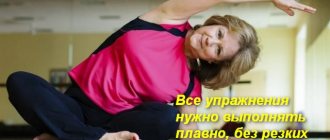
Therapeutic physical education – to help you!
Strengthening exercises for muscles will help restore the affected area. Self-selection of classes is not recommended. The doctor, taking into account the individual characteristics of the patient, will prescribe rehabilitation, the course of which will last from a month to a year. At first, it is better for a patient with a degenerative clavicular lesion to carry out exercises under the supervision of a specialist in a clinic.
Gymnastic exercises can be performed every day and at home. Examples of gymnastics:
- walking in place, arms moving along the length of the hips;
- add circular rotational movements with your arms back and forth, raise your shoulders, holding them in this position for several moments;
- clasp the hands together and gently pull them up.
In this way, the inflamed tendon is stimulated, since the load falls on the fibers of the healthy limb, and it, like a rope, pulls the diseased one along with it
Folk remedies - to help the patient
Together with medications, joint pathology is treated with traditional methods - compresses and lotions made from medicinal herbs.
For osteoarthritis, when the disease has affected the periarticular areas of the bone, ligaments, and periarticular muscles, bandages made from:
- Scrolled horseradish. Recipe: Wrap the rhizome pulp in gauze and place in a saucepan with water, let it boil. The warm composition is wrapped in canvas cloth, previously soaked in the solution in which the shavings were boiled. The lotion is applied to the painful area for an hour. The daily course lasts two weeks. This folk remedy will remove swelling, and blood will flow more actively to the joint damaged by arthrosis.
- At night, prepare a compress from medical bile, 5 percent iodine, colorless syrupy glycerin liquid, flower honey and ammonia. The ingredients are combined in equal proportions. Before use, the mixture is stored for 10 days in a dark place. Shake, heat slightly in a water bath and apply a damp cloth, securing with a bandage.
- Slightly crumpled burdock or cabbage leaves are tied to the shoulder and the pain will go away.
If you have arthrosis of the collarbone, you should eat right up to six times a day. You will have to adhere to a special diet. Spicy, salty, peppery, fatty foods are excluded from the diet. Plant foods, milk-based products, jellied meat, and jelly are welcome. Portions should be small to limit calorie intake.
Attention!
If the clavicular joint is affected, physical activity should be avoided. For preventive purposes, give up bad habits and avoid hypothermia. Author: K.M.N., Academician of the Russian Academy of Medical Sciences M.A. Bobyr
Humeroscapular periarthritis
Humeral periarthritis is an inflammatory process accompanied by degenerative changes in the periarticular tissues that take part in the functioning of the shoulder. Ligaments, muscles, tendons, and synovial bursae suffer from glenohumeral periarthritis.
The humeroscapular type of pathology in the general system of periarthritis is more common than others. It accounts for up to 80% of the total number of rheumatic inflammations of the shoulder. About 10% of the entire world population experience symptoms of this disease in one way or another. Such a wide spread of the disease is due to the fact that the muscle tendons surrounding the shoulder joint are under tension almost all the time. As a result, degenerative processes develop there very early. Most often, glenohumeral periarthritis is diagnosed in women who have crossed the age limit of 55 years, although its symptoms may begin to bother you at an earlier age.
Patients most often complain of right-sided periarthritis, since the load on the right limb is usually higher. However, the development of left-sided and bilateral glenohumeral periarthritis cannot be ruled out.
Causes of glenohumeral periarthritis
Scientists consider two main reasons that can lead to the development of glenohumeral periarthritis:
- Neurodystrophic changes occurring in the tendons, which manifest against the background of diseases of the musculoskeletal system in the cervical spine (osteochondrosis, spondylosis, vertebral displacement). In this case, the nerve roots become pinched, the vessels at the reflex level are compressed, and the normal blood supply to the shoulder joint begins to suffer. The result is the development of inflammation and the manifestation of dystrophic processes in the tendons of the shoulder girdle.
Injury to the soft structures of the shoulder girdle. A person can get injured when performing cyclical stereotypical actions that load the shoulder joint, or when an emergency occurs (falling on an outstretched arm, receiving a strong blow to the shoulder, dislocating the joint, etc.). In this case, the tendons are torn, the integrity of the shoulder cuff is disrupted, the tissues responsible for shoulder movements swell, the normal blood supply system fails, and inflammation develops.
Sometimes the reasons for the development of glenohumeral periarthritis cannot be determined.
It is impossible not to mention the risk factors that increase the likelihood of manifestation of periarthritis in the glenohumeral region:
- The person is over 40 years old.
Hypothermia, both local and of the entire body as a whole.
Spending a long time in damp conditions.
The presence of diseases of the musculoskeletal system in a person: arthrosis, sciatica, arthritis.
Cervical spondylosis with radicular syndrome is combined with glenohumeral periarthritis in 80% of cases.
The presence of congenital developmental anomalies in the glenohumeral region.
Neuropsychic disorders, including those caused by traumatic brain injury. Brain tumors and Kinsonism are also dangerous.
Coronary heart disease. Periarthritis can manifest itself both at the peak of an angina attack and during its decline.
Previous myocardial infarction. Periarthritis is observed on average in 10-15% of patients.
Hemiplegia (complete unilateral paralysis of the arm), which occurs after a stroke, or against the background of other lesions of the spinal cord and brain.
Surgeries that impair the blood supply to the shoulder joint, such as mastectomy.