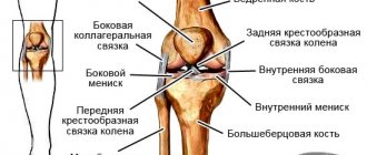
A rib bruise is damage to the soft tissues of the chest as a result of a strong blow. At the same time, the integrity of the skin and bones is preserved.
As for the severity of such an injury, the purpose of the ribs is precisely to protect the internal organs from damage. However, such a bruise is always particularly painful, and can sometimes be accompanied by the development of a serious illness.
General information
Bruising is a fairly common occurrence and not always harmless.
It occurs as a result of direct mechanical influence. The resulting blow leads to damage to soft tissue cells. Most often, with such an injury, the superficial tissues are not ruptured, only rupture of small vessels is noted. However, a very severe injury can lead to serious consequences and complications in the future. Chest injuries are especially dangerous in this context, since vital organs are located in this area, damage to which can occur as a result of a bruise. ICD-10 code for chest contusion is (S20-S29. Chest injuries). A chest injury, as a rule, does not go away without a trace, and if the blow is too strong, a life-threatening condition can develop. At the moment of a very severe bruise or after it, breathing and the heart may stop, since due to a strong blow to the chest area, the functions of two important systems - the respiratory and cardiovascular systems - are sometimes disrupted.
Often a chest bruise causes sharp pain, which intensifies during a deep breath. In the place where the damage occurred, damage and swelling develop. Severe bruising of the ribs can lead to their fracture . Therefore, injuries that cause pain and discomfort in the sternum are indications for hospitalization. The patient may need urgent care and even surgery, which can only be performed in a specialized surgery department. More details about what the consequences of such injuries can be and how to act correctly to help the victim will be discussed in this article.
Complications of rib fractures
Rib fractures, especially multiple ones, are often complicated by hemothorax, closed and valve pneumothorax, and subcutaneous emphysema.
Hemothorax
Hemothorax is the accumulation of blood in the pleural cavity, which leaked from damaged muscles or intercostal vessels, when wounded by fragments of the rib of the parietal pleura. There is less bleeding when the lung parenchyma is damaged, but then, as a rule, hemothorax is combined with pneumothorax, i.e. hemopneumothorax occurs. Depending on the degree of bleeding, hemothorax can be small - it occupies only the pleural sinus (100-200 ml of blood), medium, and does not reach the level of the lower angle of the scapula (300-500 ml). Total hemothorax (1-1.5 l) is extremely rare.
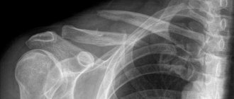
The level of hemothorax is determined by percussion and x-ray with the patient in an upright sitting position. During percussion, the upper limit of dullness of the percussion sound is especially clearly demarcated against the background of the box sound of pneumothorax. On the radiograph, the hemothorax area is darkened with a pronounced horizontal upper border. Under local anesthesia, the diagnosis is clarified by puncture of the pleural cavity. If the hemothorax is small, sometimes it is not possible to suction the blood from the sinus.
Symptoms . A small hemothorax has no special signs, and the clinical symptomatology is dominated only by signs characteristic of rib fractures. But the dynamics of hemothorax need to be monitored, since it can increase. Moderate, especially total, hemothorax compresses the lung, hypoxia, shortness of breath, sometimes hemodynamic disturbances, etc. appear. With hemothorax, body temperature predominantly increases (38-39 ° C).
Treatment . Considering that hemothorax is one of the complications of rib fractures, the patient is treated comprehensively. As for hemothorax, with minor hemorrhage into the pleural cavity, the blood gradually resolves, although puncture is done to minimize the amount of blood. Due to reactive inflammation of the pleura and blood residues, the pleural cavity becomes obliterated over time.
In case of significant hemothorax, blood from the pleural cavity is immediately sucked out with a puncture needle, since after some time it can settle into a clot, and then it is necessary to perform an operation.
If after the puncture blood appears again, which should be regarded as unstoppable bleeding from damaged vessels, the patient undergoes a thoracotomy - a surgical intervention to stop the bleeding. But before that, a puncture and a Ruvilois-Gregoire test are performed to determine whether the blood is fresh. Fresh blood obtained in a test tube in air quickly settles into a clot, but stale blood does not settle. Then you can limit yourself to repeated puncture.
There are cases when exudative pleurisy develops after hemothorax. Then the diagnosis is clarified by puncture and conservative treatment is carried out (repeated punctures, drug therapy, etc.).
Closed and valve pneumothorax
If the visceral pleura and parenchyma are damaged during inspiration, air from the lung enters the pleural cavity, where normally there is negative pressure (0.039-0.078 kPa, 4 8 mm water. in.).
The elastic lung tissue contracts and the lung collapses - a closed pneumothorax is formed. If, in addition to air, blood from damaged intercostal vessels or lung parenchyma enters the pleural cavity, hemopneumothorax is formed.
There are cases when the lung is injured so that pleural or lung tissue hangs over the rupture site. Then, when you inhale, air enters the pleural cavity, and when you exhale, this tissue, like a valve, closes the opening to the lungs and does not allow the air to escape - a valve pneumothorax is formed.
With each inhalation, the amount of air in the pleural cavity increases, its pressure rises sharply (tension pneumothorax), which leads to compression of the lung and displacement of the mediastinum. Disorders of gas exchange and hemodynamics appear quite quickly. The general condition of the patient becomes severe, severe shortness of breath, cyanosis of the skin and mucous membranes, and tachycardia occur. As a result of sudden suffocation, the patient develops fear and severe psychomotor agitation.
The presence of pneumothorax is determined by percussion by the characteristic box sound, comparing it with the healthy half of the chest. On auscultation, breathing is weakened, and with a collapsed lung, it cannot be heard. The radiograph shows a clear contour of the collapsed lung against the background of clearing of the pneumothorax area. Puncture of the pleural cavity clarifies the diagnosis; moreover, with valvular tension pneumothorax, air comes out through the needle under pressure.
Treatment . In case of closed pneumothorax, regardless of its degree, air is immediately sucked out from the pleural cavity. This, firstly, improves the general condition of the patient, and, secondly, with prolonged pneumothorax the lung becomes rigid, and then it is more difficult to straighten it.
If for hemothorax the chest is punctured in the lower section, then for pneumothorax it is punctured in the upper, mainly in the second intercostal space along the midclavicular line. The air is sucked out using a Janet syringe or a triampulle system. If the pressure in the pleural cavity becomes negative, then the triampulle system is excluded. Lung expansion is controlled by percussion and x-ray.
The general condition of a patient with a closed valve pneumothorax is so severe that he should immediately, directly at the scene of the accident, perforate the chest wall (with a thick injection needle) - convert the closed pneumothorax into an open one. After the puncture, air is immediately released from the pleural cavity under pressure. And then the pressure in the cavity is equalized with atmospheric pressure, and the patient’s general condition improves. Choking is significantly reduced. After a few hours, with collapsed lungs, the “valve” can be stuck with fibrin, and a regular closed pneumothorax is formed. In these cases, the air from the pleural cavity is sucked out using a triampulle system. If the lung has expanded, then the triampulle system is not excluded, but negative pressure is maintained in the cavity and monitored for a day or two. The system is turned off only when they are sure that the valve has closed and there is no air in the pleural cavity. This is confirmed by percussion, auscultation and x-ray.
If the amount of air sucked out exceeds the nominal volume of the pleural cavity, this indicates that air continues to flow from the damaged lung. In this case, the pleural cavity is drained using the Bulau method .
Execution technique . A finger of a surgical glove is hermetically secured to one end of a sterile rubber tube (diameter 5 mm and length 60-70 cm), the top of which is cut along the length by 1.5-2 cm. Thoracentesis is performed and the second end of the tube is inserted into the pleural cavity, fixed, sealed skin wound with a suture. The finger is dipped into a sterile jar filled with an aqueous solution of an antiseptic substance (furacilin (1: 500), etacridine lactate (1: 1000), etc.).
During inhalation, the tip of the finger collapses in the solution and closes the hole in it, preventing the solution from being sucked into the tube. When you exhale, the chest collapses and air escapes through the tube into the jar. This is how suction drainage works. After a day or two, when the valve in the lungs closes, negative pressure is created in the pleural cavity, and the lung expands, the drainage stops working, and after a day it is removed.
If the valve does not close after a few days, this indicates significant damage to the lung, and the patient is operated on. After elimination of pneumothorax, patients with rib fractures are treated according to general principles.
Subcutaneous emphysema
If there is pneumothorax and damage to the parietal pleura or mediastinum, then air from the pleural cavity through the wound enters the soft tissues of the chest or mediastinum, moves through the interfascial spaces into the subcutaneous tissue of the shoulder girdle, neck and face. Subcutaneous emphysema is especially pronounced with valvular pneumothorax.
Characteristic signs of subcutaneous emphysema: swelling in the area of air accumulation, and upon palpation - a specific crunch in the subcutaneous tissue (“walking in the snow”) due to the rupture of bubbles and movement of air. By percussion you can feel the difference in the percussion sound above the emphysema. Air in the soft tissues is also visible on a chest x-ray.
Subcutaneous emphysema gradually decreases, the air is absorbed and no special treatment is required. Only in case of excessive emphysema, when air accumulated under the skin of the neck compresses the veins or trachea, small fasciocutaneous openings with drainage are made above the collarbone, through which the air escapes.
Pathogenesis
When the chest is contused, damage occurs in which the integrity of the tissue is not compromised. The mechanism of development of chest contusion varies. Such injuries occur during a fall, after being hit by a hard object, or when a person’s body is pinched on both sides. The severity of the injuries and the mechanism of their development depend on the characteristics of the injury and the type of tissue that was impacted (skin, muscles, bones, adipose tissue, internal organs). It is in the chest that vital organs are located - the lungs and the heart. Therefore, as a result of a blow to the sternum, not only external hematomas , but also very serious damage - rupture of the diaphragm or lung, heart contusion, etc. In such conditions, immediate medical attention is required.
Symptoms
With a bruise of the soft tissues of the chest, a dull pain appears with a maximum at the time of injury. Typically, pain increases with movement, coughing, speaking, or trying to breathe deeply.
When small vessels are crushed, blood “saturates” the subcutaneous tissue, causing a bruise. The latter is visible through the skin in the form of a purple spot, which over time changes color to blue and green (which is why it is called a “bruise”). The skin over the bruise is warm, and the surrounding tissue is swollen. In this case, the height of the edema occurs not at the time of injury, but an hour or two after.
Lung contusion
Bruised chest wounds due to strong blows or prolonged compression of the chest may be accompanied by contusion of the lungs. The injury is caused by the “bending” of the ribs during the impact and subsequent damage to the lungs.
Usually there is deep pain in the area of injury, which is difficult to distinguish from superficial pain due to soft tissue bruises. The pain intensifies with deep breaths, bending over and pressing on the chest. A frequent clinical manifestation is hemoptysis - the separation of bloody sputum. Respiratory failure increases in patients: shortness of breath appears, heart rate increases and blood pressure drops. The skin first turns pale, after which diffuse cyanosis appears.
Another option for lung injury due to chest contusion is closed pneumothorax. The latter develops when the ribs are fractured and the lungs are ruptured by bone fragments. In this case, air from the injured lung tissue accumulates under the pulmonary membrane (pleura), which leads to subsequent collapse of the lung (atelectasis).
Patients usually complain of severe pain spreading to the neck and upper limbs. Shortness of breath, lack of air and a dry cough appear. The heartbeat quickens, blood pressure drops, the skin of the lips turns blue, and cold sweat appears on the face. Patients often take a forced position: they sit with their hands supporting them on the bed.
A characteristic manifestation is the accumulation of air in the skin tissue - subcutaneous emphysema. Visually, the skin of the chest, neck and face swells (up to the closure of the eyelids and the disappearance of the voice), and when palpated, a crunching sound is detected (like dry snow).
In rare cases, the damage is complicated by thromboembolism - blockage of the pulmonary artery by a blood clot. The patient experiences respiratory failure, followed by cessation of both breathing and heartbeat. This condition has a high risk of death.
Heart bruise
With a bruised wound to the heart, various parts of the organ may be primarily affected. Thus, with a closed injury of the myocardium and conduction tracts, interruptions in the functioning of the heart and pain syndrome such as angina pectoris (“angina pectoris”) are observed.
When the valves are bruised, their insufficiency appears, leading to pulmonary edema. The clinical picture of the latter includes shortness of breath, lack of air, and the release of bloody and foamy sputum. The patient's skin is bluish, sticky, and the pulse is rapid.
Trauma to the coronary vessels threatens with detachment of their inner lining followed by thrombosis. This condition is complicated by myocardial infarction: intense pain in the heart area, spreading to the left side of the body and arm.
However, isolated contusions to individual cardiac structures are rare. More often bruised injuries are combined, i.e. combine different types of damage. They occur in two main types: angina (when the main clinical symptom is chest pain) and infarction-like (with a combination of shortness of breath, fever and agitation).
Classification
Like other types of bruises, bruises of the sternum, depending on the damage received, can be divided into several degrees.
- First , minor damage is noted. There is practically no pain; abrasions and scratches may remain. After a few days, all unpleasant manifestations disappear without treatment.
- The second is pain, muscle tissue is damaged, resulting in swelling and hematomas.
- Third , in addition to external damage, internal damage to organs may also occur.
- The fourth is a serious injury, causing serious damage to internal organs and threatening health.
First aid for bruised sternum
In case of severe chest injury, you should call an ambulance. Before the team arrives, you need to do the following:
- arrange the victim in a semi-sitting position, ensure him peace;
- apply a cold compress to the damaged area, which will help reduce swelling and hematoma;
- if the pain is severe, give the patient an anesthetic and be sure to inform the arriving doctors about this.
Next, the patient should be treated by a traumatologist. An ambulance employee will decide on the spot whether hospitalization is necessary or recommend contacting a specialist for treatment.
The appointment is conducted by traumatologists
Denis Ivanovich Burmakin - Deputy Director for Medical Affairs, orthopedic traumatologist. Extensive experience in emergency traumatology, pediatric orthopedics, and rehabilitation after injuries.
Ask a question Read more...
Bryukhanov Anatoly Valentinovich - microsurgeon.
Ask a question Read more...
Bryukhanov Vladimir Innokentievich - traumatologist-orthopedist, highest qualification category, Honored Doctor of the Russian Federation, work experience of more than 35 years.
Ask a question Read more...
Zotov Vyacheslav Viktorovich - orthopedic traumatologist.
Ask a question Read more...
Pospelov Yuri Vladimirovich - orthopedic traumatologist.
Ask a question Read more...
Klimov Vladimir Aleksandrovich - orthopedic traumatologist. Operating traumatologist-orthopedist.
Ask a question Read more...
Dmitry Anatolyevich Shcherbovich - orthopedic traumatologist, chiropractor, physiotherapist, first medical category, 13 years of work experience. Head of the rehabilitation department.
Ask a question Read more...
Causes
A chest contusion occurs from a blow to a hard object, a blow from a fallen object, or compression between two hard objects.
Medical statistics show that chest contusions are a relatively rare occurrence. They make up no more than 15% of all cases of bruises treated by doctors. Most often, such injuries occur due to the following reasons:
- Car accidents - as a result of road accidents, drivers who hit their chest on the steering wheel most often suffer from such a bruise.
- Falling from height.
- Fight - bruise can occur as a result of a blow to the chest with a blunt object or fist.
- Household reasons.
- Sports activities.
- Industrial injuries.
Kinds
The classification of types of thoracic bruise consists of several elements. These include:
- external and open injuries;
- rib fractures;
- vascular injury;
- crushing of the sternum;
- injury to the heart muscle;
- dislocation, curvature of the capsular-ligamentous apparatus;
- damage to nerve endings, spinal cord;
- deformation of organs located next to the injury (abdominal wall trauma).
According to the degree of localization, contusion of the soft tissues of the chest can be of two types. These should include:
- Contusion of the chest on the right - can cause damage to the lung, resulting in a risk of organ rupture with internal bleeding.
- Left-sided contusion - in particularly difficult cases, can cause injury to the heart muscle, even leading to cardiac arrest with a fatal outcome.
Symptoms of chest contusion
The characteristic signs of any bruise are, first of all, hemorrhage in the tissue without damage to the skin. But the complex of symptoms of sternum injury depends on the severity of the injuries received. Therefore, the stronger the blow, the more pronounced and intense the symptoms will become.
Signs of a chest contusion may include the following:
- Pain in the place where the blow occurred - the pain can be aching, throbbing, or dull. If you experience throbbing pain, this may indicate damage to the nerve endings, as well as a heart injury. Pain can be bothersome both during movement and at rest. There is often an increase in pain when breathing, sneezing, coughing, or talking.
- Hematoma - a bruise forms in the place where the soft tissue of the sternum was damaged, since upon impact small capillaries are damaged and bleeding occurs into the subcutaneous tissue. The hematoma may be larger than the affected area. Minor lesions may resolve without bruising.
- Swelling – fluid accumulates at the site of injury and swelling forms.
However, such trauma is rarely isolated. Due to the anatomical proximity of the lungs, diaphragm, and heart, the risk of their damage is very high. Most often, a bruise of the sternum is accompanied by:
- lung contusion;
- bruised ribs, which may result in a rib crack or fracture (complete or partial);
- breast injury;
- heart bruise.
If such complications occur, then the symptoms after the blow will be more pronounced. In this case, the following symptoms are likely:
- Severe pain when breathing, touching, palpating.
- The appearance of breathing problems (they indicate damage to the lung or injury to the ribs). Sometimes the pain is so severe that it is difficult for a person to breathe air.
- If the injury causes damage to the lungs or pleura, the victim may develop pneumothorax or hemothorax . Pneumothorax is a condition in which a large amount of air collects in the chest cavity, causing compression of the lung. With pneumothorax, there are serious breathing problems, decreased pulse and increased blood pressure , and the development of emphysema under the skin. The man is in serious condition. Hemothorax - when large blood vessels located in the sternum, lungs or abdominal cavity are damaged, blood enters the pleural cavity and thickens there. In this condition, serious increasing breathing problems, severe weakness, headache and other general symptoms are observed. Both of these conditions are very dangerous to health and require urgent medical attention.
Symptoms of bruised ribs immediately manifest as sharp pain and redness of the skin. Later, swelling and hematoma develop. The victim feels a constant dull aching pain, and it increases with sudden movements, coughing, laughing, and deep breathing. The same are the signs of cracked ribs. If a person feels a crunch inside when moving, this may be evidence of a rib fracture. Pain from a chest injury may not go away for a long time.
Principles of treatment
The treatment and rehabilitation program for each patient with a chest contusion is selected individually. For superficial injuries, the victim is treated on an outpatient basis. In case of severe injuries, the patient must be hospitalized in a hospital.
Drug therapy
The main goal for bruises is to reduce pain, relieve inflammation and restore the structure of soft tissues. The patient is prescribed:
- Painkillers.
- Anti-inflammatory drugs - Ibuprofen, Nise.
- Enzyme preparations - Flogenzy or Wobenzym. These medications have anti-edematous and anti-inflammatory effects. When they are used, the pain goes away faster and the resorption of bruises on the sternum improves. Enzymes are well tolerated and have virtually no contraindications for use.
If a child has a chest injury, drug treatment is selected based on his age.
The use of external preparations is also indicated. Ointments Indovazin, Capsicam, Viprosal, Dikul balm relieve pain and promote the resorption of compactions and swelling. For hematomas, it is recommended to use Spasatel, Sinyak-Off, Girudolgon balm. The area of the bruise can hurt for up to several weeks; the use of ointments speeds up the disappearance of discomfort.
A tight bandage placed on the chest helps to ease breathing. In the absence of contraindications, physiotherapeutic methods of influence are prescribed.
Full recovery for mild chest contusions takes up to three weeks. For concomitant internal injuries, treatment can last two to three months.
If the pain does not go away or decrease within 5-7 days after the prescribed treatment, then you should tell your doctor about it. Depending on the patient’s condition, the doctor will prescribe a re-examination or select stronger analgesics.
Traditional methods of treatment
Treatments prescribed by a doctor can be combined with traditional methods of treatment. At home it is recommended:
- Use cold compresses for the first two days after the stroke. The procedure is carried out up to 5 times a day, applying cold to the injured area for 15-20 minutes.
- On the third day, cold compresses are replaced with warm ones. You can start using various lotions, absorbent rubbing. The bruise site will hurt less when using compresses.
- Do breathing exercises. Its implementation will reduce congestion and normalize lung function.
An ointment made from honey and aloe juice has a resolving effect. The ingredients are mixed in equal proportions, gauze folded several times is moistened in the resulting mixture, after which the compress is applied to the bruised area for one hour. The procedure can be repeated up to three times a day.
For bruises, you can also use a warming alcohol tincture. It is prepared from half a liter of vodka and one hundred grams of medicinal herbs; you can use yarrow, eucalyptus, oregano. The mixture is infused for three days, after which a therapeutic compress is made from it on the chest. You can keep it on all night.
You should not take a steam bath in the first days after an injury.
Hot air increases blood circulation, which increases the load on the heart and lungs. It is advisable to plan a trip to a sauna or steam bath two to three weeks after a fall or blow. In severe cases, you can only take a steam bath after your doctor’s permission.
Sleeping position
During sleep, pain may intensify, since the body does not control movements at this time. To avoid discomfort, you should lie with your back slightly elevated. You can use pillows or raise the head of the bed. It is advisable to apply a tight bandage while you sleep, which also reduces the likelihood of increased pain.
Tests and diagnostics
A person who has suffered a chest injury needs to be examined by a traumatologist or surgeon. Confirming a bruise is not difficult. But at the same time, it is important to exclude the presence of injuries to the chest organs and ribs, and to promptly detect life-threatening conditions. The doctor uses the following diagnostic methods:
- Questioning, medical history - the doctor must find out how the injury was sustained.
- Inspection - allows you to get a general impression of the consequences of the injury. The doctor must pay attention to the presence of cyanosis , which is evidence of hypoxia caused by respiratory failure. The presence of subcutaneous emphysema is also determined as evidence of damage to the lung or bronchus, respiratory sounds, etc.
- Palpation - performed to determine whether a rib fracture has occurred. Sequential palpation of the ribs and sternum is carried out. The doctor pays attention to the symmetry of the sternum, the nature of breathing, the condition of the neck veins (non-pulsating swollen veins are a sign of cardiac tamponade), etc.
- Chest X-ray – allows you to evaluate the condition of the bones of the chest cavity.
- MRI - performed if there is suspicion of damage to internal organs. Allows you to assess the condition of blood vessels and soft tissues, and determine internal hematomas.
- CT scan allows you to assess the condition of bone structures in more detail.
- Electrocardiogram – to determine the condition of the heart. Blood pressure and pulse .
If necessary, other studies are also carried out. It is very important to conduct a complete and comprehensive examination so as not to miss serious internal injuries.
Rib fractures
Single rib fractures, as a rule, occur as a result of direct trauma - at the point of application of force (impact, pressing against a certain object). Double rib fractures occur. When the chest is compressed in the anteroposterior direction, several ribs break along the axillary line, and in the lateral direction, along the paravertebral and midclavicular lines. Multiple bilateral rib fractures occur in severe road traffic injuries, rubble, etc. Sometimes a sharp fragment of a rib can damage intercostal vessels, perforate the parietal pleura and even injure the lung.
Symptoms . The patient complains of sharp pain at the fracture site, which increases with inspiration. The general condition of the patient depends on the severity of the injury (number of damaged ribs, degree of lung failure, hypoxia, blood loss, pleuropulmonary shock, etc.).
With single rib fractures, the patient's general condition remains satisfactory. The patient spares the chest and breathes shallowly. Due to the pain, he cannot cough up the mucus that accumulates in the upper respiratory tract, and therefore gurgling appears, and over time, pneumonia can develop. Hemoptysis indicates lung damage.
During palpation, the points of maximum pain are determined. If you squeeze the chest lightly, local pain increases, and the patient points to the fracture site. With double fractures of the ribs (fenestrated fracture), when inhaling, this area sinks, and when exhaling, it levels out. Such flotation of the chest wall with each breath is very painful, which affects the nature of breathing, the function of the organs of the mediastinum, which also floats, and the general condition of the patient.
Multiple and especially bilateral rib fractures cause severe respiratory distress, hypoxia and traumatic pleuropulmonary shock. The examination of the patient includes chest x-ray, percussion and auscultation to identify rib fractures and possible complications - hemothorax, pneumothorax, etc.
Treatment for uncomplicated rib fractures
If individual ribs are damaged, treatment is limited to pain relief, improvement of breathing conditions and prevention of pneumonia.
The patient is placed in a semi-sitting position in bed. A local or paravertebral blockade is performed with a 1% novocaine solution, and analgesics are prescribed. After anesthesia, chest excursion improves, and breathing becomes smooth and deep, the patient can even cough up sputum, which prevents the occurrence of pneumonia. The blockade is repeated 2-3 times. In addition, patients are prescribed breathing exercises and symptomatic therapy. Fractured ribs heal in 3-4 weeks, performance is restored in 5-6 weeks.
In case of multiple rib fractures (four or more), complex treatment is carried out, which is determined by the severity of the patient’s condition. In order not to disturb a seriously ill patient with repeated blockades and to maintain constant pain relief, a thin tube (vascular catheter) is inserted into the paravertebral area through a needle, which is left, glued with an adhesive plaster to the chest wall, and its second end (catheter cannula) is brought out to the shoulder girdle. When pain occurs, without moving the patient, 15-20 ml of a 0.5% novocaine solution is injected into the catheter (4-5 times a day).
Patients with severe breathing disorders are also treated with cervical vagosympathetic blockade according to A.V. Vishnevsky and undergo intensive therapy, and sometimes resuscitation measures (intubation, mechanical breathing, etc.).
In case of double fenestrated fractures of the ribs, in order to eliminate flotation, under local anesthesia, the ribs are fixed with Kirschner wires passed percutaneously, or they are applied to the sinking area of extraction (stitching behind the soft tissues and periosteum of the middle rib with a coarse Mylar thread or using bullet forceps). Ribs fixed using the following methods fuse in normal time. Open osteosynthesis of ribs is used extremely rarely.
Complex treatment also includes oxygen therapy, suction of mucus from the trachea, antibiotic therapy, etc.
Treatment with folk remedies
You can use traditional methods aimed at rapid healing of injuries only if a slight bruise has occurred and the absence of serious injuries has been confirmed by a doctor. You can prepare folk remedies for a compress. The selected product should be used every day for 10-14 days.
- Herbal tincture for compress. Mix 20 g each of horsetail, bearberry, hernia, bean pods, horsetail, birch buds, cornflower flowers, knotweed and meadowsweet. Pour all the herbs into 0.5 liters of vodka and leave for three days. Use the product as a daily compress.
- Tincture of wild rosemary and nettle. Take 100 g of each herb and pour 100 ml of vodka. After 4 days, pour 200 ml of pomegranate juice into the product and leave for another day. Use for compresses.
- Tincture of birch buds. Take 100 g of birch buds and 50 g of barberry, mix with 100 ml of vodka and leave for several days. Make compresses every day.
- Tincture of St. John's wort and oak bark. Take 20 g of oak bark and marshmallow, 30 g of St. John's wort. Grind the herbs and pour in 0.5 liters of vodka. Leave for 4 days, strain. Use for daily compresses.
First aid
To minimize the negative consequences of an injury, immediately after the incident you must act as follows:
- Apply cold to the affected area by unbuttoning your clothes. A water bottle, a cold heating pad, or a wet towel will do.
- If cracked ribs are suspected, a bandage may be applied to the chest. It will reduce breathing movements, which will relieve some of the pain.
- If you suspect serious damage, you should call an ambulance.
- If a person is unconscious, he needs to be given ammonia to sniff and brought to his senses. If vomiting occurs, tilt the patient's head to the side.
- If breathing stops, artificial respiration must be performed immediately.
- It is important to understand that even with minor injuries you need to consult a doctor. External signs are not always evidence of the true condition of the victim.
Features of treatment for rib bruises
After receiving an injury, the victim must be provided with rest and be kept in bed for the first few days so as not to provoke an increase in pain. It is recommended to apply cold immediately to stop internal bleeding and reduce swelling.
If there is complete confidence that there is no fracture or crack in the rib, then a bandage can be applied. A person without relevant experience is unlikely to be able to do this correctly, so many turn to a doctor.
After about two days, you should replace the cold with warm compresses, which will promote the resorption of the hematoma. You can use regenerating, analgesic and antipyretic drugs if necessary.
Only after the pain has decreased should you gradually begin to perform stretching exercises.
All these measures are appropriate if the injury was mild. Otherwise, for example, after a strong blow, when the pain does not stop for a long time, the temperature persists, the victim has difficulty breathing, or unnatural dents and protrusions are noticeable at the site of the injury, it is better to contact a medical facility.
The specialist will conduct an examination, refer the patient for an x-ray, and then prescribe a set of treatment. Perhaps the fears will be in vain, but it will certainly be possible to eliminate serious consequences.
Consequences and complications
The consequences of a chest injury can be very serious. As a result of the blow, such serious consequences as a ruptured lung, ruptured diaphragm, pulmonary hematoma (hemorrhage), and heart contusion are possible.
Lung rupture often occurs due to injury from a broken rib fragment. The causes of lung rupture can be associated with both a strong blow and tissue tension. The consequences of such an injury can be eliminated if you consult a doctor in time.
With a mild injury, the consequence is a hematoma and pain, which sometimes may not disappear for a long time.
Because sternum injuries cause severe pain, patients are sometimes forced to make limited breathing movements (rigidity). A complication of this phenomenon may be hypoxemia or pneumonia .
If hemothorax is poorly drained, a purulent intrathoracic infection is likely.
Consequences
Possible consequences of a bruise in the thoracic area occur immediately or after some time. In this case, the risk of the following dangerous conditions increases:
- Fracture of the ribs, which entails damage to blood vessels and soft tissues.
- Violation of the integrity of the pleural tissue - pneumothorax, which leads to air getting between the lung and the pleural layer.
- Midline fracture of the sternum.
- Compression of the lung when blood enters the pleural cavity is hemothorax. The condition is caused by rupture of large vessels.
For women, a bruise is dangerous not only because of the above-mentioned consequences. The fair half risks damaging the mammary glands, as a result of which the risk of lumps increases. Fibrous hematomas can provoke the development of malignant tumors. Regardless of gender, bruises can cause stroke, myocardial infarction, and sudden cardiac arrest.
List of sources
- Bagnenko, S. F. Medical care for mechanical trauma of the chest and abdomen at the prehospital stage / S. F. Bagnenko, Yu. B. Shapot, A. N. Tuchunov // Bulletin of Surgery named after. I.I. Grekova - 2007. - No. 2. -S. 47-51.
- Tatyanchenko, V.K. Clinical and anatomical aspects in the surgical treatment of chest wall injuries. Educational and methodological manual / V. K. Tatyanchenko. — Rostov-n/D. : Publishing house of Rostov State Medical University, 2006. - 21 p.
- Seleznev S.A., Bagnenko S.F. Traumatic illness and its complications. St. Petersburg 2004.