A computed tomography (CT) scan of the shoulder provides cross-sectional images of the shoulder structures through the use of specialized X-ray cameras. This type of scanning makes it possible to visualize both the bony structures of the shoulder and soft tissues and identify morphological changes. CT scans also help diagnose blood clots and tumors.
A CT scan of the shoulder joint can be performed with or without contrast. The contrast agent allows for better visualization of vascular structures and some pathologies that cannot be detected without contrast.
Indications
The most common reason for ordering a CT scan of the shoulder is to assess the integrity of the shoulder structures after injury. This may be necessary both in case of acute injury and in the presence of instability and frequent dislocations. A CT scan can help your doctor pinpoint the presence of a fracture or fissure.
Main indications for a CT scan of the shoulder:
- Diagnosis of blood clots
- Detection of space-occupying processes or tumors
- Diagnosis of infections
- Detection of muscle, tendon or ligament tears
- Detection of inflammatory processes in the joint
- Diagnosis of structural integrity violations or dislocations after shoulder injuries
- Preparation for surgical treatment
- Monitoring the dynamics of treatment
Diseases that can be detected using a CT scan of the shoulder joint:
- Rotator cuff tear
- Rotator cuff tendonitis
- Bursitis
- Arthritis
- Dislocation (dislocation) or subluxation of the shoulder
- Calcific shoulder tendinitis
- AC rupture
- Foreign body
- Inflammation in the shoulder joint
- Infection in the shoulder
- Bone cancer, primary and metastatic
Also, a CT scan of the shoulder joint may be prescribed by a doctor to determine the causes of symptoms such as pain, stiffness, especially if MRI is contraindicated (for example, if the patient has a pacemaker).
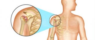
What does a CT scan of the shoulder joint show?
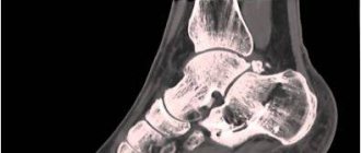
Photo of computed tomography of the shoulder joint
The shoulder joint is the most mobile joint in the human body. The huge range of movements leads to its instability, so dislocations and injuries are often recorded. Dislocation of the articular head from its seat increases the likelihood of damage, which often leads to a degenerative process in which the tissues surrounding the joint gradually break down and cease to function normally. What can be found on tomograms? A CT scan of the humerus demonstrates the complexity of the fracture, the displacement of the fragments, and the angle formed by their position. The ability to visualize images in the axial, sagittal and coronal planes, as well as in three-dimensional format, assists in the interpretation of the lesion and preoperative planning.
CT scan of the shoulder allows you to diagnose the following pathological processes:
- infectious damage to the articular apparatus;
- thrombus formation in the vessels of the shoulder joint;
- systemic connective tissue diseases involving the shoulder joint;
- osteoarticular tuberculosis;
- ruptures of tendons, muscles, ligaments;
- post-traumatic injuries (dislocation, subluxation, fracture, sprain, bruise) and complications (osteonecrosis, scar deformities, etc.);
- dystrophic changes in the area under consideration;
- tumor process (including metastatic lesions) in the joint.
The diagnosis is finally verified taking into account the totality of the results of all studies. The effectiveness of therapy is higher after complete diagnosis, and the treatment regimen is directly related to the pathogenetic factor. A CT scan of the shoulder joint allows the doctor to evaluate the bones, soft tissues, and osteoarticular joints and detect abnormalities.
Risks
A CT scan of the shoulder carries very few risks.
The contrast used in some cases may cause an allergic reaction or cause kidney damage, especially if the patient has a history of kidney disease. But new contrast agents developed recently can reduce the risk to the kidneys.
CT scans use X-rays and there is a risk of exposing the body to ionizing radiation, especially when it comes to a developing fetus. Therefore, the presence of pregnancy significantly reduces the indications for a CT scan.
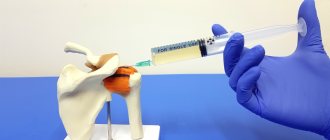
Contrasting
Injection of contrast into a vein through a catheter
Contrast significantly improves the clarity of tomograms and allows you to examine the smallest details that are not shown by a simple (native) computer scan, radiography and ultrasound of the shoulder joint. In most cases, intravenous enhancement is not required and its use is reserved for pathological processes suspicious for tumor or bone abscess. If the procedure involves the introduction of a contrast agent, there are certain nuances that you need to know:
- Intravenous injection of a radiopharmaceutical on an empty stomach increases the likelihood of developing vegetative symptoms: dizziness, nausea, drooling. Therefore, before the examination, 30-45 minutes before the examination, you need to have a light snack.
- If you have previously had an allergic reaction to contrast, tell your doctor. The feeling of hot flashes, rapid heartbeat, mild dyspeptic disorders, which disappeared after a few minutes on their own, are considered normal and are not contraindications to contrast.
- Metformin should be discontinued for diabetes mellitus 36 hours before an enhanced CT scan; discontinuation of the drug should be discussed with an endocrinologist.
- Before a contrast examination, even if there has never been kidney disease, a blood test for creatinine is required. Results are valid for 10 days.
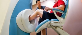
How is a CT scan of the shoulder performed?
- CT scans are performed in the radiology department of the clinic or on an outpatient basis. After the patient removes their jewelry and changes into hospital scrubs, the technologist will ask the patient to lie down on the scanner table.
- If contrast is used, it is administered intravenously through a catheter.
- The technician will ask the patient to lie in a specific position during the procedure. Pillows or straps can be used to fix the position of the body during scanning, since the correct position allows you to obtain high-quality images. In addition, the patient may hold their breath during the procedure.
- During the procedure, the X-ray tube rotates around the patient.
- After the scan cycle, it may take some time for the radiologist to assess the quality of the images.
- Once the scan is completed, the patient can return to their normal lifestyle.
- A CT scan of the shoulder joint usually takes 30-45 minutes.
Who is indicated for a CT scan of the shoulder joint?
Doctors prescribe a similar diagnosis for patients with:
- intra-articular fractures, as well as clavicle fractures;
- dislocation or subluxation of the shoulder joint;
- arthritis (inflammatory joint diseases) and arthrosis (degenerative-dystrophic diseases of the joints);
- congenital abnormal development of the shoulder joint;
- neoplasms in the area of bones and cartilage, which are benign or malignant;
- osteochondropathy (aseptic necrosis of cancellous bone);
- accumulation of fluid in the joints;
- joint hemorrhage;
- acute pain, periodic crunching and stiffness in the shoulder joint.
The doctor can also prescribe a similar diagnosis to a patient who needs surgery or after it. This will allow you to see the results of the surgical intervention.
Advantages and disadvantages of the procedure
- CT scan of the shoulder joint is a simple and relatively safe research method, since the effect of X-ray radiation is local and the harmful effect of ionizing radiation on the body is much less than with X-rays.
- The CT scan procedure does not take much time and is absolutely painless. The procedure clearly visualizes bone structures, blood vessels and nerves.
- CT is much worse than MRI at visualizing soft tissues (cartilage, ligaments, tendons).
- In addition to MRI, if it is necessary to clarify the diagnosis, research methods such as ultrasound, diagnostic arthroscopy, and fluoroscopy can be used.
- The results of a CT scan of the shoulder joint can be used by doctors of various specialties (traumatologists, orthopedists, oncologists, etc.)
Indications and contraindications
Computed tomography has its indications and contraindications
If alternative imaging methods do not provide comprehensive information or there are contraindications, MRI is a good alternative for establishing a diagnosis. Computer scanning of the shoulder is usually used to assess the condition of the joint after prosthetics, fractures, and dislocations. The doctor may prescribe this study if the patient is bothered by pain in the shoulder, characteristic clicking, limited range of motion, swelling and redness of the skin over the joint.
As with any x-ray, there is some radiation exposure during a CT scan. Its level is absolutely safe for adults if the diagnosis is carried out using a modern high-quality tomograph. During pregnancy, lactation and in childhood under 14 years of age, radiation exposure can have an adverse effect on the body, so it is preferable to do an MRI or ultrasound of the shoulder joint. Computer scanning for this category of people can be performed for health reasons. A woman who does not rule out a desired pregnancy in the early stages should refrain from any x-ray examinations.
There are also restrictions on the patient’s weight: CT is not possible for technical reasons if the body weight exceeds 150 kg.
Contraindications for contrast CT
Modern radiopharmaceuticals have a minimal risk of side effects, but for some patients, enhanced CT scans are not prescribed. Absolute contraindications to the administration of contrast are:
- hypersensitivity reactions (polyvalent allergy, urticaria, Quincke's edema, anaphylactic shock) to the radiopharmaceutical.
- chronic renal failure in the sub- and decompensation stage.
- condition after kidney transplantation, nephrectomy.
In these cases, the question of the possibility of performing computed tomography with the introduction of contrast is decided on an individual basis.
At the Magnit diagnostic center (St. Petersburg), you can do CT and MRI of the shoulder at a time convenient for you. There is a flexible system of discounts, we work 24/7. At night, examination prices are lower. A modern tomograph, high quality images, experienced specialists, an individual approach to each patient - everything that is necessary to determine the cause of the disease.
Indications for tomography
Examination of the shoulder helps determine visible changes in the structure of the tendons, bones, blood vessels, nerve fibers and other parts of the joint. In this case, computed tomography is used as a clarifying diagnosis after traditional examinations.
It is worth remembering that soft tissues are visualized a little worse during scanning than with MRI, so the appropriateness of diagnosis should be determined by the attending doctor.
Your doctor may recommend a CT scan of your shoulder in the following situations:
- limited mobility;
- pain in this area;
- swelling of unknown etiology;
- suspected tissue damage due to systemic diseases;
- shoulder injuries;
- preoperative preparation;
- control of the healing process.
Degrees of arthrosis
Clinical classification defines three degrees of arthrosis of the shoulder joint:
- 1st degree. The patient complains of a slight crunching sound that appears when moving. There is no pain syndrome. Discomfort is felt when moving the arm to the extreme position.
- 2nd degree. Pain occurs when raising the limb above shoulder level. The range of motion is reduced. After significant exertion, the patient feels pain even at rest.
- 3rd degree. Mobility in the joint is severely limited. The pain syndrome is almost constant.
Features of diagnostics
Best materials of the month
- Coronaviruses: SARS-CoV-2 (COVID-19)
- Antibiotics for the prevention and treatment of COVID-19: how effective are they?
- The most common "office" diseases
- Does vodka kill coronavirus?
- How to stay alive on our roads?
Computer diagnostics is one of the techniques that does not require special preparation of the patient. If the procedure will be carried out using a contrast agent, then doctors ask you not to drink or eat 4 hours before the diagnosis.
During the procedure, the patient should not wear anything unnecessary, namely accessories and jewelry.
The patient is placed on a special table, the radiologist controls the machine and moves the patient into his chamber. At this time, the patient should not make sudden movements, otherwise the x-ray will result in an inaccurate image.
During the diagnostic procedure, an X-ray device moves over the patient's body and scans the required area. The procedure using a contrast agent gives more informative results. Contrast is introduced into the human body orally or by injection.
Diagnosis of arthrosis of the shoulder joint
The doctor must not only make a correct diagnosis, but also determine the cause of the pathology. Treatment of the underlying disease significantly improves the patient's well-being and slows down cartilage degeneration.
Manual examination
The first stage of diagnosis is a consultation with an orthopedic traumatologist. The doctor examines the diseased joint for swelling and severe deformation. As arthrosis develops, the muscles may partially atrophy - this is visible to the naked eye.
During a manual examination, the doctor evaluates the function of the joint according to several criteria:
- Ability to make voluntary hand movements.
- Thickening of the edges of the articular surfaces (large osteophytes can be detected by palpation).
- The presence of a crunching, “clicking” sound that can be heard or felt by the hand during shoulder movement.
- Joint jamming in the presence of free chondromic bodies.
- Pathological movements in the shoulder.
Radiography
To detect signs of arthrosis of the shoulder joint, radiography is performed in two projections, which allows you to assess the degree of narrowing of the joint space, the condition of the bone surfaces, the size and number of osteophytes, the presence of fluid, and inflammation of surrounding tissues.
Ultrasound examination (ultrasound)
A non-invasive method that allows you to examine joints in pregnant women and young children. Using a sonogram, the doctor determines the thickness of the cartilage and the condition of the synovial membrane. The method well visualizes osteophytes and enlarged lymph nodes in the periarticular space.
Magnetic resonance imaging (MRI)
The MRI machine takes images in successive slices. The images clearly show not only the joint, but also the adjacent tissues. Today, magnetic resonance imaging is one of the most informative methods in the diagnosis of arthrosis.
Lab tests
As part of a comprehensive examination, the following is prescribed:
- General blood analysis. Based on the results, the doctor can judge the presence and severity of the inflammatory process. The test also helps assess your overall health.
- Analysis of urine. Kidney pathologies often cause secondary arthrosis deformans. Analysis is necessary for accurate diagnosis.
- Blood chemistry. The data helps determine the cause of inflammation. Biochemical tests are also performed to monitor complications and side effects during therapy.
In what cases is the procedure contraindicated?
Experts do not prescribe computed tomography of the shoulder joints to patients with:
- the presence of foreign metal structures;
- mental disorders and fear of closed spaces (claustrophobia);
- exacerbation of cardiovascular pathologies;
- diseases of the endocrine system;
- renal and hepatic dysfunctions;
- plaster on the examined area.
Also, doctors do not conduct such diagnostics for women during pregnancy and breastfeeding, children under 16 years of age, and unconscious patients.
If computer diagnostics are carried out using a contrast agent, then it is not allowed for patients with severe iodine intolerance.
Diagnosis cannot be carried out on patients whose body weight exceeds 180 kilograms.
Symptoms of arthrosis of the shoulder joint
Changes in cartilage and bone tissue begin long before the first signs of arthrosis appear. Joint structures have a great potential for self-healing, so pathologies are rarely diagnosed at a young age, when all metabolic processes are quite active. As the body ages, recovery processes give way to degeneration. The first signs of destruction may appear after 40-50 years, and with the deforming type of the disease, patients notice changes as early as 16-18 years.
Symptoms of arthrosis of the shoulder joint:
- Crunching of the joint during movement.
- Pain, especially severe after physical activity.
- Stiffness of movement, expressed after sleep or long rest.
- Increased pain during weather changes.