Endoprosthetics is one of the most effective methods of restoring the functionality of joints (knee, hip, shoulder, temporomandibular, etc.). Being the most effective in the practice of traumatology and orthopedics, it nevertheless has its drawbacks, including aseptic instability of the endoprosthesis (joint implant).
Aseptic instability is the failure of endoprostheses caused by disruption of the dynamics of compression and stretching of the bone, and provoking a number of negative consequences:
- metabolic disorders in bone tissue;
- slowing down the recovery (natural regeneration) of bone;
- dysfunction of the reconstructed hip joint;
- loosening of the implant stem with limitation of its function;
- local osteoporosis.
Instability of the endoprosthesis can result from both total and unipolar endoprosthetics. In the first case, we are talking about a complete replacement, in the second - about replacing only part of the joint.
Problems of endoprosthetics
joint replacement surgeries are performed . In Russia, the result is several times lower – up to 40–50 thousand manipulations. Although the need for such treatment significantly exceeds this number.
The high demand for surgery to install an implant and the activity of endoprosthetics is explained by the effectiveness of this treatment method. Remaining radical (surgical intervention), in many cases it becomes life-saving when non-surgical treatment is impossible.
But the surgeon’s responsibility is not limited to the invasion itself (the procedure for replacing part or the entire joint). In addition to the operation, monitoring the patient’s condition during the rehabilitation stage and after its completion is extremely important. The need for such control is explained by the aseptic instability of the endoprosthesis or its parts in the short or long term.
Failure of the endoprosthesis after endoprosthetics , as one of the complications of surgical treatment, is the main, but not the only problem:
- with all its achievements, orthopedics and traumatology have not yet found an ideal material for an implant that is guaranteed not to cause any damage to the human body after many years of use;
- even the most reliable materials begin to deteriorate over time - their smallest parts get into the tissue around the implant, causing inflammation, disruption of local blood supply and even cell death;
- against the background of inflammation, the prosthesis itself becomes loose, which accelerates its destruction and leads to infection with all the ensuing consequences, fractures of the hip bone, including one of the largest bones of the skeleton - the femur.
Two-stage audit
At the first stage, the infected endoprosthesis is removed, and after cleaning the wound, a temporary articular spacer is installed in its place. The latter is similar to the primary endoprosthesis, but is encased in a shell of acrylic cement with a high concentration of antibiotic. Cement allows you to fill all bone defects and creates a high local concentration of the antibiotic. This allows you to reduce the course dose of postoperative intravenous antibiotic therapy. The use of an articular spacer helps to restore the patient's walking function with full load on the joint. After 3-6 or more months, in the absence of clinical and laboratory signs of infection, the second stage of the operation is performed - removing the spacer and replacing it with a revision endoprosthesis. When using two-stage revision tactics, the success rate increases to 90% of cases.
Return to Dr. Kalganov's page
Causes
Aseptic instability of the endoprosthesis is observed more often due to the high load on the musculoskeletal system. Its causes are usually:
- incorrect localization of the implant;
- abnormal load distribution or perception;
- torque;
- infection of the implantation site;
- the small contact area of the surfaces of the prosthesis structure is another common reason for the failure of endoprostheses;
- An increase in body weight and, accordingly, load often leads to instability of the knee endoprosthesis .
Even before the appointment of endoprosthetic surgery, the likelihood of aseptic instability of the endoprosthesis is increased by osteoporosis, aseptic necrosis of the femoral head (AFH), and significant abrasion of the diseased joint.
The failure of endoprostheses itself can manifest itself in the form of:
- wear or complete destruction of the polyethylene liner;
- fracture of the endoprosthesis leg;
- aseptic loosening of the elements of the head of the femoral bone tissue (acetabular or femoral component);
- suppuration of the inflammation focus;
- femur fracture;
- irritation or pinching of the sciatic nerve;
- dislocation or crushing of the endoprosthesis head.
The above list of complications of aseptic instability of the endoprosthesis is a good reason not to delay contacting a specialist if any of its symptoms are detected. Especially considering the fact that failure of endoprostheses is not the only possible consequence of the operation (infectious infection of the endoprosthesis site, pulmonary fat embolism syndrome, and thrombosis may also be observed).
Septic loosening of the endoprosthesis
Infection of the surgical wound and prosthesis is a serious postoperative complication. Therefore, the requirements for sterility during endoprosthetics operations are the highest in orthopedic surgery. The principles of preventing purulent complications must be strictly observed by operating personnel. Despite all the precautions taken, according to world statistics, infection accounts for 1% to 5% of endoprosthetics complications. Infectious complications are divided into acute and chronic.
Symptoms
Instability of the endoprosthesis components can be successfully treated with timely consultation with a doctor. Symptoms for which you should seek advice:
- constant aching pain in the installation area, in some cases worsening at night (during a person’s sleep);
- general weakness in the legs (often observed when the process of implantation of the hip joint endoprosthesis is underway);
- fatigue while walking;
- lack of support for the implant.
The patient must be told in advance about such manifestations, as well as the possible consequences of the operation, in preparation for surgical treatment. Having information on the results of a comprehensive diagnosis, the surgeon must anticipate the most likely complications and instruct the patient in detail regarding the rules of behavior to prevent these complications.
In addition, the service life of the implanted structures largely depends on the doctor who will perform the manipulation. If the material is chosen incorrectly, the latter may not last even 5 years.
Operation.
If conservative treatment is unsuccessful, the only option to solve the problem is to replace the knee joint with an artificial one. To do this, regional anesthesia or general anesthesia is performed, access to the knee joint is made, worn-out articular surfaces are removed, then the femur and tibia are prepared with special tools for the installation of endoprosthetic components. Next comes the actual implantation. The range of motion, the stability of the endoprosthesis and the tension of the periarticular ligaments are tested. A drainage is installed and the surgical wound is sutured.
During the preoperative planning process, the profile and type of fixation of the endoprosthesis is selected. This occurs after careful analysis of all risks and benefits, age and bone condition of the patient. The endoprosthesis is attached to the prepared and cleaned bone using special orthopedic glue (cement). There are several main options for endoprostheses.
Total knee replacement with posterior cruciate ligament preservation.
If during treatment of the tibia it is possible to preserve the attachment site of the posterior cruciate ligament, it is preferable to use an endoprosthesis that presupposes its preservation. Currently, this is the standard solution for joints without severe bone deformation.
Total knee replacement with posterior cruciate ligament replacement.
In cases of severe arthrosis of the knee joint, preservation of the posterior cruciate ligament is impossible. In this case, it is necessary to use a posteriorly stabilized knee endoprosthesis. It should be noted that the long-term results of using endoprostheses without posterior stabilization and posteriorly stabilized ones do not differ.
Minimally invasive knee arthroplasty, unicondylar endoprostheses.
In recent years, the technique of knee replacement surgery has undergone significant changes. If possible, we use a special instrument to access the knee joint through a very small skin incision, with minimal damage to the periarticular soft tissue. The patient benefits from faster rehabilitation and cosmetic results (smaller skin incision).
Combining this minimally invasive technique with the placement of a unicondylar endoprosthesis when possible can often preserve the natural biomechanics of the knee joint. This is especially important in young patients. You will receive comprehensive information about this option during your personal conversation with your surgeon.
Diagnostics
Diagnosis is carried out not only by detecting symptoms of aseptic instability of endoprostheses. An experienced traumatology or surgical orthopedics doctor will definitely prescribe a series of preventive diagnostic procedures in the near future after installation.
In general, the program for detecting the first signs of endoprosthesis instability includes:
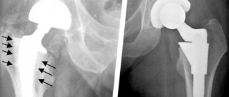
- X-ray;
- 2-energy X-ray absorptiometry (densitometry or DXA) with monitoring of bone density in seven Gruen zones;
- checking bone metabolic markers.
The first diagnosis is usually carried out in the coming months after surgery. The higher the risk of developing aseptic instability of the endoprosthesis (for example, the patient had osteoporosis before treatment), the sooner the doctor will conduct a follow-up examination.
Treatment
The symptoms of aseptic instability of the hip endoprosthesis largely replicate ANFH, which makes it possible to use proven courses of therapy for necrosis of the femoral head for its treatment. The program includes:
- vitamin therapy;
- unloading of the limbs (the patient moves for some time with the help of crutches);
- drug treatment (selected individually, taking into account the age, general condition of the patient, clinical picture before and after implantation);
- therapeutic diet (including those with the addition of bioactive complexes);
- a set of special exercises to prevent instability of the hip joint endoprosthesis ;
- electrical stimulation of areas of the legs or pelvis.
To visualize progress in the complex treatment of aseptic instability of endoprostheses, patients are required to keep a daily diary of their well-being. As a rule, this is enough for successful rehabilitation, normalization of the processes of resorption (destruction of bone tissue cells) and bone formation, and normal implant healing. In some cases, a new surgical procedure (revision endoprosthetics) is performed.
Acute infection or suppuration of a surgical wound
Acute infection, as a rule, develops in the superficial soft tissues of the surgical wound without penetration into the deeper layers and without involving the installed endoprosthesis in the infectious process. Its development becomes possible when the patient’s immunity is weakened and infection prevention measures are not followed, sterility is violated and microbial contamination of the wound surface. Staphylococcus aureus (Staphylococcus aureus) is usually cultured from wound discharge. After determining the sensitivity of microbes to antibiotics, an intravenous course of antibiotic therapy is prescribed. The duration of treatment takes from several days to a month.
If antibiotic therapy is ineffective, surgical cleansing of the wound is performed, necrotic tissue is removed, while the endoprosthesis remains in place. At the same time, a new antibiotic or their combination is selected. If the treatment tactics are chosen correctly, the infection is completely eliminated and the endoprosthesis is preserved. If treatment is unsuccessful, an acute infection can become chronic.